Original Article / Neurology and Clinical Neuroscience
Needle electromyography, muscle MRI, and muscle pathology: Correlations in idiopathic inflammatory myopathies
Reika Aoki, M.D.1, Norito Kokubun, M.D., Ph D.1, Tomoko Komagamine, M.D., Ph D.1, Jun Shimizu, M.D., Ph D.2,3, Ichizo Nishino, M.D., Ph D.4, Kazuhiro Kurasawa, M.D., PhD.5 and Koichi Hirata M.D., Ph D.1
1 Department of Neurology, Dokkyo Medical University, Japan
2 Department of Neurology, the University of Tokyo Hospital, Japan
3 Department of Physical Therapy, School of Health Sciences, Tokyo University of Technology, Japan
4 Department of Neuromuscular Research, National Center of Neurology and Psychiatry, Japan
5 Department of Rheumatology, Dokkyo Medical University, Japan
Running title: EMG, MRI and pathology in myositis
Title, 97 characters; manuscript, 4143 words; abstract, 250 words; figures, 1; tables, 4; references, 27.
Corresponding author: N. Kokubun
Department of Neurology, Dokkyo Medical University
880 Kitakobayashi, Mibu, Shimotsuga, Tochigi, 321-0293, Japan Tel.: +81-282-86-1111; Fax: +81-282-86-5884
e-mail: [email protected]
e-mail addresses of each author
R. Aoki, [email protected]
T. Komagamine, [email protected] I. Nishino, [email protected]
J. Shimizu, [email protected]
K. Kurasawa, [email protected] K. Hirata, [email protected]
Author contributions
Study concept and design: NK; analysis and interpretation: RA and NK; drafting the manuscript: RA, TK, and NK; and critical revision of the manuscript for important intellectual content: JS, IN, KK, and KH.
All of the authors have read and approved the final version of the manuscript.
Ethical standards
This study was approved by the ethics committee of Dokkyo Medical University Hospital (No. R-19-5J).
Financial disclosure statement
This work was supported by JSPS KAKENHI Grant Number JP19K07956 (J. Shimizu).
ABSTRACT
Background: On needle electromyography (EMG), abnormal spontaneous activity is considered a feature of active myositis. However, the correlation between needle EMG and muscle pathology is not always clear. Moreover, the changes in EMG findings after corticosteroid therapy are not fully
understood.
Aim: To investigate the correlations among muscle pathology, needle EMG and muscle MRI findings in patients with idiopathic inflammatory myopathies (IIMs).
Methods: The clinical features, laboratory results, needle EMG, muscle MRI findings and pathological features of 50 consecutive patients with IIMs who underwent muscle biopsies were reviewed. We also describe the changes in these findings between before and after corticosteroid treatments.
Results: The most common IIM was dermatomyositis (40%). On needle EMG, fibrillation/positive sharp waves (Fib/PSWs) were observed in 72% of the patients. MRI abnormalities were observed in 86% of the patients.In the pathology study, 88% of patients had inflammatory markers. The incidence of Fib/PSWs did not differ between pretreatment patients (79%) and steroid-treated patients (62%). In the pathology study, perimysium/perivascular cell infiltration also did not differ between before and after treatment.
However, endomysium cell infiltration was significantly less frequent in steroid-treated patients (0%) than in pretreatment patients (29%) (P< 0.05).
Conclusions: Our study confirmed that Fib/PSWs on needle EMG and muscle MRI were sensitive guides for diagnosing IIMs, and Fib/PSWs might be correlated with muscle fiber injuries or segmental necrosis rather than inflammatory cell infiltration in the affected muscles. Before treatment and within at least one month after starting corticosteroid treatment, Fib/PSWs could be detected by needle EMG.
Keywords: myositis, idiopathic inflammatory myopathy, needle electromyography, MRI, muscle pathology
1. Introduction
Since the establishment of diagnostic criteria for dermatomyositis (DM)/polymyositis by Bohan and Peter in the 1970s, needle electromyography (EMG) has been believed to be an important supportive
examination for diagnosing myositis 1,2. The concept of “myositis” has changed to idiopathic inflammatory myopathies (IIMs) during these two decades 3,4. DM, polymyositis, necrotizing
immune-mediated myopathy, sporadic inclusion body myositis, and nonspecific myositis are recognized as major subgroups of IIMs based on muscle pathological findings. DM without evidence of skeletal muscle involvement are defined as amyopathic DM 5. In addition, the clinical entity of anti-aminoacyl transfer RNA synthetase (ARS) antibody syndrome is associated with IIMs and has also been recently proposed as a specific clinical subgroup 5. After the induction of a pathology-based classification, the incidence of polymyositis was markedly reduced 5,6. In clinical practice, the diagnosis of IIMs is made by the combination of clinical features, laboratory findings, needle EMG, muscle MRI, and muscle
pathologic findings. While the pathological analysis is becoming the standard consensus for diagnosing and classifying IIMs 4, needle EMG is still positioned as a supplemental test and has been used to select the appropriate muscle for biopsy.
On needle EMG, abnormal spontaneous activities such as fibrillation potential and positive sharp waves (Fib/PSWs) are considered features of active myositis 7,8. However, only a few studies have
evaluated the correlation between EMG findings and muscle pathology in the current classification of IIMs, and most of the studies included a small number of patients 9-12. Although the correlation between EMG findings and muscle pathology may provide information relating to the pathophysiology in the affected muscle, whether abnormal spontaneous activities on needle EMG originate from muscle membrane irritability, muscle fiber splitting due to segmental necrosis, or inflammatory cell infiltration has not yet been fully explored.
The aim of this study was to evaluate whether abnormal spontaneous activities on needle EMG correspond to pathological findings in the biopsied muscles, muscle MRI findings, or serum CK levels in each subgroup of IIM. We also investigated the changes in abnormal spontaneous activities in patients before and after corticosteroid treatment.
2. Patients and Methods
2-1. Patients
The clinical, neurophysiological and histopathological records of 50 consecutive adult patients with IIMs who underwent needle EMG and muscle biopsy between January 2013 and December 2018 at Dokkyo Medical University Hospital were reviewed. The clinical features were evaluated at the time of needle EMG; to analyze the serum creatine kinase (CK) level, the maximum value during the course of the illness (max CK level) and the most recent data before the needle EMG (recent CK level) were collected.
The malignancy associated with myositis was defined when the disease was diagnosed within 3 years of the onset of myositis or related symptoms. We also compared the results between patients who did not receive any kind of immune treatment and patients who received corticosteroids with or without other immune treatments at the time of the examinations. This procedure was approved by the ethics committee of Dokkyo Medical University Hospital.
2-2. Need EMG evaluation
Needle EMG was performed in two or more muscles. To record abnormal spontaneous potentials, one or two separate penetrations of the needle electrode through the skin were made in each muscle, and 10 or more insertions were performed at each site. Fib/PSWs were classified into five grades according to Oh’s criteria: 0 = no Fib/PSWs could be recorded; 1+ = one abnormal spontaneous potential persisting for at least 400 ms in at least two sampling sites or one abnormal spontaneous potential that persisted long
enough to be manually captured on the screen without any difficulty; 2+ = two or more continuously active spontaneous potentials, filling 1/3 of the screen and noted in three or more sampling sites; 3+ = continuously active abnormal spontaneous potentials filling half of the screen and easily found in most sampling sites; and 4+ = many continuously active abnormal spontaneous potentials nearly filling the entire screen in all sampling sites 13. In this study, we did not include the morphology of motor unit potentials to judge myogenic changes to avoid subjectivity. Neurogenic changes were not recorded in all patients.
2-3. MRI
Muscle MRI of the bilateral thighs or unilateral upper arm was performed in 49 patients, and the
sequences included T1-weighted imaging (T1WI), T2-weighted imaging (T2WI), and short tau inversion recovery (STIR) sequences. The bright abnormal intramuscular T2 signals on the STIR image was divided into four grades: 0 = none; 1 = subtle; 2 = easily detectable; and 3 = intense 14. We defined the presence of MRI abnormalities in patients as grade 1 or higher. In addition, intramuscular edema and perimuscular fascial edema were evaluated.
2-4. Muscle biopsy and histopathologic evaluation
For all patients, the muscle biopsy was mainly performed in the vastus lateralis or biceps brachii muscles, which were on the contralateral side of the needle EMG site. If the patients did not have any muscular symptoms or signs, the vastus lateralis muscle was selected. The histopathological analyses of the muscle specimens were performed at the Department of Neurology, the University of Tokyo Hospital (JS) and Dokkyo Medical University, or Department of Neuromuscular Research, National Center of Neurology and Psychiatry (IN). The mononuclear inflammatory cell infiltration into the perimysium/perivascular or endomysium and the existence of necrotizing/regenerative/degenerative fibers were quantitatively
assessed by hematoxylin and eosin (H&E) staining. In addition, the expression of MHC class I molecules in the nonnecrotic muscle fiber membrane was assessed in 42 patients. In this study, we divided the individual features as presence or absence.
2-5. Classification of myositis subgroups
Based on the diagnostic criteria of the 119th European Neuromuscular Centre (ENMC) international workshop and those of Needham and Mastaglia, the patients were classified into the following six subgroups through the combination of clinical manifestations and the muscle pathology features: DM, polymyositis, immune-mediated necrotizing myopathy, inclusion body myositis and nonspecific myositis
15,16. Furthermore, the patients with myositis symptoms and serum anti-ARS antibodies were considered to have anti-ARS antibody syndrome 17. The anti-ARS antibody assay was conducted for 34 patients and measured antibodies against Jo-1, PL-7, PL-12, EJ, and KS. In 46 patients, an additional 10 antibodies including five anti-synthetase antibodies (anti-Jo1, -PL7, -PL12, -EJ and -OJ antibodies), other
myositis-specific antibodies (anti-Mi-2 and -SRP antibodies) and myositis-associated antibodies (anti-PM/Scl 75, -PM/Scl 100, -Ku and -Ro-52 antibodies) were examined using Euroline Myositis Profile 3 (Euroimmun, Lübeck, Germany). Anti-TIF1-γ were measured in 13 patients (MESACUP anti-ARS test, MBL, Nagoya, Japan). Serum anti-3-hydroxy-3-methylglutarylcoenzyme A
reductase (HMGCR) antibodies were measured in one patient by the method reported elsewhere18. In addition, the patients who met the DM criteria set by the 119th ENMC international workshop15, except for the pathological findings, were diagnosed with clinically amyopathic dermatomyositis (CADM). In the statistical analysis, amyopathic dermatomyositis and CADM were combined into one CADM subgroup.
2-6. Statistical analysis
Differences in the frequencies between groups were compared by the Fisher exact test (two-tailed).
Differences in medians were examined by the Mann-Whitney U test. The Jonckheere–Terpstra trend test was used to analyze CK levels and Fib/PSW grades. The analyses were performed with BellCurve software for Excel (Social Survey Research Information Co., Ltd. Tokyo, Japan), which is add-in
software to Excel for statistical evaluations, and IBM SPSS, version 25 (IBM SPSS, Armonk, NY, USA).
A P<0.05 was considered statistically significant.
3. Results
3-1. Clinical features of the patients
The clinical information of the patients is shown in Table 1. DM was the most common IIM (40%), followed by anti-ARS antibody syndrome (24%). Four patients (8%) had CADM. Two patients had polymyositis, five had immune-mediated necrotizing myopathy, one had inclusion body myositis, and six had nonspecific myositis. Interstitial pneumonia was observed in 54% of the patients, and the incidence was high, especially in patients with anti-ARS antibody syndrome (100%). Eventually, malignancies associated with myositis were found in 10 patients (20%), and five (10%) died because of exacerbated interstitial pneumonia or cancers. Of those with anti-ARS antibody syndrome, six of 12 patients (50%) had anti-Jo-1 antibodies, three (25%) had anti-PL-12, and two (17%) had anti-EJ antibodies. The remaining patient had anti-KS antibodies. Anti-MDA5 antibodies were found in nine patients with DM, one patient with CADM and one patient with nonspecific myositis patient. Six patients with DM had anti-TIF1-γ antibodies, and one patient with immune-mediated necrotizing myopathy had anti-HMGCR antibodies.
3-2. Needle EMG, muscle MRI and pathology study results
The numbers of muscles in which needle EMG was performed was as follows: in the trapezius (n=8), deltoid (n=5), biceps brachii (n=37), iliopsoas (n=14), rectus femoris (n=2), vastus lateralis
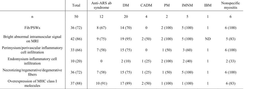
(n=48), lumbar paraspinal (n=1) and other muscles (n=7). The results of the needle EMG, muscle MRI and pathology study are shown in Table 2. On needle EMG, Fib/PSWs were observed in 36 of 50 patients (72%). MRI abnormalities anywhere in the muscles were found in 42 patients (86%). In a pathological study of muscle specimens from 50 patients, perimysium/perivascular and endomysium inflammatory cell infiltration was observed in 33 (66%) and ten (20%) patients, respectively. In total, 38 of 42 (90%) specimens that underwent immunohistochemical analysis were found to overexpress MHC class I molecules. Eventually, 44 patients (88%) showed any type of pathological abnormality that supported a diagnosis of myositis. Of the 44 patients with pathology-proven IIMs, 34 patients (77%) showed Fib/PSWs, 40 patients had MRI examinations including muscle biopsy, and 30 patients (75%) had MRI abnormalities in the biopsied muscles. Of the six patients with negative pathology results, one patient showed Fib/PSWs on the contralateral side of the biopsied muscle, and one showed MRI
abnormalities in the biopsied muscle.
In the individual myositis subgroups (Table 2), none of the four CADM patients showed Fib/PSWs on needle EMG, whereas one showed endomysium inflammatory cell infiltration and two showed bright abnormal intramuscular T2 signals on MRI.
3-3. Correlation between serum CK levels and needle EMG findings
The maximum CK levels and Fib/PSW results are shown in Figure 1. Although a high median serum CK level was significantly correlated with high-grade fibrillation or positive sharp waves (P<0.01), the highest CK level was recorded in patients with grade 1 Fib/PSWs, which was the lowest grade.
3-4. Correlation between EMG findings and MRI or pathology study results
The existence of Fib/PSWs was significantly correlated with bright intramuscular T2 signals (P<0.05) but was not correlated with intermuscular or perimuscular fascial edema on MRI. Eight patients with bright intramuscular T2 signals on MRI did not show Fib/PSWs, whereas eight patients without Fib/PSWs showed bright intramuscular T2 signals. The existence of Fib/PSWs was associated with
perimysium/perivascular inflammatory cellular infiltration and the existence of
necrotizing/regenerative/degenerative fibers (P<0.01). However, no correlations were observed between Fib/PSWs and endomysium inflammatory cell infiltration or the overexpression of MHC class I
molecules.
3-5. EMG findings before and after corticosteroid therapy
In our study, 29 of 50 patients did not receive any immune treatments at the time of needle EMG (pretreatment patients), whereas 21 received corticosteroid treatments with or without other immune therapies (steroid-treated patients). The modified Rankin scale scores did not differ among the groups.
The clinical background of the patients is shown in Table 3. The steroid-treated patients frequently had interstitial pneumonia and developed unfavorable prognoses.
The positivity rate of Fib/PSWs in the pretreatment patients was 79% and that of steroid-treated patients was 62% (Table 4), which was not a significant difference. Of the DM patients, ten were treated with steroids, and ten were pretreatment patients; the incidence of Fib/PSWs did not differ between these two groups of patients. Three patients showed Fib/PSWs more than 4 weeks after receiving corticosteroid therapy. The dose of prednisolone for these patients ranged from 20 to 60mg/day, and the duration of treatment ranged from 29 days to five years. Two of the patients also received other
immune-suppressive treatments, such as oral administration of cyclosporine, tacrolimus or methotrexate or intravenous cyclophosphamide pulse therapy.
3-6. Pathological findings before and after corticosteroid therapy
At the time of muscle biopsy, 35 patients did not receive any immune treatments, whereas 15 received corticosteroid treatments. Perimysium/perivascular inflammatory cell infiltration was found in 63% of the pretreatment patients and 73% of the steroid-treated patients (N.S). Endomysium inflammatory cell infiltration was observed in 10 of 35 (29%) pretreatment patients but zero of 15 (0%) steroid-treated patients (P< 0.05) (Table 4). Of those with DM, nine of 11 pretreatment patients (81%) and six of nine
(67%) of steroid-treated patients had perimysium/perivascular inflammatory cell infiltration. In contrast, endomysium inflammatory cell infiltration was observed in two (18%) pretreatment patients and zero (0%) steroid-treated patients, but the difference was not significant.
3-7. MRI features before and after corticosteroid therapy
In total, 40 patients underwent muscle MRI before steroid therapy, whereas 9 underwent steroid therapy.
Of the 40 pretreatment patients, 34 (85%) showed bright intramuscular T2 signals in any of the muscles.
Of the nine steroid-treated patients, eight (89%) showed bright intramuscular T2 signals. The existence of MRI abnormalities was not significantly different between these patients.
4. Discussion
In our study, 48% of the patients had DM or CADM, and expectedly, only 4% had polymyositis. In total, 88% of the patients had evidence of myositis in the pathological study. A total of 77% of the
pathology-proven IIM patients showed Fib/PSWs on needle EMG. All subgroups, except for CADM, showed Fib/PSWs. A total of 64% patients showed abnormal results on all examinations, including needle EMG, MRI and pathological studies, whereas four (8%) of the patients showed normal results in all examinations (two with CADM and two with anti-ARS-antibody syndrome). All four CADM patients did not show Fib/PSWs on needle EMG, suggesting that needle EMG can be useful in predicting negative muscle pathology.
Our EMG results were similar to those of classical studies. Bohan et al. identified Fib/PSWs in 74% of 153 polymyositis/DM patients 19. Another study showed that 45% of 98 patients with
polymyositis/DM showed Fib/PSWs and polyphasic-short duration motor unit potentials 20. In the current myositis classification 15, 70% of our DM patients and 67% of those with anti-ARS antibody syndrome showed Fib/PSWs. In our study, the main IIM subgroup was DM. However, our results were similar to those of previous studies that used current diagnostic criteria and included mainly patients with inclusion
body myositis and polymyositis 9,11. Therefore, the incidence of positive needle EMG findings is not very different among individual IIM subgroups.
While the combination of serum CK level, needle EMG, MRI and muscle pathology are routinely used to diagnose IIM, evidence on which examination should be prioritized has not been reported. For various myopathies, including IIMs, the correlation between needle EMG and muscle pathology findings was recently reported, and the absence of Fib/PSWs had a highly negative predictive value for inflammation, muscle fiber splitting, or vacuolar changes in the muscle fibers 9. Our observation of absent needle EMG findings in CADM confirmed the previous report. One study that investigated the correlation between MRI and EMG findings in IIMs was available. The apparent diffusion coefficient on MRI was associated with needle EMG findings in seven patients with IIMs 12. To the best of our
knowledge, the correlations among clinical findings, serum CK levels, MRI and EMG findings in IIMs have rarely been described, and our study might provide useful information for diagnosing IIMs.
Fib/PSWs may diminish or disappear within a few weeks after successful steroid treatment 21. Fib/PSWs in myositis are believed to originate from muscle fiber splitting due to segmental necrosis.
However, Fib/PSWs may be caused by other possibilities, such as irritation of the muscle fiber membrane by inflammatory cell invasion. No comprehensive studies that evaluate needle EMG changes in myositis after treatment exist. In the present study, patients with life-threatening conditions, such as interstitial pneumonia, received corticosteroids before needle EMG or muscle biopsy. The serum CK levels,
modified Rankin scale scores and incidence of Fib/PSWs did not differ between patients before and after treatment. In contrast, in the pathological evaluation, endomysium inflammatory cell infiltration was significantly less frequent in post-steroid treatment patients, which might indicate that the Fib/PSWs in IIMs do not correlate with active inflammation but are the result of muscle fiber injuries orsegmental necrosis. Therefore, based on our results, we considered that Fib/PSWs can be recorded for at least 4 weeks after the administration of corticosteroids. Conversely, the highest serum CK level was recorded in
patients with minimal grade Fib/PSWs (Figure 1). We also considered that overwhelming muscle fiber damage could reduce the generation of abnormal spontaneous potentials. We should not hesitate to provide corticosteroid therapy before needle EMG for patients with severe conditions; however, we should perform EMG before excessive muscle fiber damage occurs.
Amyopathic DM has been recognized for at least the past 25 years 22. Amyopathic DM is defined by a typical and pathologically confirmed skin rash for DM without muscle weakness, an elevated serum CK level, EMG abnormalities and pathological evidence of myositis 15. In our four CADM patients, no patients showed Fib/PSWs. However, two patients showed MRI abnormalities, and one had endomysium inflammatory cell infiltration and overexpression of MHC class I molecules in the pathology study. A previous study showed that 13% of CADM patients develop muscle weakness after six months 23. Our study confirmed that amyopathic DM and typical DM share a continuous spectrum and that amyopathic DM might be a subtype of DM with far fewer muscle lesions. Our study also suggests that needle EMG is not useful for differentiating true amyopathic DM from clinically amyopathic DM.
Previous reports showed that the incidence of abnormal MRI findings in the muscles was 80 to 90% in patients with IIMs 24,25. Our study confirmed these results and showed an incidence of 86% for abnormalities anywhere in the muscles. However, MRI abnormalities were found in the biopsied muscles of 75% of our patients with pathologically-proven IIMs. In contrast, one of five pathologically-negative patients showed MRI abnormalities in the biopsied muscles. Therefore, MRI may underestimate or overestimate the muscle lesions. MRI is a noninvasive examination that has a broad scanning area
compared with needle EMG and muscle biopsy and has the advantage of visualizing deeper muscle layers
26. MRI has marked clinical superiority as a screening examination. However, a long T2 signal on STIR images can theoretically be detected in muscles with inflammation and edema, as well as in denervated muscles or even in normal muscles after exercise 27. Therefore, we consider that needle EMG can increase the certainty of an MRI diagnosis.
The limitations of our study were as follows: 1) our study had a retrospective design; 2) the myositis subgroups included a small number of patients; 3) the paraspinal muscles, which have been previously reported as the most frequently affected muscles in myositis 8, were not included in the needle EMG of most patients in this study; 4) when assessing pre- and post-treatment patients, patients with more severe conditions were included as steroid-treated patients than as pretreatment patients; and 5) the dose and duration of treatment with corticosteroids and immune-suppressants could have
influenced the severity of myositis in post-treatment patients. In addition, afundamental inherent limitation exists since needle EMG cannot be performed in biopsied muscle. The incompatibility between pathology studies and needle EMG findings being performed on contralateral sides cannot be avoided. A larger prospective study is required.
In conclusion, our study confirms that Fib/PSWs on needle EMG and muscle MRI are sensitive guides for diagnosing IIMs and that Fib/PSWs might be correlated with muscle fiber injuries or
segmental necrosis rather than inflammatory cell infiltration in affected muscles. Before treatment and for at least one month after starting corticosteroid treatment, Fib/PSWs could be detected by needle EMG.
Acknowledgments
This work was supported by JSPS KAKENHI (grant number JP19K07956).
Conflict of interest
The authors declare no conflict of interest for this article.
References
1. Bohan A, Peter JB. Polymyositis and dermatomyositis (second of two parts). N Engl J Med 1975;292:403-407.
2. Bohan A, Peter JB. Polymyositis and dermatomyositis (first of two parts). N Engl J Med 1975;292:344-347.
3. Dalakas MC. Inflammatory Muscle Diseases. N Engl J Med 2015;373(4):393-394.
4. Lundberg IE, Tjarnlund A, Bottai M, et al. 2017 European League Against Rheumatism/American College of Rheumatology classification criteria for adult and juvenile idiopathic inflammatory myopathies and their major subgroups. Ann Rheum Dis 2017;76:1955-1964.
5. De Bleecker JL, De Paepe B, Aronica E, et al. 205th ENMC International Workshop: Pathology diagnosis of idiopathic inflammatory myopathies part II 28-30 March 2014, Naarden, The Netherlands. Neuromuscul Disord 2015;25:268-272.
6. van der Meulen MF, Bronner IM, Hoogendijk JE, et al. Polymyositis: an overdiagnosed entity.
Neurology 2003;61:316-321.
7. Mechler F. Changing electromyographic findings during the chronic course of polymyositis. J Neurol Sci 1974;23:237-242.
8. Streib EW, Wilbourn AJ, Mitsumoto H. Spontaneous electrical muscle fiber activity in polymyositis and dermatomyositis. Muscle Nerve 1979;2:14-18.
9. Sener U, Martinez-Thompson J, Laughlin RS, Dimberg EL, Rubin DI. Needle electromyography and histopathologic correlation in myopathies. Muscle Nerve 2019;59:315-320.
10. Dardiotis E, Papathanasiou E, Vonta I, Hadjigeorgiou G, Zamba-Papanicolaou E, Kyriakides T. A correlative study of quantitative EMG and biopsy findings in 31 patients with myopathies. Acta Myol 2011;30:37-41.
11. Blijham PJ, Hengstman GJ, Hama-Amin AD, van Engelen BG, Zwarts MJ. Needle electromyographic findings in 98 patients with myositis. Eur Neurol 2006;55:183-188.
12. Meyer HJ, Emmer A, Kornhuber M, Surov A. Associations between apparent diffusion coefficient
and electromyography parameters in myositis-A preliminary study. Brain Behav 2018;8:e00958.
13. Oh SJ. Principles of electromyography. Case studies. Baltimore, Lippincott Williams & Wilkins, 1998; pp97-98.
14. Elessawy SS, Abdelsalam EM, Abdel Razek E, Tharwat S. Whole-body MRI for full assessment and characterization of diffuse inflammatory myopathy. Acta Radiol Open
2016;5:2058460116668216.
15. Hoogendijk JE, Amato AA, Lecky BR, et al. 119th ENMC international workshop: trial design in adult idiopathic inflammatory myopathies, with the exception of inclusion body myositis, 10-12 October 2003, Naarden, The Netherlands. Neuromuscul Disord 2004;14:337-345.
16. Needham M, Mastaglia FL. Immunotherapies for Immune-Mediated Myopathies: A Current Perspective. Neurotherapeutics 2016;13:132-146.
17. Satoh M, Tanaka S, Ceribelli A, Calise SJ, Chan EK. A Comprehensive Overview on
Myositis-Specific Antibodies: New and Old Biomarkers in Idiopathic Inflammatory Myopathy.
Clin Rev Allergy Immunol 2017;52:1-19.
18. Kadoya M, Hida A, Hashimoto Maeda M, et al. Cancer association as a risk factor for
anti-HMGCR antibody-positive myopathy. Neurol Neuroimmunol Neuroinflamm 2016;3:e290.
19. Bohan A, Peter JB, Bowman RL, Pearson CM. Computer-assisted analysis of 153 patients with polymyositis and dermatomyositis. Medicine (Baltimore) 1977;56:255-286.
20. DeVere R, Bradley WG. Polymyositis: its presentation, morbidity and mortality. Brain 1975;98:637-666.
21. Kimura J. Electrodiadnosis in disease of nerve and muscle. Principle and practice. 4th ed.
NewYork, Oxford University Press, 2013; pp863-889.
22. Callander J, Robson Y, Ingram J, Piguet V. Treatment of clinically amyopathic dermatomyositis in adults: a systematic review. Br J Dermatol 2018;179:1248-1255.
23. Gerami P, Schope JM, McDonald L, Walling HW, Sontheimer RD. A systematic review of
adult-onset clinically amyopathic dermatomyositis (dermatomyositis sine myositis): a missing link
within the spectrum of the idiopathic inflammatory myopathies. J Am Acad Dermatol 2006;54:597-613.
24. Maurer B, Walker UA. Role of MRI in diagnosis and management of idiopathic inflammatory myopathies. Curr Rheumatol Rep 2015;17:67.
25. Fraser DD, Frank JA, Dalakas M, Miller FW, Hicks JE, Plotz P. Magnetic resonance imaging in the idiopathic inflammatory myopathies. J Rheumatol 1991;18:1693-1700.
26. Pipitone N. Value of MRI in diagnostics and evaluation of myositis. Curr Opin Rheumatol 2016;28:625-630.
27. Fleckenstein JL, Canby RC, Parkey RW, Peshock RM. Acute effects of exercise on MR imaging of skeletal muscle in normal volunteers. AJR Am J Roentgenol 1988;151:231-237.
Figure legends
Figure 1. The maximum CK levels during the course of the disease and abnormal spontaneous activities observed on needle EMG.
The highest CK value was recorded in patients with minimal fibrillation potentials and/or positive sharp waves. Fib/PSWs; fibrillation potential and positive sharp waves. Max CK level indicates the maximum CK value observed during the course of the illness.
Table 1. Clinical characteristics of the patients
ab; antibody, DM; dermatomyositis, CADM; clinically amyopathic dermatomyositis, PM; polymyositis, IMNM; immune-mediated necrotizing myopathy, IBM; inclusion body myositis, M; male, F; female, mRS; modified Rankin scale, MRC; Medical Research Council, CK; creatine phosphokinase. Data are given as a number (percentage) unless otherwise indicated.
Total Anti-ARS ab syndrome DM CADM PM IMNM IBM Nonspecific myositis
n 50 (100) 12 (24) 20 (40) 4 (8) 2 (4) 5 (10) 1 (2) 6 (12)
Sex (M/F) 22/28 3/9 12/8 2/2 0/2 0/5 1/0 4/2
Mean onset age (range) 60 (31 to 85) 61 (44 to 78) 63 (37 to 77) 65 (60 to 76) 52 (36 to 68) 67 (31 to 85) 60 54 (43 to 68)
Complicated malignancy 10 (20) 1 (8) 8 (40) 0 0 0 0 1 (17)
Death 5 (10) 0 (0) 3 (15) 2 (50) 0 0 0 0
Interstitial pneumonia 27 (54) 12 (100) 9 (45) 2 (50) 0 1 (20) 0 3 (50)
mRS grade 3 or higher 13 (26) 1 (8) 6 (30) 1 (25) 0 4 (80) 0 1 (17)
Typical rash 31 (62) 6 (50) 20 (100) 4 (100) 0 0 0 1 (17)
Median MRC sum score 52 60 56 53 48 53 45 56
(range) (0 to 60) (56 to 60) (44 to 60) (46 to 60) (48 to 48) (40 to 56) (52 to 60)
Median max CK 640 463 1021 78 3622 4776 380 803
(IQR) (322 to 2599) (187 to 1239) (383 to 2896) (55 to 104) (2131 to 5112) (2175 to 6708) (507 to 1265)
Median max CRP 1.00 2.21 1.00 2.85 0.51 0.15 0.24 2.73
(IQR) (0.23 to 3.29) (0.56 to 4.37) (0.21 to 1.84) (0.99 to 7.44) (0.28 to 0.73) (0.13 to 0.21) (0.78 to 4.33) Myositis-specific
autoantibodies 30 (60) 12 (100) 15 (75) 1 (25) 0 1 (20) 0 1 (17)
Table 2. Summary of the needle EMG, MRI and histopathological features
ab; antibody, DM; dermatomyositis, CADM; clinically amyopathic dermatomyositis, PM; polymyositis, IMNM; immune-mediated necrotizing myopathy, IBM; inclusion body myositis, Fib/PSWs; fibrillation potentials and/or positive sharp waves, ND; not done, EMG; electromyography. Data are given as a number (percentage).
Total Anti-ARS ab
syndrome DM CADM PM IMNM IBM Nonspecific
myositis
n 50 12 20 4 2 5 1 6
Fib/PSWs 36 (72) 8 (67) 14 (70) 0 2 (100) 5 (100) 1 6 (100)
Bright abnormal intramuscular signal
on MRI 42 (86) 9 (75) 19 (95) 2 (50) 2 (100) 5 (100) ND 5 (83)
Perimysium/perivascular inflammatory
cell infiltration 33 (66) 7 (58) 15 (75) 0 1 (50) 3 (60) 1 6 (100)
Endomysium inflammatory cell
infiltration 10 (20) 0 2 (10) 1 (25) 2 (100) 2 (40) 1 2 (33)
Necrotizing/regenerative/degenerative
fibers 36 (72) 7 (58) 15 (75) 1 (25) 1 (50) 5 (100) 1 6 (100)
Overexpression of MHC class I
molecules 37 (88) 10 (91) 17 (89) 2 (50) 1 (100) 1 (100) 1 6 (83)
Table 3. Clinical background of pretreatment and corticosteroid-treated patients that underwent needle EMG and muscle biopsy
EMG; electromyography, mRS; modified Rankin scale, CK; creatine phosphokinase. Max CK level indicates the maximum serum CK value during the course of illness, and the recent CK level indicates the most recent data before the needle EMG. Data are given as a number (percentage) unless otherwise indicated.
For needle EMG Pretreatment patients Steroid-treated patients P
n 29 21
mRS grade 3 or higher 9 (31) 5 (24) n.s.
Death 1 (3) 4 (19) n.s.
Interstitial pneumonia 10 (34) 17 (81) P < 0.05
Max. CK level (IQR) 855 (47 to 8950) 581 (300 to 1394) n.s.
CK level at examination (IQR) 358 (30 to 7228) 204 (59 to 534) n.s.
For muscle biopsy Pretreatment patients Steroid-treated patients
n 35 15
mRS grade 3 or higher 10 (29) 3 (20) n.s.
Death 1 (3) 4 (27) P < 0.05
Interstitial pneumonia 14 (40) 13 (87) P < 0.05
Max. CK level (IQR) 1003 (367 to 3218) 487 (255 to 513) n.s.
Recent CK level (IQR) 356 (128 to 2204) 128 (57 to 513) n.s.
Table 4. EMG and pathology results before and after corticosteroid treatments
EMG; electromyography, Fib/PSWs; fibrillation potentials and/or positive sharp waves, DM; dermatomyositis. Data are given as a number (percentage).
Days after corticosteroid administration
Needle EMG Pretreatment patients Steroid-treated patients P 2 to 7 days 8 to 14 days 15 to 28 days > 28 days
All patients 29 21 6 7 5 3
Fib/PSWs 23 (79) 13 (62) n.s. 6 (100) 3 (43) 1 (20) 3 (100)
DM patients 10 10 1 3 3 3
Fib/PSWs 9 (90) 5 (50) n.s. 1 (100) 1 (33) 0 (0) 3 (100)
Pathology
All patients 35 15 5 2 6 2
Perimysium/perivascular
inflammatory cell infiltration 22 (63) 11 (73) n.s. 4 (80) 1 (50) 5 (83) 1 (50)
Endomysium inflammatory cell
infiltration 10 (29) 0 (0) P < 0.05 0 (0) 0 (0) 0 (0) 0 (0)
DM patients 11 9 5 0 2 2
Perimysium/perivascular
inflammatory cell infiltration 9 (82) 6 (67) n.s. 4 (80) 1 (50) 1 (50)
Endomysium inflammatory cell
infiltration 2 (18) 0 (0) n.s. 0 (0) 0 (0) 0 (0)