Fukushima Medical University
福島県立医科大学 学術機関リポジトリ
This document is downloaded at: 2021-11-08T00:08:20Z
Title Multiple and incidentally found cancers detected by 18F-FDG positron emission tomography/CT at one examination
Author(s)
Takekawa, Shoichi; Kodaira, Yasunaga; Baba, Maiko; Igarashi, Yasuhiro; Konno, Akiyoshi; Teranishi, Yasushi; Nakazawa, Toshihiro; Sakuma, Hideo; Watanabe, Kazuo; Miyazaki, Makoto; Shishido, Fumio
Citation Fukushima Journal of Medical Science. 53(2): 109-118
Issue Date 2007-12
URL http://ir.fmu.ac.jp/dspace/handle/123456789/215
Rights © 2007 The Fukushima Society of Medical Science
DOI
Text Version publisher
Fukushima ]. Med. Sci., Vol. 53, No.2, 2007
[Case Report]
MULTIPLE AND INCIDENTALLY FOUND CANCERS DETECTED BY
lBF-FDG
POSITRON EMISSION TOMOGRAPHY/CTAT ONE EXAMINATION
SHOICHI TAKEKAWN), YASUNAGA KODAIRN)7), MAIKO BABN), YASUHIRO IGARASHP), AKIYOSHI KONN02), YASUSHI TERANISHP),
TOSHIHIRO NAKAZAWN), HIDEO SAKUMN), KAZUO WATANABE6), MAKOTO MIY AZAKIB) and FUMIO SHISHIDOB)
Departments of llRadiology, 2)Oto-rhino-laryngology, 3)Surgery, 4) Gastroenterology, 5)Pathology and 6)Neurosurgery, Southern Tohoku General HoSPital, Koriyama, 963-8563, japan, 7)Department of Radiology, Banbuntanehotokukai Hospital, Fujita Health University, Nagoya, 8)Department of Radio·
logy, Fukushima Medical University, Fukushima, 960-1295, japan (Received January 22, 2007, accepted June 25, 2007)
Abstract: During the initial 8 months period of lBF-FDG PET/CT examination in our institution eleven cases of double cancers were detected. Eight cases were simultaneous second cancers and 3 cases are consecutive cancers. All cases are clinical ones and were referred from both outside hospitals and our own hospital.
lBF-FDG PET/CT examination were utilized either to determine the extent of tumor or to stage the cancer or to detect recurrent tumors during the follow-up period.
During the 8-months period 964 cases were studied. Therefore, the detection rates of simultaneous and consecutive cancers are 0.83% and 0.31% respectively.
All together the detection rate of double cancer was 1.14%.
To gain the general conception of double cancers the authors reviewed the autopsy registry of Japanese Society of Pathology during the four years from 2000 through 2003, and tabulated the combination of primary and second cancers.
Frequently found combination of cancers were cancers of the thyroid, lung, stomach, liver, biliary tract, colon, rectum, and prostate.
lBF-FDG PET/CT examination seems to be very useful in the management of cancer patients in terms of whole patient care.
Key words: Positron Emission Tomograpy, PET, Double cancer, Multiple cancer, PET/CT
¥.rJlljE-,
IJ\ljZ~7.k, ~~Jftt{T,1i+Jil.l3t5L.,
~!l!fpgft, ~fffi ~, ~jRlii!c*, ~~rdl**,ill:if-*,
'8~ 1{, ~.P)'c~Corresponding author: Shoichi Takekawa E-mail: [email protected] 109
110
s.
T AKEKA W A et al.INTRODUCTION
Double cancers or multiple cancers are well known to the pathologists and clinical oncologists. They are usually found incidentally or during the follow-up of cancer patients in clinical practice. Pathologists find multiple cancers at the time of autopsies.
Small cancers, however, are not easily found by conventional high technology modalities such as CT, MRI, US and Gallium scintigraphy.
Recently 18F-FDG Positron Emission Tomography (hereafter, PET) became available clinically, and it is being utilized even for mass screening of cancer
1-6)because of its capability to detect smaller cancers in its earlier stage than before. It is said that even a tumor as small as 5 mm in diameter could be found in some cases.
PET is utilized clinically to assertain the extent of tumor or staging in cancer management.
Normal and pathologic FDG uptake is discussed by Kostakoglu et at
7 )and Nakamoto and his associates8).
The combined machine of PET and CT (hereafter, PET/CT) enhanced the accuracy in diagnosis because of its exactly fused images of both PET and CT, enabling easy localization of lesions
9•10).We found eight simultaneous double cancers and three consecutive cancers during the initial evaluation of the first cancer or follow-up study of the initial cancer or evaluation of malignancy of the first tumor at one examination of 18F - FDG PET /CT during the initial eight months after the installation of the PET /CT in our institution.
Herein we report double cancer or second cancer found by whole body screening using 18F-FDG PET/CT, and also review the results of annual autopsy registry of double cancer gathered by Japanese Society of Pathology18).
MATERIALS AND METHOD
In
2004five units of PET scanners were installed in our institution. Two of five scanners are PET /CT scanners (Discovery LS, GE Medical Systems, Milwaukee, Wis.). Other three scanners are Advance NXi. Clinical studies were carried out by PET/CT scanners. Namely, PET/CT scanners are used to assess the metabolic activity of malignant tumor or extent of a tumor.
The patients were prepared by fasting for more than six hours.
PET imaging was performed
60minutes after the administration of
210to
230MBq of FDG. The patients were requested to lie quietly on a reclining couch for
about
60minutes without moving any muscles as much as possible after the adminis-
tration of FDG. Blood glucose levels were checked before the administration of
FDG.
DOUBLE CANCER DETECTED BY PET/CT 111
The initial technical parameters for CT of the PET ICT are as follows: The CT of PET ICT is a 4-channel multi detector row CT. The beam pitch is 1.5 : 1 (equiva- lent to the slice pitch of 6 : 1 in a single helical CT). The gantry rotation time of 0.5 seconds, table speed of 30 mm per gantry rotation, 120 k V p, and 300 mA. The technique was then changed from July, 2004 as follows: Auto mA control system was introduced and kVp was elevated to 140 kVp and the tube current ranged from 50 to 169 mA depending on the thickness or density of the part of the body. The gantry rotation time and the table speed are same as they were before.
PET images and fused PET -CT images were evaluated regarding malignant nature and extent of the tumor. SUV of tumors were measured on the PET image whenever possible. SUV stands for the "standardized uptake value." We mea- sured SUV by encircling the entire FDG high uptake area (so-called hot spot) in the MIP images and the value was expressed in the maximum values, as Jinnouchp
1)recommended. Both 3 dimensional (3D) black and white images and color images were evaluated by certified radiologists.
RESULTS
During the period of April through November, 2004 the second cancers were found in 11 patients. Those second malignant tumors in 8 cases were found during the assessment of the first tumors. Cases were referred from both outside hospitals and our own hospital for the assessment of extent of tumor or confirmation of malignancy. Two tumors were suspected to have the possibility of malignancy, so FDG PET examination was carried out. They turned out to be submandibular tumor and parotid tumor, but the second incidentally found tumors were cancer of the colon and rectum. Two concurrent tumors were found during the search of suspected malignancy (Table 1).
As to the detection time of a second cancer, simultaneous detection was in 8 cases, and consecutive detection was in 3 cases. During the period of initial 8 months in PET ICT studies 964 cases were examined. Therefore, the detection rate of simultaneous double cancer was 0.83%, and the detection rate of consecutive cancer was 0.31%. The overall detection rate of double cancer was 1.14%.
Eight cancers were found by PET alone, but the remaining three patients were diagnosed as cancer by CT of PET-CT, contrast-enhanced (CE) CT or cystoscopy and biopsy.
False negative cases on FDG PET are a case of thyroid cancer, a case of HCC and a case of the urinary bladder. The combination of PET study and CT were very useful in detection of cancers, which were not revealed by that time.
A few cases of incidentally found cancers will be illustrated.
Case 1. A 61-year-old man presented with the chief complaint of backache.
X -ray of the thoracic spine showed suspected metastasis in the thoracic vertebrae.
The patient was then referred to the PET Center for evaluation of malignancy.
112 S. T AKEKA W A et al.
Table 1. Survey of Double Cancer or Incidental Second Cancer
Case First Cancer or Second Cancer/ + Modality of positive Note: Detectability
Age Sex Dis. + or - on by PET, (Pathology
#
I.
2.
3.
4.
5.
6.
7.
8.
9.
10.
II.
PET (SUV) or - on PET (SUV) finding for 2nd Ca of 1st. Cancer) M a l i g n a n t
61 M Ca thyroid/ + lymphoma, lower PET Concurrent detec- (SUV 16.5) thoracic spine tion of two cancers
region/
+
(20.4)FMH testis & LN SUV (10.6 at Rt.
75 M Ca tongue/ + (6.6) meta/ + (11.1, testis; PET Buttock, 6.2 at Lt.
16.9, Rt. ing. LN) fern. LN) F/U of P.O. Ca Ca (TCC) of blad-
70 F tongue (w / d der /- (missed detec- CT & Cystoscopy, FN on PET reo ca
SCC)/-, No tion) TUR-BT TCC bladder
recurrence
Ca of Rt. Hypo· Follicular Ca thy· CT of PET-CT, FN on PET reo ca 79 F pharynx/ +(12.9) roid/-(not detect- Low density mass ed) on CT thyroid
Ca of Lt. Hypo· HCC/-(not detect· CT of PET-CT and FN on PET reo
73 M pharynx/ +(30.1) ed) CECT HCC, + Lt. cer-
vical LN (16.0) 73 M Ca (SCC)/ + (8.6) Lt. lung Ca sigmoid (Ca in adenoma)/(lI.6) PET
Ca lung (SCC)/
+
Ca (SCC) of esopha· Endoscopy68 M (15.9) gus/+(5.5) PET Advanced ca esoph·
agus F /U of PO Ca lar· Ca of colon (ascend·
79 M ynx/-No recur· ing)/+(lI.9) PET renee
F/U of PO Ca Rt Ca of colon (ascend- 79 F breast/-No ing)/
+
(23.5) PETrecurrence
Lt. Subman· Ca sigmoid (Pleomorphic
79 M dibular tumor/
+
colon/+
(8.5) PET Adenoma submand.(5.1) gland.)
Rt. Parotid ( C h r o n i c
63 M tumor/ +(4.2) Ca rectum/+(25.7) PET Sialoadenitis of Rt.
Parotis) FN: False negative. Ca: Carcinoma. re: regarding. SUV is expressed in maxi·
mum value. F/U: Follow-up
18F-FDG PET revealed high uptake of FDG in the region of thoracic vertebrae (SUV 20.4) and also in the thyroid gland (SUV 16.5) (Fig. lA and IB).
Aspiration biopsy of the thyroid gland showed adenocarcinoma of the thyroid gland. Pathology of the lesion after thyroidectomy was papillary adenocarcinoma.
The lesion in and around the lower thoracic vertebrae turned out to be malignant lymphoma.
Case 2. This 73-year-old man with carcinoma of the left lung was examined
with FDG PET
ICTto assess the extent of the lesion and staging. FDG PET
revealed high uptake of FDG in the sigmoid colon in addition to the high uptake in
the bilateral hilar lymph nodes. The SUV values of the left hilar node and the
DOUBLE CANCER DETECTED BY PET/CT
Fig.lA Fig.lB
Fig.1. Carcinoma of the thyroid gland and malignant lymphoma.
lA: Frontal image. Note the very high uptake of FDG in the right lobe of the thyroid (SUV 16.5) and in the region of the lower thoracic spine (SUV 20.4).
IB: Lateral image. The high uptake of FDG in the region of the lower thoracic spine projects further anteriorly and posteriorly from the thoracic vertebra.
Fig 2A Fig.2B
Fig. 2. Carcinoma of the left lung with bilateral hilar node metastases and carcinoma of the sigmoid colon. This 73-year-old man presented with the complaint of cough and wheezing. Chest X -ray showed atelectatic lesion in the left upper lobe.
2A: Frontal image. FDG PET showed high uptake in bilateral hilar nodes and sigmoid region.
2B: Oblique image. The high uptake of FDG in the sigmoid region seen in the frontal image stays in the same sigmoid location.
113
114 S. T AKEKA W A et C11.
Fig.3A Fig. 38
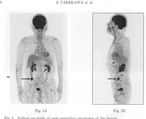
Fig. 3. Follow-up study of post-operative carcinoma of the larynx.
No FDG uptake was noted in the laryngeal region at this time.
3A: Frontal image. Cancer of the ascending colon (SUV 11.9) was incidentally found by FDG-PET. Pathological diagnosis was moderately differentiated adenocarcinoma of the ascending colon.
38: Lateral image. The high uptake of FDG in the ascending colon region is again identified in the same location.
sig moid lesion were 8.7 and 1l.6 respectively (Fig. 2A and 2B). Bronchofiberscopy revealed narrowing of the orifice of B1 +
2 of the left upper lobe and brushingof the bronchus revea
ledClass V, k
eratinizing squamouscell carcinoma. Endoscopy of the sigmoid colon and bi opsy of a polypoid tumor reveakd carcin oma in adenoma.
Case 3. This is a 79-year
-oldman with recurrent carci noma of the lar
ynxafter initi al operation in another instituti on. The rec urrent tum or was resected in our h ospit al and he was examined
withFDG PET as a fo
llow-up study. FDG PET revealed
norecurr ent
laryngeal cancer but revea led cancer of the asce nding colon.
The SUV of t
hisnew second cancer was 1l.9.
Namely a second cancer was detected b y FDG PE T (Fig. 3A and 3B). Pathology of t he colon lesio n was moderately different ia ted adenocarcin oma.
DISCUSSION
As stated above multiple cancers or double cancers were occasionally fo
undat
the time of aut opsy by pathologists. Clini cal oncolog ists also encounter second or
multiple cance rs. Up to now, however, no
concrete single examination moda li ty
existed to show mul tip le cancers at one exa minati
on.F DG PET is now poss ible to
DOUBLE CANCER DETECTED BY PET ICT 115
survey the almost whole body except the very distal portion of the legs and is capable to detect cancers as if it is a one-stop shopping. This fact is often quoted and the slogan of "PET first in cancer management" is being spread. Clinical usefulness is reported in severalliteratures
12- 16).In our initial experience of FDG PET ICT in 8-month-period we found 11 second cancers. This fact is important to manage cancer patients because we have to treat the whole body of patients.
In order to find the frequent combination of cancers in one individual patient, the registry of multiple cancers at Japanese Society of Pathology was consulted. We reviewed the registry of four fiscal years; between years 2000 and 2003. Because of numerous sites of cancers we categolized to one group in some disease groups.
They are as follows. The site of neurologic tissue included cerebrum, cerebellum, brain stem, spinal cord, meninges, and so forth. Pharynx: epipharynx, mesophar- ynx, hypopharynx, nasopharynx, pharyngoesophageal junction. Lung: lung, tra- chea, bronchus. Liver and biliary tract: liver, bile duct, gallbladder, papilla vater.
Colon and rectum: colon, rectum, cecum, appendix. Bone marrow and lymphatic system: bone marrow, spleen, lymph node.
The number of cases of each cancer group in four years was summed up and tabulated (Table 2). This table illustrates numerous combination of cancers, but there are several heavily tied combination of cancerous diseases. Some of second cancers, such as carcinoma of the prostate, seemed prone to occur in aged patients.
The frequently found second cancers in patients with cancers of lung, liver group, bone marrow group and pancreas are cancers of the thyroid, stomach, colon group and prostate. The concurrent double cancers of the first cancer of the colon and rectum noted at the registry of Pathology are cancers of the thyroid gland, stomach and prostate.
FDG PET reflects the metabolic activity of a disease. It picks actively growing cancer in its earlier stage and therefore is very useful to determine the malignant nature and the extent of a disease. However, FDG PET shows inflammatory lesions also as does the Gallium scintigraphy.
Another interesting point of differential diagnosis is that atheroma can uptake FDGl7). The exact localization of FDG will help in the differential diagnosis.
The definitive diagnosis depends on the tissue diagnosis by pathologists, but the FDG PET leads the selection of further examination or treatment methods. It detects hidden second cancer also. This is beneficial also to the patients to consider and select treatment methods by patients themselves.
CONCLUSION
Eleven second cancers were incidentally found by FDG PET ICT during the
period of eight months in our institution. FDG PET is very useful in the manage-
ment of cancer patients in terms of whole patient care.
Table 2. Double Cancer, From the Registry of Double Cancer Diagnosed by Autopsy. Japanese Society of Pathology. Period: Year 2000 through 2003
~
Sitel Thyroid Paranasal Neurologic Tongue Oral Liver and Small Colon and marrow, Bone Kidney, Ureter, Uterine UterinePharynx Larynx Esophagus Lung Bt:east Stomach Renal Pancreas Bladder. Prostate Ovary
Primary sinuses tissue cavity Biliary tract intestine Rectum Lymph. pelvis Urethra cervix body
Site 2 System
Thyroid 4 8 4 7 4 2 9 136 37 84 137 7 49 109 39 47 12 20 11 8 18
Paranasal 0 0 0 0 0 0 0 2 0 0 1 0 0 0 0 0 0 1 0 0 0
sinuses
Neurologic 1 0 0 0 0 0 0 2 1 2 2 1 1 4 0 3 0 1 0 0 0
tissue
Tongue 0 0 0 3 0 0 7 5 0 1 2 0 0 2 0 0 0 1 0 0 0
Pharynx 0 0 0 0 0 0 16 11 1 5 2 0 2 2 1 2 1 0 0 0 0
Oral cavity 0 0 0 3 2 0 9 15 0 5 3 0 2 4 0 0 0 0 0 0 0
Larynx 2 1 0 1 1 0 11 22 0 9 8 0 5 7 2 1 2 4 0 0 0
Esophagus 3 1 3 0 21 2 3 46 3 35 22 0 9 11 3 27 7 6 1 1 1
Lung 13 0 3 1 3 3 9 31 12 72 70 3 29 74 13 31 18 22 3 1 4
Breast 3 1 2 1 2 2 0 5 41 31 38 0 14 30 2 17 0 0 4 7 13
Stomach 8 4 8 4 12 1 4 107 281 15 225 2 69 98 18 65 15 51 7 3 6
Liver and
5 0 1 1 4 2 1 21 80 12 72 5 31 63 13 36 8 14 2 1 3
Biliary tract
Small intestine I 0 0 0 0 0 0 5 8 3 2 9 3 8 1 2 0 0 0 0 0
Colon and 13 2 4 0 7 1 5 50 177 11 174 159 10 119 20 69 21 35 8 8 9
Rectum Bone marrow,
2 0 3 1 3 1 1 7 42 9 44 27 1 7 4 15 2 15 5 0 3
Lymph. System Kidney, Renal
4 1 3 1 1 2 0 4 53 2 34 40 1 25 33 20 20 16 0 1 0
pelvis
Pancreas 1 2 1 0 2 0 1 6 12 4 23 20 0 7 14 1 8 9 1 0 2
Ureter, Bladder. 1 0 2 1 1 0 0 4 57 2 28 24 4 15 17 37 10 13 1 0 0
Urethra
Prostate 9 5 4 6 11 9 5 41 331 0 193 177 4 76 165 35 83 49 0 0 0
Uterine cervix 3 0 1 0 0 1 0 2 13 6 9 26 0 10 14 5 6 7 0 1 1
Uterine body 0 0 1 0 0 0 0 0 2 3 2 5 0 1 3 0 1 2 0 0 6
Ovary 0 0 0 0 0 0 0 0 4 4 4 5 0 8 2 1 4 1 0 1 2
Otbers 5 0 5 1 3 1 0 3 38 9 32 23 2 9 36 9 8 8 12 4 1 0
...
...
0'>
fIl
~
~
~
DOUBLE CANCER DETECTED BY PET ICT 117
ACKNOWLEDGMENT
The authors gratefully acknowledge Mr. Yasutaka Suzuki and Ms. N oriko Yaginuma for their invaluable assistance
inthe preparation of the tables and manuscript.
REFERENCES
1. Yasuda S, Ide M, Fujii H, Nakahara T, Mochizuki Y, Takahashi W, Shohtsu A.
Application of positron emission tomography to cancer screening. Brit J Cancer, 83 : 1607-1611, 2000.
2. Ide M. Cancer screening using FDG-PET (in Japanese). Jpn J Diag Imaging (Gazo- Shindan), 23: 1190-1197, 2003.
3. Uno K. Cancer screening. In: Ito M, et aI, eds. Frontiers in high technology practice of cancer diagnosis and therapy. CAFI, Tokyo, 69-75, 2004.
4. Jinnouchi S. Data of PET screening. In: Jinnouchi S, ed. FDG-PET Manual. P' ed.
Inner Vision, Tokyo, 62, 2004.
5. Moriyama N. Evaluation of cancer screening and expectation in future: Summary of results at Research Center for Prevention and Screening of Cancer at National Cancer Center one year after its opening (in Japanese). New Medicine in Japan (Shin-iryou), No. 374, 82-84, Feb., 2006.
6. Chen YK, Kao CH, Liao AC, Shen YY, Su CT. Colorectal cancer screening in asymptomatic adults: The role of FDG PET scan. Anticancer Res, 23: 4357-4362,2003.
7. Kostakoglu L, Hardoff R, Mirtcheva R, Goldsmith SJ. PET-CT fusion imaging in differentiating physiologic from pathologic FDG uptake. RadioGraphics, 24: 1411-1431, 2004.
8. Nakamoto Y, Tatsumi M, Hammoud D, Cohade C, Osman MM, Wahl RL. Normal FDG distribution pattern in the head and neck: PET ICT evaluation. Radiology, 234: 879- 885, 2005.
9. Gutzeit A, Antoch G, KUhl H, Egelhof T, Fischer M, Hauth E, Goehde S, Bokisch A, Debatin J, Freudenberg L. Unknown primary tumors: Detection with dual-modality PET/CT-Initial experience. Radiology, 234: 227-234,2005.
10. Hany TF, Steinert HC, Goerres GW, Buck A, Schulthess GK. PET Diagnostic accu·
racy: Improvement with in-line PET-CT system: Initial results. Radiology, 225: 575- 581, 2002.
11. Jinnouchi S. Method of PET screening. In: Jinnouchi S, ed. FDG-PET Manual. P' ed. Inner Vision, Tokyo, 17, 2004.
12. Pannu HK, Bistow RE, Cohade C, Fishman EK, Wahl RL. PET-CT in recurrent ovarian cancer: Initial observations. RadioGraphies, 24: 209-223, 2004.
13. Kazama T, Faria SC, Varavithya V, Phongkitkarun S, Ito H, Macapinlac HA. FDG PET in the evaluation of treatment for lymphoma: Clinical usefulness and pitfalls.
RadioGraphies, 25: 191-207, 2005.
14. Ito M, et aI., eds. Frontiers of Clinical PET. Published by Research Institute of Frontier Medical Technologies, Tokyo, August, 2004.
15. Ito M, et aI., eds.: Frontiers of Practice of Diagnosis and Therapy of Cancer by high technologies: From PET to Particle Radiotherapy. Published by CAFI, Tokyo, Decem·
ber, 2004.
16. Watanabe K, Matsuzawa T, Ishii K, Ito M, eds. Progress in Researches and Clinical Practice of PET; Cancer and Brain. Published by Southern Tohoku Research Institute
118 S. T AKEKA W A et at.
for Neuroscience, Koriyama, Japan, March, 2005.
17. Tatsumi M, Cohade C, Nakamoto Y, Wahl RL. Fluorodeoxyglucose uptake in the aortic wall at PET jCT: Possible finding for active atherosclerosis. Radiology, 229:
831-837, 2003.
18. Japanese Society of Pathology: Registry of Double Cancer. Year 2000 through 2003 In Autopsy Registry of Japanese Society of Pathology.