Fukushima Medical University
福島県立医科大学 学術機関リポジトリ
This document is downloaded at: 2021-11-07T23:57:06Z
Title Incidence of organizing pneumonia after whole-breast
radiotherapy for breast cancer, and risk factor analysis( 本文 )
Author(s) 佐藤, 久志
Citation
Issue Date 2018-09-28
URL http://ir.fmu.ac.jp/dspace/handle/123456789/724
Rights © The Author(s)
DOI
Text Version ETD
1
TITLE
Incidence of organizing pneumonia after whole-breast radiotherapy for breast cancer, and risk factor analysis
(乳癌に対する乳房温存術後の放射線治療による放射線肺臓炎の発生頻度とリス ク要因についての解析)
NAME
Hisashi Sato1, Junko Ebi1, Tomoaki Tamaki1, Ami Yukawa1, Masaharu Nakajima1, Tohru Otake2, Yoshiyuki Suzuki1
1Department of Radiation Oncology, Fukushima Medical University, School of Medicine, 1 Hikarigaoka, Fukushima-City, Fukushima 960-1295, Japan
2Department of Breast Surgery, Fukushima Medical University, School of Medicine, 1 Hikarigaoka, Fukushima-City, Fukushima 960-1295, Japan
2
ABSTRUCT
Radiation-induced organizing pneumonia (OP) reportedly occurs in ~2% of patients who receive whole-breast radiotherapy (WBRT). Though there are several reported risk factors, they remain unclear and controversial. We analyzed the incidence of and risk factors for OP after WBRT at our institution. We analyzed 665 breast cancer patients (with WBRT of 679 breasts) who underwent WBRT from October 2007 to September 2012 at our institution and were followed up for more than 1 year after completion of WBRT.
Factors included in the analysis were age, the side affected, central lung distance (CLD), radiation dose, concurrent endocrine therapy, and chemotherapy.
The median age was 56 years (range, 23-89 years). The sides affected were left, right and bilateral in 342, 309 and 14 patients, respectively. The median CLD was 1.1 cm (range, 0-3.0 cm). Concurrent endocrine therapy was performed in 236 patients, and chemotherapy was given in 215 patients; of these, 4 received concurrent chemotherapy.
OP developed in nine patients (1.4%). The median time taken to develop OP after the completion of WBRT was 4 months (range, 2-12 months). All nine patients were treated with steroids, and symptoms promptly improved, except in two patients who relapsed.
Statistical analysis revealed that only CLD (≥1.5 cm) was significantly associated with the development of OP (P = 0.004).
In conclusion, the incidence of OP after WBRT was 1.4%, and CLD was a significant risk factor. In these patients, OP was controlled with steroid administration.
ABBREVIATIONS
OP : Radiation-induced organizing pneumonia WBRT : whole-breast radiotherapy
3
CLD : central lung distance fr. : fraction
V20 : lung volume irradiated at 20Gy
FEC : 5-fluorouracil+epirubicin+cyclophosphamide TC : docetaxel+cyclophosphamide
CMF : cyclophosphamide+methotrexate+5-fluorouracil EC : epirubicin+cyclophosphamide
5-FU : 5-fluorouracil AI : aromatase inhibitor TAM : tamoxifen
4
INTRODUCTION
Historically, until the 1980s, based on the Halsted theory which stated that the prognosis of breast cancer patients was dependent on the maximal excision of the breast and lymphatic pathways, the main surgical approach had been the extensive excision of the breast including whole breast and lymphatic pathway and large pectoralis muscle and pectoralis muscle [1]. Radiation therapy of this age had been the irradiation of the chest wall, axillary, supraclavicular, parasternal lymph region using a Co60 source, and the side effects such as dermatitis and OP were strong.
Around 1990s, based on the Fisher theory that breast cancer is a systemic disease at the time of diagnosis and, therefore, the difference in surgical therapy does not affect prognosis, reduction surgery such as pectoral muscle preserving or breast conserving surgery has become the mainstream surgery is well accepted that the local therapy can lead to the eradication of the disease [2]. The Early Breast Cancer’ Collaborate Group reported that the local control and the overall survival at 15 years were significantly improved by the addition of WBRT after breast-conserving surgery [3]. In Japan, the combination of breast conserving surgery and WBRT is the current standard treatment and performed in more than 80% of early breast cancer cases [4].
WBRT has been performed as standard therapy for early-stage breast cancer after breast- conserving surgery. Recently, endocrine therapy and chemotherapy have been commonly used with WBRT, concurrently or sequentially. Although the most frequent adverse effect of WBRT is acute dermatitis, OP is a well-known adverse effect that is rare but significant.
Radiation-induced OP is reported to occur in ~1-3% of breast cancer patients who receive postoperative WBRT [5-13].
By definition [14], OP occurs within 12 months after WBRT. Onset of OP is usually
5
reported to be within 6 months [15]. Risk factors reported in previous studies include none [6,7], age [8,9], large irradiated lung volume [10], concurrent endocrine therapy [8], and smoking [9]. However, the significance of these risk factors remains unclear and controversial because of the rarity of OP.
In this report, we studied a total of 665 breast cancer patients (with WBRT of 679 breasts) who underwent WBRT during a period of 5 years at our institution to analyze the incidence of OP and risk factors for OP after WBRT.
MATERIALS AND METHODS/PROCEDURES Ethics statement
This retrospective study was approved by the institutional review board of Fukushima Medical University Hospital, Fukushima City, Japan (IRB approval number 1606).
Anonymity of the patients was preserved.
Patient characteristics
Between October 2007 and September 2012, 686 newly diagnosed breast cancer patients received WBRT at Fukushima Medical University Hospital after breast- conserving surgery as an initial treatment. Overall, 665 patients (97%) (with WBRT of 679 breasts) were followed up for more than 12 months. Those lost to follow-up within 12 months were excluded from this analysis. The sides affected were left, right and bilateral in 342, 309 and 14 patients, respectively. Table 1 shows the patient characteristics of this study. The median age of the 665 patients was 56 years (range, 23-89 years) and the median follow up period was 643days (range, 370-2230 days).
6
Treatments Radiotherapy
All patients were treated with WBRT after breast-conserving surgery at our institution or our neighboring hospital. WBRT was primarily performed with 6-MV X-rays using two tangential photon beams or four beams with the field-in-field technique to a total dose of 50 Gy/25 fractions/5 weeks. Nine patients with lobular carcinoma were treated with 54 Gy/27 fractions. Breasts with a positive surgical margin were irradiated with an additional 10 Gy/5 fractions using an optimal electron beam. WBRT for both sides was concurrently performed in 14 patients with bilateral cancer.
Endocrine therapy and chemotherapy
The details of the endocrine therapy and chemotherapy are shown in Table 1. In this study, administration of chemotherapy within the 3 weeks before and/or after WBRT was classified as ‘concurrent use of chemotherapy’. Endocrine therapy was administered to 507 patients (74.7%), and 241 patients received concurrent endocrine therapy.
Chemotherapy was administered to 219 patients (32.3%), of whom 212 received chemotherapy before WBRT and only 4 received concurrent chemotherapy. In 2 of those 4 cases, the chemotherapy was administered a short time before WBRT or with a limited overlap time because of the patients’ difficulties in travelling to our hospital. In the other 2 cases, chemotherapy was only limited to an oral agent in relatively high-risk patients who refused standard therapy. The Japanese guideline has not recommended concurrent administration of chemotherapy and WBRT since 2011, but these 4 patients were all treated before 2011.
7
Diagnosis of OP
In general, all patients were followed up and checked for OP at 1, 3, 9, 15 and 21 months after completion of WBRT. At 3 months after WBRT, OP was checked with chest X-ray and/or chest CT. Diagnosis of OP was performed on the basis of the clinical diagnostic criteria proposed by Crestani et al. [5]: (i) radiation therapy to the breast within 12 months, (ii) general and/or respiratory symptoms lasting for at least 2 weeks, (iii) lung infiltrates outside the radiation port, and (iv) no specific cause.
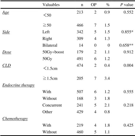
Evaluation of identifying risk factor
Factors included in the analysis were age <50 or ≥50 years, the side affected (right or left, lateral or bilateral), central lung distance (CLD; <1.5 or ≥1.5 cm), radiation dose (50 Gy or 50 Gy+boost), concurrent endocrine therapy (with or without), and chemotherapy (with or without). CLD was measured as the farthest distance between the posterior border of the irradiation field and the chest wall on approved linacgraphy.
Statistical analysis
Statistical analyses were performed using the chi-square test with SPSS 21 (SPSS, Chicago, IL). Differences were considered significant when P values were less than 0.05.
The cumulative occurrence rate of OP was calculated using the Kaplan-Meier method with SPSS 21.
RESULTS
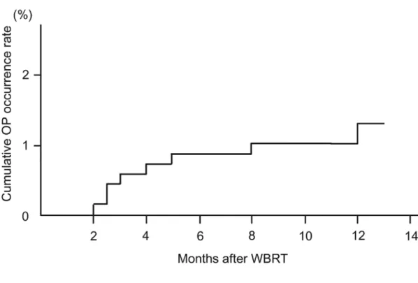
Figure 1 shows the cumulative OP occurrence rate. OP developed in 9 (1.4%) of 679 irradiated breasts (665 patients). Six of the nine patients (67%) developed OP in the first
8
6 months.
Table 1 shows the study patients’ clinical characteristics. The median CLD was 1.1 cm (range, 0-3.0 cm). Among 665 patients, 236 were treated with concurrent endocrine therapy. Chemotherapy was given to 215 patients; of these, 4 had concurrent chemotherapy.
Table 2 shows the results of univariate analysis of possible risk factors. Statistical analysis revealed that CLD was significantly associated with the development of OP (P
= 0.004), whereas other factors were not.
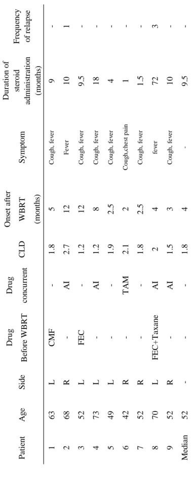
Table 3 shows the details of the nine patients who developed OP. The median time for developing OP after the completion of WBRT was 4 months (range, 2-12 months). All nine patients presented with mild fever and/or cough and were treated with steroids. Two OP patients relapsed and were treated with repeat steroid administration, and a patient who relapsed three times remained on low-dose steroid therapy 6 years after the initial onset of OP.
DISCUSSION
As previously reported, OP occurs in ~1-2% of breast cancer patients who receive WBRT. The risk factors for OP remain unclear and controversial, although several factors, such as age, endocrine therapy, and large irradiated lung volume, have been reported as risk factors in previous studies [6-9,11].
Katayama et al. reported that age (≥50 years) was a risk factor for OP in breast cancer patients treated with WBRT [8]. In addition, Murofushi et al. claimed that age was associated with OP in breast cancer patients treated with radiotherapy but not in patients treated with breast-conserving treatment [9]. Many reports [6,7,10,11], including this
9
study, did not show age to be a risk factor. However, caution should be given when treating elderly patients because older age is reportedly an important risk factor for OP in patients treated for other diseases, such as lung cancer, using modern radiotherapy techniques [16].
Murofushi et al. also reported that smoking was a risk factor for the development of OP [9]. However, in this study, none of the nine patients who developed OP had a history of smoking, and significance of smoking in the development of OP was not shown.
Concurrent use of endocrine therapy has been reported as a risk factor for OP [8], whereas other reports show no correlation [9,12]. In our current practice, we allow the concurrent use of endocrine therapy when needed because no significant correlation with the development of OP was shown in this study. Most reports have shown that chemotherapy has no significant correlation with the development of OP, and this study also found no correlation.
However, Caudia et al. reported that symptomatic OP patients had a bronchoalveolar lavage (BAL) with significantly increase in total cells compared to BAL in asymptomatic patients with predominance of T lymphocytes [17]. Naozumi at al. reported that estrogen deficiency may play a role on acceleration of organ-specific autoimmune in murine Sjögren’s Syndrome [18]. Wirsdorfer et al. reviewed the role of lymphocytes in radiotherapy-induced adverse late effects in the lung in detail [19].
Therefore, the use of drugs affecting host-immune status or immune cell activity may be related to the development or prevention of OP, and attention may be required regarding which type of chemotherapeutic agents are to be used when concurrent chemotherapy is considered.
In lung cancer patients treated with radiotherapy, the lung volume irradiated at 20 Gy
10
(V20) has been considered as a risk factor for radiation-induced pneumonia [20]. In breast cancer patients treated with WBRT, Kubo et al. reported that >1.8 cm of CLD was significantly correlated with the incidence of OP, but lung V20 was not [10]. In addition, we found that ≥1.5 cm of CLD was significantly correlated with OP in this study, although data for V20 were not available. Murofushi et al. analyzed more than 1000 breast cancer patients and reported that V20 was not a significant factor in predicting the development of OP in breast cancer patients treated with radiotherapy, including post-mastectomy radiotherapy [9]. Paradoxically, Katayama et al. reported that ≥3 cm of CLD was not a risk factor for OP after WBRT [8]. Although it remains unclear whether irradiated lung volume affects the development of OP in breast cancer patients treated with WBRT, larger irradiated lung volume is considered a strong risk factor for OP according to reports in lung and esophageal cancer patients treated with radiotherapy. Therefore, irradiated lung volume should be carefully evaluated when making a treatment plan, and patients with a large irradiated lung volume and/or CLD should be followed up carefully for possible incidence of OP. Furthermore, for patients who may have a large CLD and irradiated lung volume, such as those with funnel chest, treatment with Intensity Modurated Radiotherapy(IMRT) or special irradiation technique may be an option for avoiding the risk of OP.
As for the treatment for OP, we administered steroids in all nine OP patients, and they have taken various clinical courses. At follow up, the condition of two patients improved quickly after a short period (<2 months) of small-dose steroid administration. Conversely, six patients needed steroids for more than 6 months; of these, two patients had a relapse of OP, and one of them remained on small-dose steroid therapy 6 years after the initial onset of OP because of multiple relapses.
11
Ogo et al. reported that all 10 patients in their study improved without steroid administration within 3 months [7], and Murofushi et al. reported that only 2 of 16 OP patients required steroid treatment [9]. According to these previous reports, steroid therapy may not be for all patients. Otani et al. reported that steroid treatment increased the recurrence of radiation-induced OP after WBRT [13]. Furthermore, Oie et al. reported that four of five OP patients relapsed and that the durations of steroid administration were more than 6 months [11].
Unfortunately, these reports were not randomized studies because of the rarity of OP, and the mechanism for steroid therapy for radiation-induced OP has not been elucidated.
Although steroid administration is still the mainstay of treatment [21], symptom-oriented management as proposed by Otani et al. [14] may be a noteworthy consideration in the treatment of radiation-induced OP.
In conclusion, the incidence of OP after WBRT was 1.4%, which is similar to the findings of previous reports. OP developed within 2-12 months after WBRT, and most patients improved with steroid administration. The only risk factor for OP was CLD ≥ 1.5 cm. Our study suggests that patients treated with long CLD should be carefully observed after WBRT.
ACKNOWLEDGMENT
I wish to thank members of following facilities.
Fukushima Medical university Department of Breast Surgery
Fukushima Medical university Department of Department of Pulmonary Medicine Kita-fukusima Medical Center Center of disease of breast
Saiseikai Fukushima Hospital respiratory division/ surgery
12
Fukushima Madical University Department of Radiation Oncology
13
REFERENCES
1. Halsted WS. I. The results of operations for the cure of cancer of the breast performed at the Johns Hopkins Hospital from June, 1889, to January, 1894. Ann Surg. 1894; 20(5):
497-555
2. Fisher B, Redmond C et al. Ten-year results of a randomized clinical trial comparing radical mastectomy and total mastectomy with or without radiation. N Engl J Med.
1985; 312(11): 674-81.1.
3. Early Breast Cancer Trialists’ Collaborative Group(EBCTCG). Effect of radiotherapy after breast-conserving surgery on 10-year recurrence and 15-year breast cancer death:
meta-analysis of individual patient data for 10 801 women in 17 randomised trials.
Lancet 2011;378: 1707-16.
4. Youngjin Park, Tomoaki Kitaharaet al. Current status of therapy for breast cancer worldwide and in Japan. World J Clin Oncol. 2011 Feb 10; 2(2): 125–134.
5.Crestani B, Valeyre D, Roden S et al. Bronchiolitis obliterans organizing pneumonia syndrome primed by radiation therapy to the breast. The Groupe d’Etudes et de Recherche sur les Maladies Orphelines Pulmonaires (GERM‘O‘P). Am J Respir Crit Care Med 1998;158:1929-35.
6.Ogo E, Komaki R, Fujimoto K et al. A survey of radiationinduced bronchiolitis obliterans organizing pneumonia syndrome after breast-conserving therapy in Japan. Int J Radiat Oncol Biol Phys 2008;71:123-31.
7. Ogo E, Komaki R, Abe T et al. The clinical characteristics and non-steroidal treatment for radiation-induced bronchiolitis obliterans organizing pneumonia syndrome after breast-conserving therapy. Radiother Oncol 2010;97:95-100.
8. Katayama N, Sato S, Katsui K et al. Analysis of factors associated with radiation-
14
induced bronchiolitis obliterans organizing pneumonia syndrome after breast- conserving therapy. Int J Radiat Oncol Biol Phys 2009;73:1049-54.
9. Murofushi KN, Oguchi M, Gosho M et al. Radiation-induced bronchiolitis obliterans organizing pneumonia (BOOP) syndrome in breast cancer patients is associated with age. Radiat Oncol 2015;10:103.
10. Kubo A, Osaki K, Kawanaka T et al. Risk factors for radiation pneumonitis caused by whole breast irradiation following breast-conserving surgery. J Med Invest 2009;56:99-110.
11. Oie Y, Saito Y, Kato M et al. Relationship between radiation pneumonitis and organizing pneumonia after radiotherapy for breast cancer. Radiat Oncol 2013;8:56.
12. Takigawa N, Segawa Y, Saeki T et al. Bronchiolitis obliterans organizing pneumonia syndrome in breast-conserving therapy for early breast cancer: radiation-induced lung toxicity. Int J Radiat Oncol Biol Phys 2000;48:751-5.
13. Otani K, Nishiyama K, Ito Y et al. Steroid treatment increases the recurrence of radiation-induced organizing pneumonia after breast-conserving therapy. Cancer Med 2014;3:947-53.
14. Otani K, Seo Y, Ogawa K. radiation-induced organizing pneumonia: a characteristic disease that requires symptom-oriented management. Int J Mol Sci 2017;18:E281.
15. Epler GR, Kelly EM. Systematic review of postradiotherapy bronchiolitis obliterans organizing pneumonia in women with breast cancer. Oncologist 2014;19:1216-26.
16. Zhao J, Yorke ED, Li L et al. Simple factors associated with radiation-induced lung toxicity after stereotactic body radiation therapy of the thorax. Int J Radiat Oncol Biol Phys 2016;95: 1357–66.
15
17. Naozumi I, Kaoru S. et al. Estrogen deficiency accelerates autoimmune
exocrinopathy in murine Sjögren’s syndrome through Fas-mediated apoptosis. Am J Pathol. 1999 Jul; 155(1): 173-181.
18. Claudia L, Aneta S et al. The bronchoalveolar lavage pattern in radiation
pneumonitis secondary to radiotherapy for breast cancer. Maedica(Buchar). 2010 Dec;
5(4): 250-257.
19. Wirsdörfer F, Jendrossek V. The role of lymphocytes in radiotherapy-induced adverse late effects in the lung. Front Immunol 2016;7:591.
20. Palma DA, Senan S, Tsujino K et al. Predicting radiation pneumonitis after chemoradiation therapy for lung cancer: an international individual patient data meta- analysis. Int J Radiat Oncol Biol Phys 2013;85:444-50.
21. Kainthola A, Haritwal T, Tiwari M et al. Immunological aspect of radiation-induced pneumonitis, current treatment strategies, and future prospects. Front Immunol 2017;8:506.
16
FIGURES
Fig. 1 : Cumulative organizing pneumonia occurrence rate
The vertical axis shows the cumulative occurrence rate, the horizontal axis shows months after WBRT.
17
Table 1 : Patient characteristics Age
Male 0
Female 665
Side
Left 342
Right 309
Bilateral 14
Age Median 56y
(range 23-89)
<50 213
≧50 466
Dose
50Gy/25fr. 491
54Gy/27fr. 9
50Gy/25fr.+Boost 10Gy/5fr. 179
Central lung distance Median 1.1cm (range 0-3.0)
<1.5cm 474
≧1.5cm 205
Endocrine Therapy
Yes(concurrent) 241
Anti-estrogen 36
Aromatase inhibitor 165 LH-RH analog+Anti-estrogen 40
Yes(after WBRT) 257
Antiestrogen 90
Aromatase inhibitor 133 LH-RH analog+Anti-estrogen 30
Unknown 4
Yes(period unknown) 9
Aromatase inhibitor 7
18
Unknown 2
No 168
Unknown 4
Chemotherapy
Yes 291
before WBRT 208
Bwfore+concurrent 4
After WBRT 7
FEC+Taxiane 146
FEC 23
TC 28
CMF 10
Taxane 3
EC 1
EC+Taxane 2
5-Fu 6
No 460
19
Table 2 : Univariate analysis of variables
Valuables n OP % P value
Age <50 213 2 0.9 0.552
≧50 466 7 1.5
Side Left 342 5 1.5 0.855*
Right 309 4 1.3
Bilateral 14 0 0 0.658**
Dose 50Gy+boost 179 2 1.1 0.912
50Gy 491 6 1.2
CLD <1.5cm 474 2 0.4 0.004
≧1.5cm 205 7 3.4
Endocrine therapy
With 507 6 1.2 0.555
Without 168 3 1.8
Concurrent 241 5 2.1 0.218
Other 429 4 0.8
Chemotherapy
With 219 4 1.8 0.425
Without 460 5 1.1
*The comparisons is ‘left’ vs ‘right’.
**The comparisons is ‘unilateral’ vs ‘bilateral’.
20
Table 3 : Clinical characteristics of nine patients
DrugDrugOnset after Before WBRTconcurrentWBRT (months) 163LCMF-1.85Cough, fever9- 268R-AI2.712Fever101 352LFEC-1.212Cough, fever9.5- 473L-AI1.28Cough, fever18- 549L--1.92.5Cough, fever4- 642R-TAM2.12Cough,chest pain1- 752R--1.82.5Cough, fever1.5- 870LFEC+TaxaneAI24fever723 952R-AI1.53Cough, fever10- Median52---1.84-9.5- Frequency of relapsePatientAgeSideCLDSymptom
Duration of steroid administration (months)