はじめに
Epididymis-specific secretory proteins, epididymis protein 2 type C(EP2c)はアンドロゲン依存性蛋白で 精子に結合し,精子形成に関係している.ヒト EP2c 遺 伝子は chromosome 8(8p23)に位置し抗菌ペプチドで あるβ-defensin 遺伝子とクラスターを形成している.
Defensin は貪食細胞と肥満細胞に遊走作用を持ち,
CCL20(MIP3α)の受容体である CCR6 によって,未 分化樹状細胞の活性化は促される.それゆえ,EP2c は chemokine receptors を介して未熟な樹状細胞を活性化 させると仮定し実験を行った.
研究目的
粘膜免疫におけるキャリアとして EP2c を利用できる かどうか検討した.すなわち,抗菌作用を持つβ-defen- sin 様のペプチドである EP2c が未知の受容体を介して 免疫細胞を活性化させ,さらに EP2c と融合した抗原が MHC Class I & II を通じて粘膜免疫における抗原提示細 胞への標的とする可能性を検証する.
Materials and Methods 1.MHC Class II presentation
Wild-type BALB/c splenocytes were incubated with 0.01‑1 μg/ml murine or human EP2c peptide fused with VL315 (VL from MOPC315 plasmacytoma cells, Igλ chain) O/N. Splenocytes were washed, irradiated, and mixed with BALB/c CD4+ T cell clone 7A10B2, which recognizes processed epitope λ91‑101. IFN-γ secretion was measured after 24 h of incubation. Control spleno- cytes were incubated with VL315 fused with VH frag- ment (sFv315), with (mDF1β), or with murine macro-
phage inflammatory protein IIIα(mMIP3α).
2.MHC Class I presentation
Wild-type C57BL/6 of tibia were purified from bone marrow dendritic cells, and incubated with EP2c fusion protein and/or inhibitors. From TCR, transgenic pmel-1 mouse splenocytes were concurrently purified and co- culture with dendritic cells and splenocytes, and the IFN-γ was measured by ELISA.
3.Procedure of receptor binding assay
B6/129: mouse macrophage, RAW264.7: virus trans- formed BALB/c mouse macrophage, HEK293/mCCR6:
human adenovirus E1 transformed embryonal kidney, THP-1: human acute monocytic leukemia, CCRF-CEM:
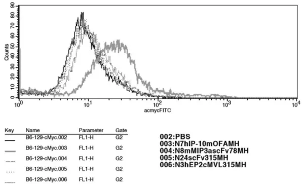
human T acute lymphoblastic leukemia, MOLT-4: hu- man T acute lymphoblastic leukemia, A20: mouse B cell lymphoma, and C57BL/6: mice bone marrow dendritic cell lines were purchased from ATCC (American Type Culture Collection). Harvest cells 0.5×10⁶/tube. Incu- bate 50 μl of 50 μg/ml of fusion protein or control pro- tein on ice. Incubate 10% goat serum/PBS/0.1% azide on ice. Add 1:300 Anti c-myc Ab on ice. Add 1:50 Anti mouse FITC-IgG Ab on ice. Analyze by FACS.
4. Chemokine fusion vaccination elicits antitumor re- sponses in BALB/c mice
Eight or 10 mice per group were electroporate immu- nized three times over a two-week interval with mEP- 2cOFApCMVE or PBS. Four days before the last im- munization, the mice were challenged subcutaneously with A20 (TIB-208; BALB/cAnN B cell lymphoma)
cells.
Tumor growth was subsequently assessed, and mice with tumors greater than 400 mm2 were euthanized.
結 果
1. EP2c は細胞表面にある受容体に結合し,腫瘍抗 原を MHC Class II へ提示する
Epididymis-specific secretory proteins, epididymis 住友 賢哉
連絡先:住友 賢哉
〒783‑8509 高知県南国市明見字中野 526‑1 JA 高知病院内科
(E-mail: [email protected])
protein 2(EP2)はアンドロゲン依存性蛋白ファミリー で精子と結合し精子の成熟に関係していると考えられて いる.ヒト EP2 遺伝子は染色体 8 番(8p23)に位置し 抗菌ペプチドであるβ-defensin 遺伝子と一群を形成して いる.EP2 遺伝子は副睾丸に強く発現し,spliced form である EP2c はβ-defensin 様の特徴と関連があると考え られている.しかし EP2c の免疫細胞での役割は不明で ある.最近我々は,β-defensin が chemokine receptor である CCR61)を介して免疫細胞の誘導を行い,未熟な 樹状細胞が TLR-42)を介して成熟することを見いだした.
さらに免疫原性のない自己の腫瘍抗原がβ-defensin や chemokine との融合蛋白により免疫原性が回復するこ とを示し1),マウス同系腫瘍モデルに対する予防的治療 的効果を示した1)3).chemokine の融合蛋白は,chemo- kine receptorを介して抗原を効率的に細胞内へ取り込み,
MHC Class I & II を通じて抗原提示を行い,CD4+ CD8+ T 細胞を反応させる1)4)5).これらの方法は chemoattrac- tant の種類に依存して免疫反応の調整を可能とする.た とえば成熟樹状細胞を chemoattract する恒常性 chemo- kine である SLC,SDF1 ではなく,未熟樹状細胞を標 的とするβ-defensin 2 や炎症性 chemokine の融合組換え 体は中等度の humoral responses を引き起こすが,強い 予防的治療的抗腫瘍効果を発揮する.対照的に,いわゆ る Th2 chemokine は細胞傷害性 T cell 反応を誘導する ことはできないが,抗体の産生には優れている1).その ため EP2c ペプチドは,粘膜の抗原提示細胞に抗原を提 示する能力があると仮定した.すなわちβ-defensin に類 似する EP2c は chemokine receptor を介して抗原提示 細胞に結合し MHC Class I & II を通じて抗原処理を効 率的に行うことができると考えた.加えて腫瘍抗原であ る VL315 や plasmacytoma MOPC315 由 来 variable light (VL)-chain fragment(Fig. 1)と融合した EP2c(ヒ ト,マウスともに)は,特異的に VL315 を MHC Class
II 経路へ運び CD4+ T 細胞を刺激することを示した(Fig.
2).それゆえ,これらの結果から EP2c は細胞表面の受 容体に結合することができ,抗原提示細胞に抗原を発現 し抗原提示を促進すると考えられた.
2. EP2c は腫瘍抗原の MHC Class I processing を促 進する(cross-presentation)
殺傷性 CD8+ T 細胞反応の誘導は,腫瘍や慢性炎症疾 患の治療の鍵である.CD8+ T 細胞反応は抗原を処理し MHC Class I に提示する.外因性の抗原は MHC Class I 処理経路にはほとんど到達しない,これを cross-pre- sentation と呼んでいる.我々の最近の結果にもかかわ らず,抗原が融合体としてβ-defensin やβ-defensin に類 似する EP2c を輸送するとしたならば,cross-presenta- tion 経路が効率的に利用されるだろう.Cross-presenta- tion を研究するため gp100 と融合した EP2c を作製した
(Fig. 3).gp100 は melanoma/melanocyte 分 化 自 己 抗 原である.我々は共同研究者である National Cancer In- stitute の Dr. Nicolaus Restiffo から,Vα1Vβ13 T cell re- ceptor を発現している transgenic mice pmel-1 を提供し ていただいた.この T cell receptor は mouse and hu- man gp100 epitope を MHC Class I: H-2Db特異的に拘束 する.我々の予備実験の結果から,EP2gp100 で処理し た抗原提示細胞は pmel-1 mice 由来の CD8+ T 細胞を刺
Fig. 1 Schema of constructs to study MHC Class II
presentation. Human or mouse EP2c was linked in frame with VL315, a plasmacytoma MOPC315 immu- noglobulin-delivered variable light (VL) -chain frag- ment, which contained CD4+ T cell epitope λ91‑101.
Control construct is single chain antibody (scFv), which is contained in frame fusions of both VH and VL fragments of Ig from MOPC315.
Fig. 2 Both human and murine EP2c can deliver anti-
gens to MHC Class II processing pathway. Murine splenocytes from BALB/c mice were incubated with 1‑0.01μ
g/ml murine or human EP2c fused with VL315 (mEP2 and hEP2, respectively) overnight.The splenocytes were then washed and irradiated and mixed with CD4+ T cells, which recognized that processed epitope λ91‑101. IFN-γ secretion was mea- sured after 48 h of incubation. Control splenocytes were incubated with VL315 fused with VH fragment
(sFv315), with mDF1β, or with mMIP3α.
抗原提示細胞の未知の受容体に特異的に結合し抗原を MHC Class I & II 処理経路に輸送することが示された.
EP2 は粘膜で産生される.それゆえ EP2 は粘膜の抗原 提示細胞を標的とし,ワクチンキャリアとして使用でき ることが示唆された.
を測定することにより抗原提示能を検討した.EP2c は chemokine receptor に結合すると考え,受容体は G 蛋 白質共役型かどうか検討した(Fig. 5).その結果 PTX や sucrose で抗原提示が阻害されることが示され,その 受容体はG蛋白質共役型であることが示唆された.また,
当初から EP2c はβ-defensin と同様,chemokine recep- tor である CCR6 に結合することにより抗原提示を行う ことが示唆されていた.そのため FACS を用いて結合 能を検討したが,結合は確認できなかった.そのほか CCR4 に結合する受容体である TARC も検討したが同 じく結合は確認されなかった.さらに未知の受容体があ ると考え,いくつかの細胞において結合の有無を検討し たが確認できなかった(Fig. 6〜8).
4. での EP2c の DNA ワクチンとしての働き マ ウ ス の モ デ ル を 用 い,EP2c 融 合 蛋 白 も し く は DNA を皮下もしくは筋肉注射で投与し全身性 T cell 反
Fig. 3 Schema of constructs to study MHC Class I
presentation. Human or mouse EP2c was linked in frame with gp100, a melanoma specific antigen that contained H-2Db-restricted mouse gp100 epitope. Con- trol protein consisted of unlinked gp100 alone.
Fig. 4 Human and murine EP2c/gp100 peptide can both deliver antigens to MHC Class I pro-
cessing pathway. Murine immature DCs from C57BL/6 mice were incubated with 1‑0.01 μg/ml murine or human EP2c fused with gp100 (mEP2gp100 and hEP2gp100, respectively) over- night, and then DCs were washed and irradiated and mixed with CD8+ T cells from pmel-1 mice, which recognized processed H-2Db-restricted epitope. IFN-γ secretion was measured af- ter 48 h of incubation. Control DCs were incubated with unlinked gp100 alone or DCs pulsed with irrelevant peptide (MOPC315 peptide).
Fig. 5 MHC Class I presentation by EP2c is mediated by G-coupled receptor endocytosis.
Fig. 6 Receptor-binding assay. Human T cells do not bind murine and human EP2c.
Fig. 7 Receptor-binding assay. CCRF-CEM (human peripheral leukemia acute lymphoblastic) cells bind
hTARC, but do not bind murine and human EP2c.Fig. 8 Receptor-binding assay. Mouse MIP3α and murine EP2c show little difference compared to single-
chain Fv315. Single chain is negative control.応の有無を調べた.同時に腫瘍を皮下投与し,予防的抗 腫瘍免疫能を検討した.
腫瘍径や生存曲線を検討した.その結果,治療群は有 意に生存曲線,腫瘍径の改善を認め,抗腫瘍効果を期待 できる結果となった(Fig. 9).
結 語
我々の究極の目標は,理想的なワクチンキャリアの作 製と粘膜免疫の誘導にある.EP2c の生物学や免疫学,
すなわち EP2c の抗菌ペプチド作用,粘膜免疫細胞の活 性化に注目することとなる.EP2c を介した MHC Class I & II 抗原提示のメカニズムの解明は,臨床応用に重要 な意味を持つであろう.腫瘍や性行為感染症(HPV,
HBV,HCV,HIV),呼吸器感染症,腸管感染症の治療 的予防的ワクチンの開発にも重要である.
引用文献
1)Biragyn A, et al. Mediators of innate immunity that
target immature, but not mature, dendritic cells in- duce antitumor immunity when genetically fused with nonimmunogenic tumor antigens. J Immunol 2001; 167: 6644‑53.
2)Biragyn A, et al. Toll-like receptor 4-dependent ac- tivation of dendritic cells by beta-defensin 2. Sci- ence 2002; 298: 1025‑9.
3)Biragyn A, et al. Genetic fusion of chemokines to a self tumor antigen induces protective, T-cell depen- dent antitumor immunity. Nat Biotechnol 1999; 17:
253‑8.
4)Schiavo R, et al. Chemokine receptor targeting effi- ciently directs antigens to MHC class I pathways and elicits antigen-specific CD8+ T-cell responses.
Blood 2006; 107: 4597‑605.
5)Biragyn A, et al. Chemokine receptor-mediated de- livery directs self-tumor antigen efficiently into the class II processing pathway in vitro and induces protective immunity in vivo. Blood 2004; 104: 1961‑9.
a
b