Polarizes HURP Near Chromosomes
Author Kenta Tsuchiya, Hisato Hayashi, Momoko Nishina, Masako Okumura, Yoshikatsu Sato, Masato T. Kanemaki, Gohta Goshima, Tomomi Kiyomitsu
journal or
publication title
Current Biology
volume 31
number 1
page range 115‑127.e3
year 2020‑11‑12
Publisher Elsevier Inc.
Rights (C) 2020 The Author(s).
Author's flag publisher
URL http://id.nii.ac.jp/1394/00001699/
doi: info:doi/10.1016/j.cub.2020.09.091
Creative Commons Attribution 4.0 International(https://creativecommons.org/licenses/by/4.0/)
Ran-GTP Is Non-essential to Activate NuMA for Mitotic Spindle-Pole Focusing but Dynamically Polarizes HURP Near Chromosomes
Graphical Abstract
Highlights
d
Using AID technology, we developed mitotic depletion assays for the Ran pathway
d
The Ran pathway is non-essential to activate NuMA for spindle-pole focusing
d
Ran-GTP is not required to target TPX2 but is required to localize HURP and HSET
d
The Ran pathway maintains HURP’s polarized spindle localization during metaphase
Authors
Kenta Tsuchiya, Hisato Hayashi, Momoko Nishina, ...,
Masato T. Kanemaki, Gohta Goshima, Tomomi Kiyomitsu
Correspondence
tomomi.kiyomitsu@oist.jp
In Brief
Tsuchiya et al. develop acute mitotic depletion assays using auxin-inducible degron technology to dissect Ran’s mitotic roles in human HCT116 cells. This study shows that Ran-GTP is non- essential to activate NuMA away from chromosomes but is required to activate HURP near chromosomes.
Tsuchiya et al., 2021, Current Biology31, 115–127
January 11, 2021ª2020 The Authors. Published by Elsevier Inc.
https://doi.org/10.1016/j.cub.2020.09.091
ll
Article
Ran-GTP Is Non-essential to Activate NuMA
for Mitotic Spindle-Pole Focusing but Dynamically Polarizes HURP Near Chromosomes
Kenta Tsuchiya,1,6Hisato Hayashi,1,6Momoko Nishina,1,6Masako Okumura,1,6Yoshikatsu Sato,1 Masato T. Kanemaki,2,3,4Gohta Goshima,1and Tomomi Kiyomitsu1,2,5,7,*
1Division of Biological Science, Graduate School of Science, Nagoya University, Chikusa-ku, Nagoya 464-8602, Japan
2Precursory Research for Embryonic Science and Technology (PRESTO) Program, Japan Science and Technology Agency, 4-1-8 Honcho Kawaguchi, Saitama 332-0012, Japan
3Department of Chromosome Science, National Institute of Genetics, Research Organization of Information and Systems (ROIS), Yata 1111, Mishima, Shizuoka 411-8540, Japan
4Department of Genetics, SOKENDAI (The Graduate University of Advanced Studies), Yata 1111, Mishima, Shizuoka 411-8540, Japan
5Okinawa Institute of Science and Technology Graduate University, 1919-1 Tancha, Onna-son, Kunigami-gun, Okinawa 904-0495, Japan
6These authors contributed equally
7Lead Contact
*Correspondence:tomomi.kiyomitsu@oist.jp https://doi.org/10.1016/j.cub.2020.09.091
SUMMARY
Spindle assembly is spatially regulated by a chromosome-derived Ran- GTP gradient. Previous work pro- posed that Ran-GTP activates spindle assembly factors (SAFs) around chromosomes by dissociating inhib- itory importins from SAFs. However, it is unclear whether the Ran-GTP gradient equivalently activates SAFs that localize at distinct spindle regions. In addition, Ran’s dual functions in interphase nucleocytoplasmic transport and mitotic spindle assembly have made it difficult to assess its mitotic roles in somatic cells.
Here, using auxin-inducible degron technology in human cells, we developed acute mitotic depletion assays to dissect Ran’s mitotic roles systematically and separately from its interphase function. In contrast to the prevailing model, we found that the Ran pathway is not essential for spindle assembly activities that occur at sites spatially separated from chromosomes, including activating NuMA for spindle-pole focusing or for targeting TPX2. On the other hand, Ran-GTP is required to localize HURP and HSET specifically at chromo- some-proximal regions to set proper spindle length during prometaphase. We demonstrated that Ran-GTP and importin-
bcoordinately promote HURP’s dynamic microtubule binding-dissociation cycle, which main- tains HURP near chromosomes during metaphase. Together, we propose that the Ran pathway acts on spin- dle assembly independently of its interphase functions in mitotic human cells but does not equivalently regu- late all Ran-regulated SAFs. Ran-dependent spindle assembly is likely coupled with additional parallel pathways that activate SAFs distantly located from the chromosomes.
INTRODUCTION
During cell division, a microtubule-based spindle structure is assembled around chromosomes to efficiently capture and segregate duplicated chromosomes into daughter cells.1,2Spin- dle assembly is dependent on a gradient of a guanosine triphos- phate (GTP)-bound form of Ran (Ran-GTP), which surrounds chromosomes in animal cells.3,4Ran-GTP is produced by regu- lator of chromosome condensation 1 (RCC1), a guanine nucleo- tide exchange factor (GEF) for Ran,5and is hydrolyzed to Ran- guanosine diphosphate (GDP) by RanGAP1, a GTPase-acti- vating protein for Ran.6As RCC1 and RanGAP1 mainly localize on chromosomes and in the cytoplasm, respectively, these opposing enzymes create a chromosome-derived Ran-GTP gradient after the nuclear envelope breaks down (Figure 1A). Af- ter mitotic exit, RCC1 still binds to chromatin although RanGAP1
localizes to the nuclear envelop and the cytoplasm. Thus, these enzymes generate different Ran-GTP concentrations in the nu- cleus and cytoplasm, which drives nucleocytoplasmic transport during interphase.4The Ran-GTP gradient has been best char- acterized inXenopusegg extracts,7,8but is also found in other meiotic and mitotic cell types.9–11Recent studies indicate that Ran-GTP is essential for acentrosomal spindle assembly in fe- male meiosis,9,12,13but the significance of Ran-GTP in mitotic spindle assembly has been debated.10,11,14
Similar to the mechanisms of nucleocytoplasmic transport,4 Ran-GTP binds to importin-bduring mitosis, thereby releasing inhibitory importins from spindle assembly factors (SAFs) near chromosomes (Figure 1A).15–18 Once activated, most SAFs interact with microtubules and spatially regulate microtubule nucleation, dynamics, transport, and cross-linking, which in turn creates specialized local structures of the spindle.3,4For
Current Biology31, 115–127, January 11, 2021ª2020 The Authors. Published by Elsevier Inc. 115
instance, nuclear mitotic apparatus protein (NuMA) recognizes the minus ends of microtubules and transports and cross-links microtubules in cooperation with cytoplasmic dynein to focus and maintain spindle microtubules at the poles in mammalian cells.19–22 The targeting protein for Xklp2 (TPX2) is required for spindle pole organization23,24 and stimulates microtubule nucleation in a Ran- and importin-a-regulated manner.25–28 Kinesin-14 human spleen, embryo, and testes expressed (HSET/XCTK2) cross-links both parallel and anti-parallel microtubules near chromosomes but preferentially cross-links parallel microtubules near the spindle poles.29–31 Hepatoma upregulated protein (HURP) accumulates on microtubules near chromosomes to form bundled kinetochore microtubules (k-fibers).32 Most SAFs, including NuMA, TPX2, and HSET, contain a nuclear localization sequence/signal (NLS),29,33,34 which is specifically recognized by importin-a (Figure 1A). On the other hand, some SAFs, such as HURP, are directly recog- nized by importin-b(Figure 1A).32
NuMA was first described as a Ran-importin-regulated SAF in Xenopusegg extracts15,16(Figure 1A) together with TPX2.17We
now know that, in mitotic human cells, NuMA localizes to the spindle poles and the polar cell cortex, where it facilitates spin- dle-pole focusing and astral microtubule capture/pulling, respectively.19,20,35 Recently, Chang et al.33 solved a crystal structure of the importin-a-NuMA-NLS complex, illustrating that NuMA’s microtubule-binding activities are inhibited by steric blockage of importin-bin vitro. However, how Ran-GTP acti- vates NuMA in cells has not been rigorously examined.
To understand the mechanisms and significance of Ran- based regulation of SAFs, it is critical to separate Ran’s mitotic roles from its interphase nucleocytoplasmic transport function.
To achieve this, we developed mitotic depletion assays for the Ran pathway in human cells by combining mitotic drugs with auxin-inducible degron (AID) technology,36which allows us to degrade mAID-tag fusion proteins with a half-life of 20 min. In contrast to the prevailing model, we found that depletion of RCC1, RanGAP1, or importin-b, even during mitosis, does not substantially affect the localization and function of NuMA at the spindle poles. However, we also found that Ran-GTP is required to localize HURP and HSET near chromosomes.
A B
C D
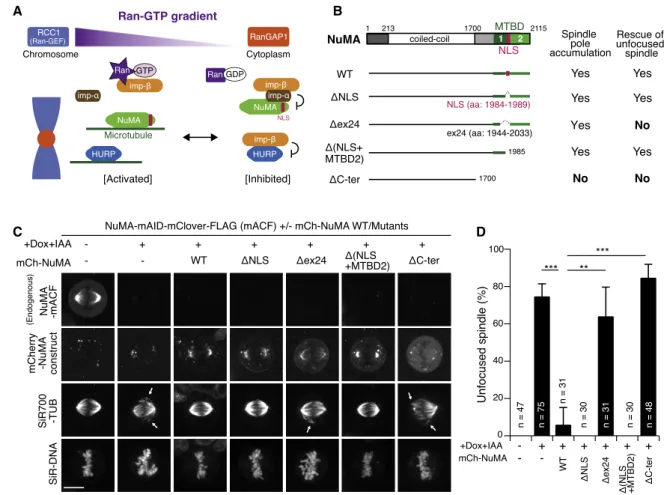
Figure 1. NuMA Acts in Spindle-Pole Focusing Using Its Conserved Microtubule-Binding Domain (A) The prevailing model of SAF inhibition and activation.
(B) Full-length NuMA and truncation fragments.
(C) Live fluorescence images of metaphase NuMA-mACF cells 24 h after treatment with Dox and IAA. Arrows indicate unfocused microtubules.
(D) Quantification of unfocused spindles for each condition in (C) from 3 independent experiments. p values were calculated using Dunnett’s multiple com- parisons test after one-way ANOVA (F(3,6) = 33.81; p = 0.0004).
See alsoFigure S1. Error bars indicate mean ± SD; *p < 0.05, **p < 0.01, ***p < 0.001, ****p < 0.0001; scale bars, 10mm.
Overall, our studies indicate that the Ran pathway acts on spin- dle assembly separately from its interphase functions but does not equivalently regulate all Ran-regulated SAFs in mitotic hu- man cells.
RESULTS
NuMA Focuses Spindle Microtubules Using Its C- Terminal Conserved Microtubule-Binding Domain NuMA has two microtubule-binding domains (MTBDs) at the C-terminal region33,37(Figure 1B) that are critical for spindle- pole focusing.19To understand which domain is required for spindle-pole focusing, we replaced endogenous NuMA with C-terminal truncation mutants (Figure 1B). Endogenous NuMA was fused with an mAID-mClover-FLAG (mACF) tag20 (Figure S1A) and depleted using the AID system following doxy- cycline (Dox) and indole-3-acetic acid (IAA) treatment.20,36In parallel, mCherry-tagged NuMA mutants were expressed from the Rosa 26 locus by Dox treatment (Figures 1B, 1C, S1B, and S1C).20Equivalent to endogenous NuMA, mCherry- tagged NuMA wild type (WT) accumulated in interphase nuclei (Figure S1C) and at mitotic spindle poles (Figure 1C) and was able to rescue pole-focusing defects caused by NuMA deple- tion (Figures 1C and 1D).20NuMA-DNLS mutants were unable to localize at nuclei in interphase (Figure S1C), but were able to accumulate at spindle poles to rescue pole-focusing defects (Figures 1C and 1D). As expected, NuMA DC-ter mutants, which lack both MTBDs, diffused into the cytoplasm during metaphase (Figure 1C) and were unable to rescue the spin- dle-pole focusing defects (Figures 1C and 1D).19 Similarly, NuMA Dex24 mutants, which lack the NLS and part of MTBD1, were unable to fully rescue focusing defects, despite localizing around the spindle poles (Figures 1C and 1D).
Although this localization appears to be slightly reduced at the poles (Figure 1C), the fluorescence intensities were not significantly different between Dex24 and DNLS mutants at either mitotic poles or interphase cytoplasm (Figures S1D and S1E). In contrast, NuMAD(NLS+MTBD2) mutants, which accu- mulate on mitotic spindle poles and interphase microtubules around centrosomes, were able to rescue the focusing defects (Figures 1C, 1D, andS1C). These results indicate that NuMA’s MTBD1, containing a well-conserved NuMA-Lin5-Mud (NLM) motif (Figure S1F),38,39is essential for spindle-pole focusing in human cells, similar to mouse fibroblasts.22
NuMA Localizes at the Spindle Poles Independently of RCC1
NuMA’s MTBD1 is adjacent to the NLS, which is recognized by importin-a (Figures 1A, 1B, and S1F).33A recent in vitro study demonstrated that NuMA’s microtubule-binding activity is sterically inhibited by the importin-a/b complex, but Ran- GTP releases the importin complex, allowing NuMA to interact with microtubules (Figure 1A).33To test this model in cells, we depleted RCC1 (RanGEF) by integrating an mAID-mClover (mAC) tag into the gene (Figures S1A andS2A; hereafter, all PCR validation results of the established cell lines are summa- rized in Figure S7).36 Surprisingly, RCC1 depletion did not substantially affect NuMA’s spindle-pole localization (Fig- ure 2A). Although NuMA intensities at the spindle poles were
slightly reduced in RCC1-depleted cells (Figure 2B), the rela- tive intensities of NuMA on microtubules slightly increased (Figure 2C) as a result of the reduction of microtubule inten- sities following RCC1 depletion (Figures 2A andS2B). RCC1 depletion also shortened the metaphase spindle (Figures 2A and 2D) and delayed mitotic progression (Figures S2C–S2E), but spindle poles were focused normally and the cells eventu- ally exited mitosis, as reported in RCC1-depleted chicken DT40 cells.14 These results indicate that, although Ran-GTP is required for some aspects of spindle assembly, it is not required to localize and activate NuMA at the spindle poles in human cells.
NuMA Participates in Spindle-Pole Focusing Independently of RCC1
To further analyze the functions of NuMA in RCC1-depleted cells, we next co-depleted RCC1 and NuMA. Following treat- ment with Dox and IAA, both RCC1-mAC and NuMA-mAID- mCherry were depleted (Figure 2E), and unfocused spindles were frequently observed (Figures 2E and 2F). In addition, some spindles were completely collapsed and did not form a bi-polar spindle structure (Figures 2E, bottom, and 2F). Unex- pectedly, co-depletion of RCC1 and NuMA further diminished the intensities of tubulin (Figures 2G andS2F). These results sug- gest that NuMA is functional in the absence of Ran-GTP to focus spindle poles and stabilize spindle microtubules.
We note that the frequency of an unfocused spindle in RCC1 and NuMA co-depleted cells (Figure 2F; ~20%) is lower than that of NuMA single-depleted cells (Figure 1D; ~74%). The milder phenotype might reflect smaller pole-splitting forces exerted in the co-depleted cells, as RCC1 depletion results in shorter meta- phase spindle formation (Figure 2D) with reduced microtubules (Figure S2B).
RanGAP1 and Importin-bDegradation Do Not Affect NuMA Localization and Function at Spindle Poles Although RCC1 is dispensable for NuMA localization and func- tion, depletion of RanGAP1 or importin-bmay cause overactiva- tion of NuMA (Figure 1A), resulting in spindle assembly defects.
To test this, we depleted either RanGAP1 or importin-b(Figures 2H–2K andS2G–S2J). RanGAP1 depletion did not affect Nu- MA’s spindle-pole localization (Figures 2H and 2I), spindle length, or mitotic duration (Figure S2H). Importin-b depletion also did not affect NuMA’s localization at spindle poles (Figures 2J and 2K), but it did result in short spindles and mitotic delay (Figure S2J).
To further test the contribution of the Ran-importin pathway to NuMA activation, we next expressed importin-a DIBB mutants that lack the importin-b-binding (IBB) domain (Figure S2K). Im- portin-a DIBB mutants are insensitive to Ran-GTP due to the lack of an IBB domain but are still able to interact with NuMA and partially inhibit NuMA’s microtubule-binding activityin vitro (Figure S2K).33 However, importin-a DIBB diffused into cyto- plasm similarly to importin-aWT, and neither affected NuMA’s spindle-pole localization or co-localized with NuMA at the spin- dle poles in our experimental conditions (Figures S2L and S2M).
Together, these results indicate that the traditional Ran pathway (Figure 1A) is dispensable for NuMA regulation and activation in cultured human cells.
Mitotic Degradation of RCC1 Does Not Affect Localization and Function of NuMA at Spindle Poles In the above experiments, RCC1, RanGAP1, or importin-bwere depleted in asynchronous culture. However, given Ran’s func- tion in interphase nuclear-cytoplasmic transport, unknown sec- ondary effects may have induced their mitotic phenotypes. In addition, because NuMA is maintained in the nucleus following RCC1 depletion in interphase (Figure S2E; t = 0:10), the
majority of NuMA may already have been liberated from impor- tins by pre-existing RCC1 and exist as an active form in the nu- cleus, thereby producing no aberrant phenotypes during mitosis.
To exclude these possibilities, we next depleted RCC1 in noco- dazole-arrested cells and analyzed the behavior of NuMA following nocodazole washout (Figure 3A).
In control cells, NuMA diffused into the cytoplasm during no- codazole arrest (Figure 3B; t = 90), but rapidly accumulated
A B C D
E
F G
H I J K
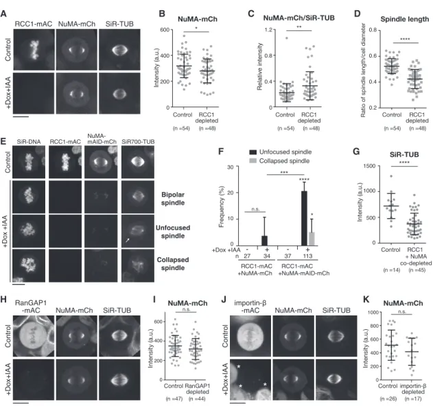
Figure 2. NuMA Functions in Spindle-Pole Focusing Independently of RCC1 (A) Live fluorescence images of metaphase RCC1-mAC cells 24 h after Dox and IAA treatment.
(B) Intensity of NuMA-mCh at spindle poles in controls (318 ± 90.86) and RCC1-depleted cells (279.6 ± 92.39).
(C) Relative intensity of NuMA-mCh/SiR-tubulin at spindle poles in controls (0.22 ± 0.14) and RCC1-depleted cells (0.33 ± 0.22).
(D) Ratio of spindle length and cell diameter in controls and RCC1-depleted cells.
(E) Live images of RCC1-mAC and NuMA-mAID-mCh double knockin cells 24 h after Dox and IAA treatment. Projected images from 5 z sections are shown. The arrow indicates unfocused microtubules.
(F) Quantification of cells in (E) from >4 independent experiments. p values were calculated using Dunnett’s multiple comparisons test after one-way ANOVA (F(3,14) = 36.40; p = 0.0001).
(G) Intensity of SiR-tubulin at spindle poles in controls (719.6 ± 242.7) and RCC1 and NuMA co-depleted cells (371.1 ± 209.2).
(H) Live images of metaphase RanGAP1-mAC cells 24 h after Dox and IAA treatment.
(I) Quantification of NuMA-mCh signals at spindle poles in controls (349.6 ± 108.3) and RanGAP1-depleted cells (318.6 ± 107.7).
(J) Live images of metaphase importin-b-mAC cells 24 h after Dox and IAA treatment. Asterisks indicate RCC1-non-depleted cells (seeSTAR Methods).
(K) Quantification of NuMA-mCh signals at spindle poles in controls (511.2 ± 223) and importin-b-depleted cells (414.8 ± 199.5) from >3 independent experiments.
See alsoFigure S2. Error bars indicate mean ± SD; *p < 0.05, **p < 0.01, ***p < 0.001, ****p < 0.0001; scale bars, 10mm.
near chromosome masses following nocodazole washout (Fig- ure 3B; t = 10;Video S1). Unexpectedly, NuMA also displayed punctate signals throughout cells (Figures 3B, t = 10, andS3A), some of which co-localized with SiR-tubulin (Figure S3B). These NuMA dots disappeared during spindle assembly, and the ma- jority of NuMA localized at the poles of metaphase spindles within 60 min (Figure 3B; t = 60). Following mitotic exit, NuMA was localized in the nucleus (Figure 3B; t = 85).
Importantly, even if RCC1 was depleted during nocodazole ar- rest, NuMA accumulated as usual at focused spindle poles.
RCC1-mAC signals were initially detectable (Figure 3C; t = 90, arrow), but were reduced to undetectable levels after addi- tion of IAA (Figures 3C, t = 0, and 3D). After nocodazole-washout, NuMA accumulated near chromosome masses (Figure 3C; t = 10) and localized to focused spindle poles (Figure 3C; t = 55;
Video S2), as observed in control cells (Figure 3E). During the
process, the number of NuMA dots appeared to be reduced (Fig- ures 3C, t = 10, andS3C), but the number of SiR-tubulin dots on chromosomes, which might represent non-centrosomal micro- tubules nucleated from chromosomes, was not significantly affected by RCC1 depletion (Figure S3D). RCC1-depleted cells entered anaphase with timing similar to that of control cells (Fig- ure S3E), but NuMA was absent from the nucleus after mitotic exit (Figure 3C; t = 80).
As observed when RCC1 was depleted in asynchronous culture (Figure 2D), the metaphase spindle became shorter when RCC1 was depleted during nocodazole arrest (Figure 3F). In addition, metaphase spindle poles were well focused, and unfocused NuMA signals were never observed in both control (n = 28) and RCC1-depleted (n = 25) cells. The frequency of mis-oriented spin- dles slightly increased in RCC1-depleted cells (Figure S3F), but the difference was not statistically significant. Taken together, A
B
C
D
E
F
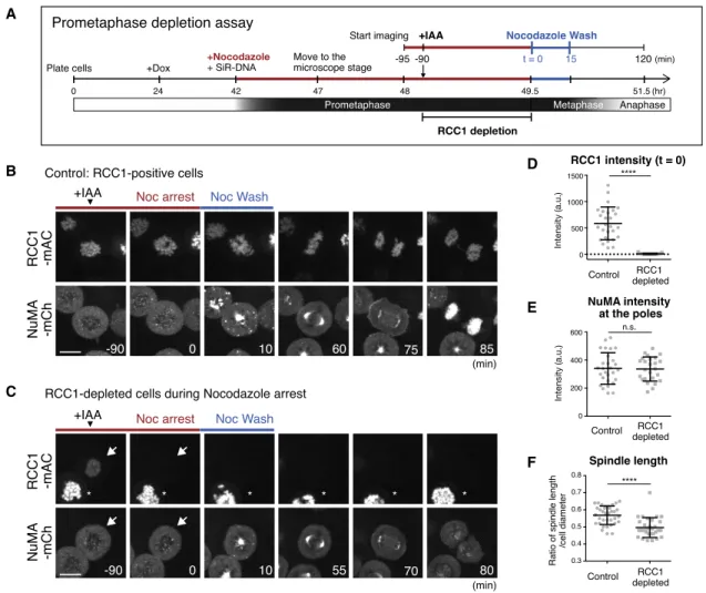
Figure 3. RCC1 Depletion during Prometaphase Does Not Affect NuMA Localization and Function at the Spindle Poles (A) Diagram of prometaphase depletion assay (seeSTAR Methods).
(B and C) Live fluorescence images in RCC1-positive control (B) and RCC1-negative cells (C) treated with nocodazole and IAA, as described in (A). Arrows and asterisks indicate RCC1-depleted and non-depleted cells, respectively.
(D) Intensity of RCC1 on chromosomes at t = 0 in controls (585.2 ± 311.8; n = 27) and RCC1-depleted cells (7.6 ± 10.3; n = 25).
(E) Intensity of NuMA at metaphase spindle poles in controls (340.6 ± 111.6; n = 28) and RCC1-depleted cells (336.0 ± 86.5; n = 25). Welch’s t test gave a p of 0.9542.
(F) Ratio of spindle length and cell diameter in control (0.57 ± 0.05; n = 35) and RCC1-depleted (0.50 ± 0.06; n = 30) cells from >3 independent experiments.
See alsoFigure S3andVideos S1andS2. Error bars indicate mean ± SD; *p < 0.05, **p < 0.01, ***p < 0.001, ****p < 0.0001; scale bars, 10mm.
these results indicate that RCC1 participates on some level in spindle assembly independently of its interphase functions, but is dispensable for NuMA localization and function at spindle poles.
RCC1 Regulates Chromosome-Proximal Localization of HURP and HSET
To elucidate Ran’s spindle assembly function, we set out to iden- tify SAFs regulated by Ran-GTP, initially focusing on the localiza- tion of 3 major SAFs: TPX2; HSET; and HURP. TPX2 co-localized
with SiR-tubulin signals in metaphase (Figure 4A, top), but its localization was virtually unaffected in RCC1-depleted cells (Figures 4A, bottom, and 4B). In contrast, HSET localized everywhere along spindle microtubules (Figure 4C, top),31 and following RCC1 depletion, its spindle localization was selectively reduced near chromosomes (Figures 4C, bottom, 4D, andS4A). Remarkably, HURP localization was affected by RCC1 depletion: HURP accumulated at kinetochore fibers (k-fibers) near chromosomes in all analyzed cells (n = 40), but A
C
E
G
B
D
F
H
I
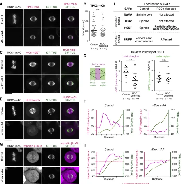
Figure 4. RCC1 Regulates Chromosome-Proximal Localization of HURP and HSET
(A, C, E, and G) Live fluorescent images of metaphase RCC1-mAC cells 24 h after Dox and IAA treatment.
(B) Intensity of TPX2-mCh at spindle poles in controls (212.7 ± 127.3) and RCC1-depleted cells (203.2 ± 119.1).
(D) Left: diagram showing the central and polar regions of the spindle. Right: relative intensity of mCh-HSET against SiR-tubulin at central regions in controls (5.68 ± 1.03) and RCC1-depleted cells (4.16 ± 1.34) and at polar regions in controls (2.48 ± 0.61) and RCC1-depleted cells (2.55 ± 0.95) is shown. Welch’s t test gave a p of 0.67.
(F and H) Line scans showing fluorescence intensities of SiR-tubulin and HURP-mCh or importin-b-mCh on k-fibers indicated as dotted lines in (E) and (G), respectively.
(I) List summarizing localization of SAFs.
See alsoFigure S4. Error bars indicate mean ± SD; *p < 0.05, **p < 0.01, ***p < 0.001, ****p < 0.0001; scale bars, 10mm.
localized weakly on spindle microtubules in all RCC1-depleted cells observed (n = 43;Figures 4E and 4F).
As we found that endogenous importin-balso accumulates at k-fibers in both living (Figures 2J andS4B) and fixed cells (Fig- ure S4C), we next analyzed importin-bin RCC1-depleted cells.
In 84% of control cells (n = 45), importin-bsignals were detected in both k-fibers and cytoplasm (Figure 4G, top). However, the k- fiber signals were not detectable in 88% of RCC1-depleted cells (n = 43;Figure 4G, bottom). Although microtubule density is reduced to 60%–70% by RCC1 depletion (Figure S2B), the k-fiber signals of importin-bshould still be detected, if they exist, as cytoplasmic intensities of importin-b were not significantly changed by RCC1 depletion (Figures 4H andS4D).
Together, these results suggest that the Ran-GTP gradient activates at least two established SAFs, HSET and HURP,
preferentially near chromosomes, but does not affect NuMA and TPX2 (Figure 4I).
HURP, but Not Importin-b, Is Required to Stabilize K- Fibers
Importin-bacts as an inhibitor of HURP (Figure 1A).32However, importin-bco-localizes with HURP at k-fibers (Figure S4C) and behaves similarly to HURP downstream of Ran-GTP (Figures 4E and 4G). To understand the relationship between HURP and importin-b, we next sought to deplete endogenous HURP using AID (Figures 5A andS5A). Importin-bwas detected on k-fi- bers in 81% of control cells (n = 49;Figures 5A and 5B), but not observed in any HURP-depleted cells analyzed (n = 43;Figures 5A and 5B). HURP depletion also reduced mitotic spindle length (Figure 5C), but it did not significantly change microtubule
A B C D
E F G
H I J K
Figure 5. HURP, but Not Importin-b, Is Required to Stabilize K-Fibers
(A) Live fluorescence images of metaphase HURP-mACF (mAID-mClover-FLAG) cells 24 h after Dox and IAA treatment.
(B) Line scans showing fluorescence intensities of SiR-tubulin and importin-b-mCh on k-fibers indicated as dotted lines in (A).
(C) Ratio of spindle length and cell diameter in control (0.64 ± 0.05) and HURP-depleted (0.52 ± 0.06) cells.
(D) Fluorescence images of HURP-mACF, TUB, and DNA (Hoechst 33342) in metaphase fixed cells treated with ice-cold medium for 20 min. Two cells with or without HURP signals were analyzed in the same field.
(E) Live images of metaphase importin-b-mAC cells 24 h after Dox and IAA treatment.
(F) Relative intensities of HURP-mCh against SiR-tubulin at the poles in control (0.5217 ± 0.5171) and importin-b-depleted (2.802 ± 2.574) cells.
(G) Fluorescence images of fixed metaphase cells treated with ice-cold medium for 20 min. Five z section images were obtained using 0.5-mm spacing; maximum intensity projection images are shown in (D) and (G).
(H and J) Live images of metaphase RanGAP1-mAC cells 24 h after Dox and IAA treatment.
(I) Relative intensities of HURP-mCh against SiR-tubulin at the poles in control (0.04708 ± 0.02666) and RanGAP1-depleted (0.1362 ± 0.0592) cells.
(K) Relative intensities of importin-b-mCh against SiR-tubulin at the poles in control (0.02695 ± 0.002289) and RanGAP1-depleted (0.05852 ± 0.03579) cells.
See alsoFigure S5. Error bars indicate mean ± SD; *p < 0.05, **p < 0.01, ***p < 0.001, ****p < 0.0001; scale bars, 10mm.
density at the poles (Figure S5B). Consistent with a previous study,32HURP localized to cold-stable microtubules (Figure 5D, top), which were subsequently disrupted by HURP depletion (Figure 4D, bottom).
We next depleted importin-b(Figure 5E). Importin-bdepletion caused a remarkable re-localization of HURP from k-fibers to spindle microtubules (Figures 5E and 5F). Although k-fiber localization of HURP was unclear in importin-b-depleted cells due to the relatively strong accumulation of HURP on spindle microtubules around spindle poles (Figure 5E, bottom), HURP was clearly detected on cold-stable k-fibers in importin- b-depleted cells (Figure 5G, bottom). These results suggest that HURP acts in k-fiber stabilization independently of importin-b.
HURP and Importin-bLocalize throughout the Spindle in RanGAP1-Depleted Cells
To better understand the mechanisms of Ran-based spatial regulation of HURP and importin-b, we next analyzed the behavior of HURP and importin-bin RanGAP1-depleted cells, in which Ran-GTP should exist evenly throughout the cell. Inter- estingly, both HURP and importin-b localized throughout the spindle with increased intensities in RanGAP1-depleted cells (Figures 5H–5K). These results suggest that HURP and impor- tin-bact together and interact with microtubules preferentially in the presence of Ran-GTP.
Fast Turnover of Chromosome-Proximal HURP in the Presence of Importin-b
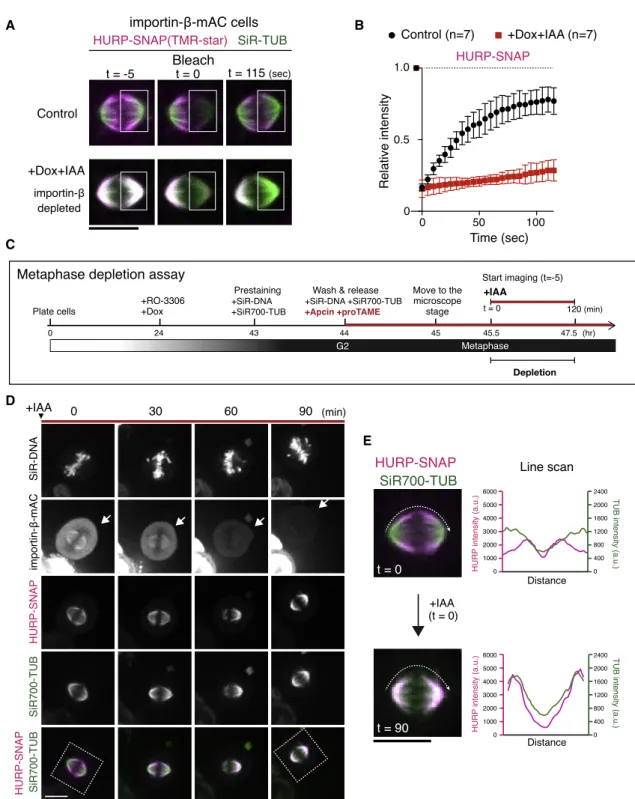
Based on our results, we developed a local cycling model for the activation and polarization of HURP (Figure S5C). In this model, importin-binhibits HURP globally, including at k-fibers, by masking HURP’s 2nd microtubule-binding domain (MTBD2).40 The resulting HURP-importin-b complex binds weakly to microtubules through HURP’s MTBD1,40 but the Ran-GTP gradient locally releases importin-b from HURP, re- sulting in the full activation of HURP near chromosomes (Fig- ure S5C). To test this model, we performed fluorescence recov- ery after photobleaching (FRAP) for HURP and analyzed its dynamics on spindle microtubules in the presence and absence of importin-b. In control cells, HURP quickly recovered at k-fi- bers near chromosomes after bleaching (Figures 6A, top, 6B, left, black, andS6A; t1/2= 20.5 s). In contrast, HURP’s recovery was hardly seen on the spindle in importin-b-depleted cells (Fig- ures 6A, bottom, 6B, red, andS6B), suggesting a stable binding to spindle microtubules.
HURP Is Dynamically Maintained at K-Fibers during Metaphase
To confirm the dynamic regulation of HURP by importin-band Ran-GTP, we next sought to deplete importin-bduring meta- phase (Figure 6C).41Following treatment with the antigen-pre- senting cell (APC)/C inhibitors, apcin and proTAME, cells were arrested at metaphase, at which point both importin-b and HURP accumulated at k-fibers near chromosomes (Fig- ure 6D; t = 0). Importantly, importin-b-mAC signals diminished to undetectable levels 60–90 min after the addition of IAA (Fig- ure 6D, arrows), and HURP relocated from k-fibers to spindle
microtubules in response to the reduction of importin-b(Figures 6D and 6E).
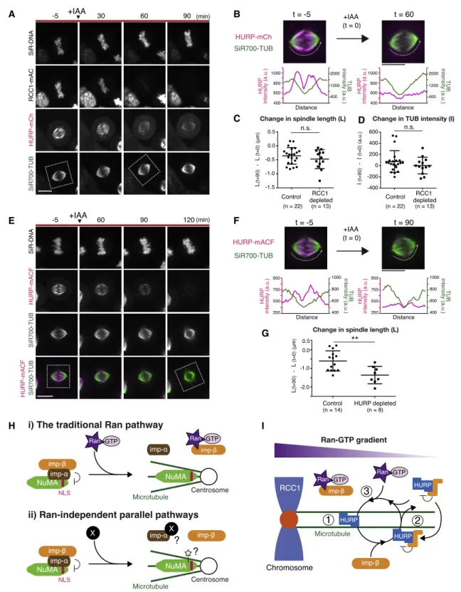
To confirm these results, we next acutely depleted RCC1 in metaphase-arrested cells. As with importin-bdepletion, HURP dissociated from k-fibers and localized weakly on the spindle in response to RCC1 depletion (Figures 7A and 7B;Video S3).
However, in contrast to the prometaphase depletion assay (Fig- ure 3F), spindle length appeared to be normal when RCC1 was depleted in the metaphase-arrested condition (Figure 7C). In addition, SiR-tubulin intensities at the poles did not significantly change when RCC1 was depleted during metaphase (Figure 7D).
To compare these phenotypes, we lastly depleted HURP via auxin, which significantly reduced HURP signals 90–120 min following treatment (Figures 7E and 7F). In contrast to RCC1 depletion, the metaphase spindle became shorter in response to the depletion of HURP (Figures 7E and 7G).
Taken together, these results indicate that HURP is dynami- cally maintained at k-fibers near chromosomes by the Ran-im- portin pathway, even in metaphase. In addition, HURP is required to maintain spindle length during metaphase (Fig- ure 7G), whereas Ran-GTP appears to contribute to spindle length control preferentially during prometaphase (Figures 3F and7C).
DISCUSSION
NuMA Is Liberated from Importins Independently of Ran- GTP for Spindle-Pole Focusing
In contrast to the prevailing model (Figure 1A), we demonstrated that the Ran-Importin pathway is dispensable for localization and function of NuMA at spindle poles in human HCT116 cells (Fig- ures 2 and 3). This is consistent with the recent observation that NuMA is less sensitive to Ran-GTP levels than to HSET/
XCTK2.30Although we do not exclude the possibility that Ran- GTP liberates NuMA from importin-a/bcomplexes near chromo- somes, we favor the idea that parallel pathways exist to activate NuMA in mitotic human cells (Figure 7H). In fact, recent studies indicate that importin-a/b-binding TPX2 can be activated not only by Ran-GTP but also by Golgi- or palmitoylation-dependent sequestration of importin-a.42,43In addition, phosphorylation of TPX2’s NLS also acts to release importins from TPX2.44Similar mechanisms might exist for NuMA around centrosomes (Figure 7H).
Although TPX2-NLS is well conserved in vertebrates, the NLS of NuMA is not well conserved in fish (Figure S1F). Furthermore, the NLS is absent in other NuMA-like proteins in lower eukary- otes.38,45,46Future research should be undertaken to under- stand how the NuMA-importin interaction is regulated in a Ran-independent manner and why NLS-dependent regulation of NuMA was acquired in higher animals.
The Ran-Importin Pathway Locally Activates HURP by Promoting Its Microtubule Binding-Dissociation Cycle Near Chromosomes
In contrast to NuMA, we demonstrated that HURP is preferen- tially regulated by the Ran-importin pathway in mitotic human cells (Figures 4E,5H, and 5J). Although HURP has been identi- fied previously as a downstream target of Ran-GTP,32we found that HURP also co-localizes with importin-b on k-fibers near
A B
C
D
E
Figure 6. HURP Dynamically Accumulates on Metaphase K-Fibers in an Importin-b-Dependent Manner
(A) Live fluorescence images of HURP-SNAP visualized with TMR-star and SiR-tubulin in control and importin-b-depleted cells. Fluorescence signals were bleached in the boxed regions at t = 0, and fluorescence recovery was monitored for 120 s.
(B) FRAP plots of means with SDs from 7 samples.
(C) Diagram of metaphase depletion assay (seeSTAR Methods).
(D) Live images of DNA, tubulin, and indicated proteins. IAA was added at t = 0. Arrows indicate a cell showing a reduction of importin-bduring metaphase.
(E) Enlarged images from (D) and line scans of HURP and SiR-tubulin intensities on spindle microtubules indicated as dotted lines in the left images, showing a re- localization of HURP from k-fibers (t = 0) to the spindle (t = 90).
See alsoFigure S6. Error bars indicate mean ± SD; scale bars, 10mm.
A B
C D
E F
G
H I
Figure 7. Models of Local Activation Mechanisms for HURP and NuMA in Mitosis (A) Live fluorescence images of DNA, tubulin, and indicated proteins. IAA was added at t = 0.
(B) Enlarged images of indicated regions in (A) and line scans of indicated microtubules, showing a reduction of HURP-mCh from k-fibers in response to RCC1 depletion.
(C) Change in spindle length in control ( 0.36 ± 0.30) and RCC1-depleted ( 0.47 ± 0.34) cells.
(D) Change in SiR-tubulin intensities in control (74.2 ± 193.4) and RCC1-depleted (2.269 ± 148.1) cells. Welch’s t test gave a p of 0.505 in (C) and 0.275 in (D).
(E) Live images of indicated proteins. IAA was added at t = 0.
(F) Enlarged images of indicated regions in (E) and line scans of indicated microtubules, showing a reduction of HURP-mACF.
(G) Change in SiR-tubulin intensities in control ( 0.60 ± 0.54) and RCC1-depleted ( 1.36 ± 0.46) cells from >3 independent experiments.
(legend continued on next page)
chromosomes (Figures S4C,5A, and 5E) and stabilizes k-fibers independently of importin-b(Figures 5D and 5G). Based on these and other results (Figures 4E,5E, 5H,6A, 6D, and7A), we pro- pose a local cycling model for the establishment and mainte- nance of HURP’s polarized localization to spindle microtubules (Figures 7I and S5C). This model nicely explains the reason why HURP, but not importin-b, stabilizes microtubules and gen- erates stable k-fibers near chromosomes (Figures 5D and 5G).This dynamic regulation is similar to that of HSET/XCTK247 and would be suitable for bundling short microtubules around kinetochores during prometaphase48and for coupling HURP’s polarized localization with microtubule flux on the metaphase spindle.
RCC1 Promotes Proper Spindle Assembly in Human Mitotic Cells
By depleting RCC1 during prometaphase, we demonstrated that RCC1 participates in proper spindle assembly independently of its interphase function in human mitotic cells (Figure 3F). Short spindles caused by RCC1 depletion could be explained by mul- tiple defects, including the lack of HURP-based k-fiber formation (Figures 5C and 5D) and HSET-dependent spindle elongation (Figure 4C).31Although HURP depletion shortened the meta- phase spindle (Figure 7E), RCC1 depletion in metaphase did not affect spindle length (Figure 7A). The HURP-importin-bcom- plex may therefore play a role in the maintenance of the estab- lished spindle structure in RCC1-depleted cells.
In addition to spindle assembly, Ran-GTP also contributes to microtubule nucleation during mitosis.28Microtubule intensities were reduced when RCC1 was depleted before mitosis (Figures 2A andS2B), but not during metaphase (Figures 7A and 7D).
Once microtubule nucleation pathways are activated in mitosis, additional Ran-based activation may not be required. Alterna- tively, the requirement of Ran for microtubule nucleation may be different between cell types. Unexpectedly, we found that Nu- MA’s punctate signals appear transiently in a Ran-GTP-depen- dent manner (Figures 3B, 3C, andS3C). The significance of the NuMA puncta is currently unclear, but these may be related to minus-end stabilization of nucleated microtubules.
A New Toolkit and Mitosis-Specific Protein-Depletion Assays to Dissect the Mitotic Roles of the Ran-Importin Pathway
Mitotic inactivation is critical to precisely analyze mitotic func- tions of the Ran pathway. Previously, tsBN2, a temperature- sensitive RCC1 mutant hamster cell line,49,50and a small-mole- cule inhibitor, importazole,51have been developed to acutely inhibit functions of RCC1 and importin-b, respectively. Here, we established many human AID-cell lines (Table S1)36 and succeeded in depleting RCC1 specifically in prometaphase (Figures 3A–3C) or metaphase (Figures 7A and 7E). As these AID cell lines and mitotic depletion assays are applicable to
other Ran-regulated proteins4,50,52 and other multi-functional proteins, such as dynein and NuMA,20,36they can be used to further advance our understanding of the mechanisms and roles of spindle assembly, maintenance, and positioning in animal cells.
STAR+METHODS
Detailed methods are provided in the online version of this paper and include the following:
d KEY RESOURCES TABLE
d RESOURCE AVAILABILITY B Lead Contact
B Materials Availability B Data and Code Availability
d EXPERIMENTAL MODEL AND SUBJECT DETAILS
d METHOD DETAILS B Plasmid Construction
B Cell Culture, Cell Line Generation, and Antibodies B Microscope System
B Immunofluorescence and Live Cell Imaging
B Prometaphase Depletion Assay and Nocodazole Washout
B Metaphase Depletion Assay B Cold Treatment Assay B FRAP
d QUANTIFICATION AND STATISTICAL ANALYSIS B Quantification of Fluorescent Intensities B Statistical Analysis
SUPPLEMENTAL INFORMATION
Supplemental Information can be found online athttps://doi.org/10.1016/j.
cub.2020.09.091.
ACKNOWLEDGMENTS
We thank Iain M. Cheeseman for critical reading of the manuscript and Yuki Tsukada, Rie Inaba, Kiyoko Murase, Taisei Kumazaki, and Susan Boerner for technical assistance. This work was supported by grants from the PRESTO program (JPMJPR13A3) of the Japan Science and Technology agency (JST) to T.K.; a Career Development Award of the Human Frontier Science Program (CDA00057/2014-C) to T.K.; KAKENHI (16K14721 and 17H05002 to T.K. and 17H01431 for G.G. and T.K.) of the Japan Society for the Promotion of Science (JSPS); NIG-JOINT (2014B-B-3, 2015-A1-19, and 2016-A1-22 to T.K.) of Na- tional Institute of Genetics (NIG); the Naito Foundation to T.K.; and JSPS and DFG (Deutsche Forschungsgemeinschaft, Germany) under the Joint Research Projects-LEAD with UKRI (UK Research and Innovation, UK) to G.G.
AUTHOR CONTRIBUTIONS
Conceptualization, T.K.; Investigation, T.K., K.T., H.H., M.N., and M.O.; Formal Analysis, T.K. and K.T.; Methodology, T.K., Y.S., and M.T.K.; Writing, T.K.; Su- pervision, T.K. and G.G.; Funding Acquisition, T.K. and G.G.
(H) Models of the traditional Ran pathway (i) and Ran-independent parallel pathway (ii). The Ran-independent parallel pathway would activate NuMA away from chromosomes.
(I) A local cycling model of HURP on k-fibers. HURP strongly interacts with microtubules through its two MTBDs (1). Importin-breduces HURP’s microtubule- binding affinity by masking one of HURP’s MTBDs (2). However, in the vicinity of chromosomes, Ran-GTP releases HURP from importin-b, resulting in local activation of HURP (3).
See alsoVideo S3. Error bars indicate mean ± SD; *p < 0.05, **p < 0.01, ***p < 0.001, ****p < 0.0001; scale bars, 10mm.
DECLARATION OF INTERESTS
The authors declare no competing interests.
Received: April 24, 2020 Revised: August 25, 2020 Accepted: September 30, 2020 Published: November 12, 2020 REFERENCES
1.Reber, S., and Hyman, A.A. (2015). Emergent properties of the metaphase spindle. Cold Spring Harb. Perspect. Biol.7, a015784.
2.Heald, R., and Khodjakov, A. (2015). Thirty years of search and capture:
the complex simplicity of mitotic spindle assembly. J. Cell Biol.211, 1103–1111.
3.Kalab, P., and Heald, R. (2008). The RanGTP gradient - a GPS for the mitotic spindle. J. Cell Sci.121, 1577–1586.
4.Forbes, D.J., Travesa, A., Nord, M.S., and Bernis, C. (2015). Reprint of
‘‘Nuclear transport factors: global regulation of mitosis’’. Curr. Opin. Cell Biol.34, 122–134.
5.Bischoff, F.R., and Ponstingl, H. (1991). Catalysis of guanine nucleotide exchange on Ran by the mitotic regulator RCC1. Nature354, 80–82.
6.Bischoff, F.R., Klebe, C., Kretschmer, J., Wittinghofer, A., and Ponstingl, H. (1994). RanGAP1 induces GTPase activity of nuclear Ras-related Ran. Proc. Natl. Acad. Sci. USA91, 2587–2591.
7.Kalab, P., Weis, K., and Heald, R. (2002). Visualization of a Ran-GTP gradient in interphase and mitotic Xenopus egg extracts. Science295, 2452–2456.
8.Kala´b, P., Pralle, A., Isacoff, E.Y., Heald, R., and Weis, K. (2006). Analysis of a RanGTP-regulated gradient in mitotic somatic cells. Nature440, 697–701.
9.Dumont, J., Petri, S., Pellegrin, F., Terret, M.E., Bohnsack, M.T., Rassinier, P., Georget, V., Kalab, P., Gruss, O.J., and Verlhac, M.H. (2007). A centriole- and RanGTP-independent spindle assembly pathway in meiosis I of vertebrate oocytes. J. Cell Biol.176, 295–305.
10.Moutinho-Pereira, S., Stuurman, N., Afonso, O., Hornsveld, M., Aguiar, P., Goshima, G., Vale, R.D., and Maiato, H. (2013). Genes involved in centro- some-independent mitotic spindle assembly in Drosophila S2 cells. Proc.
Natl. Acad. Sci. USA110, 19808–19813.
11.Hasegawa, K., Ryu, S.J., and Kala´b, P. (2013). Chromosomal gain pro- motes formation of a steep RanGTP gradient that drives mitosis in aneu- ploid cells. J. Cell Biol.200, 151–161.
12.Holubcova´, Z., Blayney, M., Elder, K., and Schuh, M. (2015). Human oo- cytes. Error-prone chromosome-mediated spindle assembly favors chro- mosome segregation defects in human oocytes. Science348, 1143–1147.
13.Drutovic, D., Duan, X., Li, R., Kalab, P., and Solc, P. (2020). RanGTP and importinbregulate meiosis I spindle assembly and function in mouse oo- cytes. EMBO J.39, e101689.
14.Furuta, M., Hori, T., and Fukagawa, T. (2016). Chromatin binding of RCC1 during mitosis is important for its nuclear localization in interphase. Mol.
Biol. Cell27, 371–381.
15.Nachury, M.V., Maresca, T.J., Salmon, W.C., Waterman-Storer, C.M., Heald, R., and Weis, K. (2001). Importin beta is a mitotic target of the small GTPase Ran in spindle assembly. Cell104, 95–106.
16.Wiese, C., Wilde, A., Moore, M.S., Adam, S.A., Merdes, A., and Zheng, Y.
(2001). Role of importin-beta in coupling Ran to downstream targets in microtubule assembly. Science291, 653–656.
17.Gruss, O.J., Carazo-Salas, R.E., Schatz, C.A., Guarguaglini, G., Kast, J., Wilm, M., Le Bot, N., Vernos, I., Karsenti, E., and Mattaj, I.W. (2001).
Ran induces spindle assembly by reversing the inhibitory effect of importin alpha on TPX2 activity. Cell104, 83–93.
18.Stewart, M. (2007). Molecular mechanism of the nuclear protein import cy- cle. Nat. Rev. Mol. Cell Biol.8, 195–208.
19.Hueschen, C.L., Kenny, S.J., Xu, K., and Dumont, S. (2017). NuMA recruits dynein activity to microtubule minus-ends at mitosis. eLife6, e29328.
20.Okumura, M., Natsume, T., Kanemaki, M.T., and Kiyomitsu, T. (2018).
Dynein-dynactin-NuMA clusters generate cortical spindle-pulling forces as a multi-arm ensemble. eLife7, e36559.
21.Gaglio, T., Saredi, A., and Compton, D.A. (1995). NuMA is required for the organization of microtubules into aster-like mitotic arrays. J. Cell Biol.131, 693–708.
22.Silk, A.D., Holland, A.J., and Cleveland, D.W. (2009). Requirements for NuMA in maintenance and establishment of mammalian spindle poles.
J. Cell Biol.184, 677–690.
23.Wittmann, T., Wilm, M., Karsenti, E., and Vernos, I. (2000). TPX2, a novel xenopus MAP involved in spindle pole organization. J. Cell Biol.149, 1405–1418.
24.Garrett, S., Auer, K., Compton, D.A., and Kapoor, T.M. (2002). hTPX2 is required for normal spindle morphology and centrosome integrity during vertebrate cell division. Curr. Biol.12, 2055–2059.
25.Petry, S., Groen, A.C., Ishihara, K., Mitchison, T.J., and Vale, R.D. (2013).
Branching microtubule nucleation in Xenopus egg extracts mediated by augmin and TPX2. Cell152, 768–777.
26.Roostalu, J., Cade, N.I., and Surrey, T. (2015). Complementary activities of TPX2 and chTOG constitute an efficient importin-regulated microtubule nucleation module. Nat. Cell Biol.17, 1422–1434.
27.King, M.R., and Petry, S. (2020). Phase separation of TPX2 enhances and spatially coordinates microtubule nucleation. Nat. Commun.11, 270.
28.Scrofani, J., Sardon, T., Meunier, S., and Vernos, I. (2015). Microtubule nucleation in mitosis by a RanGTP-dependent protein complex. Curr.
Biol.25, 131–140.
29.Ems-McClung, S.C., Zheng, Y., and Walczak, C.E. (2004). Importin alpha/
beta and Ran-GTP regulate XCTK2 microtubule binding through a bipar- tite nuclear localization signal. Mol. Biol. Cell15, 46–57.
30.Ems-McClung, S.C., Emch, M., Zhang, S., Mahnoor, S., Weaver, L.N., and Walczak, C.E. (2020). RanGTP induces an effector gradient of XCTK2 and importin a/b for spindle microtubule cross-linking. J. Cell Biol. 219, e201906045.
31.Cai, S., Weaver, L.N., Ems-McClung, S.C., and Walczak, C.E. (2009).
Kinesin-14 family proteins HSET/XCTK2 control spindle length by cross- linking and sliding microtubules. Mol. Biol. Cell20, 1348–1359.
32.Sillje, H.H., Nagel, S., Ko¨rner, R., and Nigg, E.A. (2006). HURP is a Ran-im- portin beta-regulated protein that stabilizes kinetochore microtubules in the vicinity of chromosomes. Curr. Biol.16, 731–742.
33.Chang, C.C., Huang, T.L., Shimamoto, Y., Tsai, S.Y., and Hsia, K.C.
(2017). Regulation of mitotic spindle assembly factor NuMA by importin- b. J. Cell Biol.216, 3453–3462.
34.Giesecke, A., and Stewart, M. (2010). Novel binding of the mitotic regulator TPX2 (target protein for Xenopus kinesin-like protein 2) to importin-alpha.
J. Biol. Chem.285, 17628–17635.
35.Kiyomitsu, T. (2019). The cortical force-generating machinery: how cortical spindle-pulling forces are generated. Curr. Opin. Cell Biol.60, 1–8.
36.Natsume, T., Kiyomitsu, T., Saga, Y., and Kanemaki, M.T. (2016). Rapid protein depletion in human cells by auxin-inducible degron tagging with short homology donors. Cell Rep.15, 210–218.
37.Gallini, S., Carminati, M., De Mattia, F., Pirovano, L., Martini, E., Oldani, A., Asteriti, I.A., Guarguaglini, G., and Mapelli, M. (2016). NuMA phosphoryla- tion by Aurora-A orchestrates spindle orientation. Curr. Biol.26, 458–469.
38.Siller, K.H., Cabernard, C., and Doe, C.Q. (2006). The NuMA-related Mud protein binds Pins and regulates spindle orientation in Drosophila neuro- blasts. Nat. Cell Biol.8, 594–600.
39.Du, Q., Stukenberg, P.T., and Macara, I.G. (2001). A mammalian Partner of inscuteable binds NuMA and regulates mitotic spindle organization. Nat.
Cell Biol.3, 1069–1075.
40.Song, L., Craney, A., and Rape, M. (2014). Microtubule-dependent regula- tion of mitotic protein degradation. Mol. Cell53, 179–192.
41.Sackton, K.L., Dimova, N., Zeng, X., Tian, W., Zhang, M., Sackton, T.B., Meaders, J., Pfaff, K.L., Sigoillot, F., Yu, H., et al. (2014). Synergistic blockade of mitotic exit by two chemical inhibitors of the APC/C. Nature 514, 646–649.
42.Wei, J.H., Zhang, Z.C., Wynn, R.M., and Seemann, J. (2015). GM130 reg- ulates Golgi-derived spindle assembly by activating TPX2 and capturing microtubules. Cell162, 287–299.
43.Brownlee, C., and Heald, R. (2019). Importinapartitioning to the plasma membrane regulates intracellular scaling. Cell176, 805–815.e8.
44.Eibes, S., Gallisa`-Sun˜e, N., Rosas-Salvans, M., Martı´nez-Delgado, P., Vernos, I., and Roig, J. (2018). Nek9 phosphorylation defines a new role for TPX2 in Eg5-dependent centrosome separation before nuclear enve- lope breakdown. Curr. Biol.28, 121–129.e4.
45.Lorson, M.A., Horvitz, H.R., and van den Heuvel, S. (2000). LIN-5 is a novel component of the spindle apparatus required for chromosome segrega- tion and cleavage plane specification in Caenorhabditis elegans. J. Cell Biol.148, 73–86.
46.Greenberg, S.R., Tan, W., and Lee, W.L. (2018). Num1 versus NuMA: in- sights from two functionally homologous proteins. Biophys. Rev.10, 1631–1636.
47.Weaver, L.N., Ems-McClung, S.C., Chen, S.H., Yang, G., Shaw, S.L., and Walczak, C.E. (2015). The Ran-GTP gradient spatially regulates XCTK2 in the spindle. Curr. Biol.25, 1509–1514.
48.Sikirzhytski, V., Renda, F., Tikhonenko, I., Magidson, V., McEwen, B.F., and Khodjakov, A. (2018). Microtubules assemble near most kinetochores during early prometaphase in human cells. J. Cell Biol.217, 2647–2659.
49.Nishimoto, T., Eilen, E., and Basilico, C. (1978). Premature of chromosome condensation in a ts DNA- mutant of BHK cells. Cell15, 475–483.
50.Kiyomitsu, T., and Cheeseman, I.M. (2012). Chromosome- and spindle- pole-derived signals generate an intrinsic code for spindle position and orientation. Nat. Cell Biol.14, 311–317.
51.Soderholm, J.F., Bird, S.L., Kalab, P., Sampathkumar, Y., Hasegawa, K., Uehara-Bingen, M., Weis, K., and Heald, R. (2011). Importazole, a small molecule inhibitor of the transport receptor importin-b. ACS Chem. Biol.
6, 700–708.
52.Kiyomitsu, T., and Cheeseman, I.M. (2013). Cortical dynein and asym- metric membrane elongation coordinately position the spindle in anaphase. Cell154, 391–402.
53.Schindelin, J., Arganda-Carreras, I., Frise, E., Kaynig, V., Longair, M., Pietzsch, T., Preibisch, S., Rueden, C., Saalfeld, S., Schmid, B., et al.
(2012). Fiji: an open-source platform for biological-image analysis. Nat.
Methods9, 676–682.
54.Kiyomitsu, T., Murakami, H., and Yanagida, M. (2011). Protein interaction domain mapping of human kinetochore protein Blinkin reveals a consensus motif for binding of spindle assembly checkpoint proteins Bub1 and BubR1. Mol. Cell. Biol.31, 998–1011.
55.Goshima, G., Nedelec, F., and Vale, R.D. (2005). Mechanisms for focusing mitotic spindle poles by minus end-directed motor proteins. J. Cell Biol.
171, 229–240.
STAR + METHODS
KEY RESOURCES TABLE
RESOURCE AVAILABILITY Lead Contact
Further information and requests for resources and reagents should be directed to and will be fulfilled by the Lead Contact, Tomomi Kiyomitsu (tomomi.kiyomitsu@oist.jp).
Materials Availability
Plasmids generated in this study will be deposited to Addgene.
Data and Code Availability
This study did not generate/analyze any datasets.
REAGENT or RESOURCE SOURCE IDENTIFIER
Chemicals
SiR-tubulin Spirochrome Cat# SC002
SiR-DNA Spirochrome Cat# SC007
SiR700-tubulin Spirochrome Cat# SC014
SNAP Cell 647-SiR New England BioLabs Cat# S9102S
SNAP Cell TMR-star New England BioLabs Cat# S9105S
Hoechst 33342 Sigma-Aldrich Cat# B2261
Nocodazole Sigma-Aldrich Cat# M1404
MG132 Sigma-Aldrich Cat# C2211
RO-3306 Sigma-Aldrich Cat# SML0569
Apcin Boston Biochem Cat# I-444
proTAME Boston Biochem Cat# I-440
Puromycin dihydrochloride Wako Pure Chemical Industries Cat# 160-23151
G-418 solution Roche Cat# 04727894001
Hygromycin B Wako Pure Chemical Industries Cat# 084-07681
Blasticidin S hydrochloride Funakoshi Biotech Cat# KK-400
Doxycycline hyclate Sigma-Aldrich Cat # D9891
3-Indoleacetic acid (IAA) Wako Pure Chemical Industries Cat # 098-00181
DirectPCR(cell) Viagen Biotech Cat #302-C
Antibodies
Anti-a-tubulin (clone DM1A) Sigma-Aldrich Cat# T9026; RRID:AB_477593
Rabbit polyclonal anti-NuMA Abcam Cat# ab36999; RRID:AB_776885
Rabbit polyclonal anti-RCC1 Cell Signaling Technology Cat# 5134
Mouse monoclonal anti-RanGAP1 Santa Cruz Biotechnology Cat# sc-25630
Mouse anti-importin-b GeneTex Cat# GTX22811
Rabbit anti-importin-a Novus biologicals Cat# NBP1-31098
Anti-HURP Nigg lab N/A
Sheep anti-mouse IgG-HRP GE Healthcare Cat# NA931
Donkey anti-rabbit IgG-HRP GE Healthcare Cat# NA934
Software and Algorithms
Photoshop CS5, version 12.0 Adobe Systems https://www.adobe.com
Fiji 53 https://fiji.sc/
Metamorph Molecular Devices https://www.moleculardevices.com
GraphPad Prism 6, version 6.0c GraphPad Software https://www.graphpad.com
Excel Microsoft https://www.microsoft.com/microsoft-365
EXPERIMENTAL MODEL AND SUBJECT DETAILS
Established human tissue culture cell lines, and sequence information about guide RNA and PCR primers used in this study are described inTables S1–S3, respectively.
METHOD DETAILS Plasmid Construction
Plasmids for CRISPR/Cas9-mediated genome editing and auxin-inducible degron were constructed according to protocols of Nat- sume et al.36and Okumura et al.20To construct donor plasmids containing homology arms for RCC1 (~500-bp homology arms), Ran- GAP1 (~500-bp), importin-b(~500-bp), HURP (~200-bp), TPX2 (~200-bp), and HSET (~200-bp), gene synthesis services from Euro- fins Genomics K.K. (Tokyo, Japan) or Genewiz (South Plainsfield, NJ) were used. Plasmids and sgRNA sequences used in this study are listed inTables S1andS2, and will be deposited in Addgene.
Cell Culture, Cell Line Generation, and Antibodies
HCT116 cells were cultured as described previously.20Knock-in cell lines were generated according to procedures described in Okumura et al.20To activate auxin-inducible degradation, cells were treated with 2mg/mL Dox and 500mM indoleacetic acid (IAA) for 20–24 hr. Cells with undetectable signals for mAID-fusion proteins were analyzed. In the AID system, the target proteins are not degraded in a small population of the cells even in the presence of Dox and IAA, possibly due to the heterogenous induction of OsTIR1. We took advantages of this to compare two neighboring cells with or without target proteins in the same field inFigures S2F,S3A,5D, and 5G.
Flip-In T-REx 293 cells were used inFigures S2L and S2M to express mCherry-tagged importin-aconstructs. Cell lines were created according to procedures described in Kiyomitsu et al.54To induce transgenes, cells were incubated with 1mg/mL tetracy- cline (MP Biomedicals). Cell lines and primers used in this study are listed inTables S1andS3, respectively.
Cell concentrations were determined using the BIO-RAD TC20 Automated Cell Counter (standard protocol). Cells were diluted to a final concentration of 100,000 cells/ml in medium and transferred to 6-well plates for subsequent counting at 24, 48 and 72 hr. Cells were cultured in 4 independent wells and counted twice from each well using the TC20 cell counter.
Antibodies against tubulin (DM1A, Sigma-Aldrich, 1:2,000), NuMA (Abcam, 1:1,000), RCC1 (Cell Signaling Technology, D15H6, Rabbit mAb, 1:100), RanGAP1 (Santa Cruz Biotechnology, H-180, 1:200), importin-b(GeneTex, 3E9 Mouse mAb, 1:100), and HURP (E. Nigg laboratory, 1:200) were used for western blotting. For RCC1 immunoblots, membranes were incubated with anti- RCC1 antibody overnight at 4C.
Microscope System
Imaging was performed using spinning-disc confocal microscopy with a 6031.40 numerical aperture objective lens (Plan Apol, Nikon, Tokyo, Japan). A CSU-W1 confocal unit (Yokogawa Electric Corporation, Tokyo, Japan) with five lasers (405, 488, 561, 640, and 685 nm, Coherent, Santa Clara, CA) and an ORCA-Flash 4.0 digital CMOS camera (Hamamatsu Photonics, Hamamatsu City, Japan) were attached to an ECLIPSE Ti-E inverted microscope (Nikon) with a perfect focus system. DNA images inFigures S2D and S2E orS3A were obtained using a SOLA LED light engine (Lumencor, Beaverton, OR).
Immunofluorescence and Live Cell Imaging
For immunofluorescence inFigure S1K, HURP-mACF cells were fixed with PBS containing 3% paraformaldehyde and 2% sucrose for 10 min at room temperature. Fixed cells were permeabilized with 0.5% Triton X-100 for 5 min on ice, and pretreated with PBS containing 1% BSA for 10 min at room temperature after washing with PBS. Importin-bwas visualized using anti-importin-bantibody (1:500). Images of multiple z sections were acquired by spinning-disc confocal microscopy using 0.5-mm spacing and camera binning 2. Maximally projected images from 3 z sections are shown.
For live cell imaging, cells were cultured on glass-bottomed dishes (CELLview, #627860 or #627870, Greiner Bio-One, Kremsmu¨n- ster, Austria) and maintained in a stage-top incubator (Tokai Hit, Fujinomiya, Japan) to maintain the same conditions used for cell culture (37C and 5% CO2). In most cases, three to five z section images using 0.5-mm spacing were acquired and single z section images are shown, unless otherwise specified. Microtubules were stained with 50 nM SiR-tubulin or SiR700-tubulin (Spirochrome) for
> 1 hr prior to image acquisition. DNA was stained with 50 ng/mL Hoechst33342 (Sigma-Aldrich) or 20 nM SiR-DNA (Spirochrome) for > 1 hr before observation. To visualize SNAP-tagged HURP, cells were incubated with 0.1mM TMR-Star (New England BioLabs) for > 2 hr, and TMR-Star were removed before observation. To optimize image brightness, the same linear adjustments were applied using Fiji and Photoshop.
Prometaphase Depletion Assay and Nocodazole Washout
To degrade mAID-tagged proteins during nocodazole arrest, cells were treated with 2mg/mL Dox and 3.3mM nocodazole at the indicated times (Figure 3A). Five hours after addition of nocodazole, cell culture dishes were moved to the stage of a microscope equipped with a peristaltic pump (SMP-21S, EYELA, Tokyo Rikakikai). Two z section images were acquired using 2-mm spacing at three different (X.Y) positions and at 5-min intervals, with 500mM IAA added during the first interval. After 90 min, the
nocodazole-containing medium was completely replaced with fresh medium using the peristaltic pump at a velocity of 20 s/mL for 15 min. Images were acquired for a further 2 hr and maximum intensity projection images are shown inFigures 3B and 3C. To analyze spindle orientation inFigure S3F, we took five z section images using 2-mm spacing. When both spindle poles are included within three z section images, we judged the spindle as having parallel orientation. InFigure S3D, as the largest SiR-tubulin signal in the center of the cell represents centrosomal microtubules, we defined other SiR-tubulin dots on chromosomes as chromosomal micro- tubule dots and counted the number in the RCC1 positive and negative cells.
Metaphase Depletion Assay
To degrade mAID-tagged proteins in metaphase-arrested cells, cells were treated with 50mM Apcin (I-444, Boston Biochem) and 20mM proTAME (I-440, Boston Biochem) at the indicated times (Figure 6C). Three z section images were acquired using 1-mm spacing at six different (X,Y) positions and at 5-min intervals, with 500mM IAA added during the first interval. Maximum intensity pro- jection images are shown inFigures 6D,7A, and 7E.
Cold Treatment Assay
To increase the number of cells in metaphase, cells were treated with 20mM MG132 (C2211, Sigma-Aldrich) for 90 min. To visualize SNAP-tagged HURP, cells were incubated with 0.1mM TMR-Star (S9105S, New England BioLabs) for at least 30 min. Before fixation, cells were incubated in ice-cold medium for 20 min32to depolymerize non-kinetochore microtubules.
FRAP
FRAP was conducted with a microscope (LEM 780, Carl Zeiss MicroImaging, Inc.), using a 63 x objective lens. Images were acquired every 5 s before and after photobleaching. The bleached area (BA) was set as it covers half spindle and illuminated at t = 0 using 560 nm laser (20 mW) with the following setting: speed 4.0 and iteration 1. Metaphase cells that orient parallel to the bottom cover-glass were selected. HURP (TMR-Star) intensity of BA was normalized using the intensity of non-bleached area (NBA) that covers the remaining half spindle. Corrected relative intensity at time tnwas calculated as (BAn– BGn) / (BA-1– BG-1) x (NBA-1– BG-1) / (NBAn– BGn), where t = 1 represents the first time point of image acquisition before bleaching. BG means background.55 Curve fitting and analyses shown inFigure S6were performed using Fiji.53
QUANTIFICATION AND STATISTICAL ANALYSIS Quantification of Fluorescent Intensities
To quantify fluorescent intensities of SAFs and SiR-tubulin signals, line scans were performed in Fiji. A 15-pixel-width line was drawn on 16-bit images as it passed on both spindle poles, and peak values were recorded as polar intensities after background subtrac- tion. To analyze intensities on k-fibers, segmented lines with 3-pixel-width were manually drawn on the k-fibers in tubulin images, and then the lines were transferred to SAF images to quantify their signal intensities. To quantify RCC1 intensities on prometaphase chro- mosomes inFigure 3D, chromosome regions were manually defined with SiR-DNA images, and the regions were transferred to RCC1 images to quantify the intensities.
Statistical Analysis
To determine the significance of differences between the mean values obtained for two experimental conditions, Welch’s t tests (Prism 6; GraphPad Software, La Jolla, CA) were used. One-way ANOVA was performed inFigures 1D,2F, andS7L–S7N using Prism 6. For all Figures: error bars indicate mean ± SD; * p < 0.05, ** p < 0.01, *** p < 0.001, **** p < 0.0001; scale bars = 10mm.