VI
塩類・結晶類―塩類,通常結晶類,異常結晶類,薬物結晶類―
Salts/Crystals: Salts, Normal Crystals, Abnormal Crystals, Drug Crystals
蟯虫卵 40× 無染色Enterobius vermicularis egg 40× No staining 蟯虫卵は,尿路に産卵されるものではなく,肛門周囲に産卵され た虫卵が,採尿の際に尿中へ混入したものである。色調は透明で 柿の種状の形状を示す。
Enterobius vermicularis eggs are not laid in the urinary tract. Rather, eggs laid around the anus are mixed into the urine during urine collection. The color tone is transparent, and the eggs are shaped like persimmon seeds.
Figure 3.349 糞線虫(フィラリア型幼虫) 20× 無染色
Strongyloides stercoralis (filariform larva) 20× No staining
糞線虫は,本来は小腸の粘膜内に寄生するが,免疫力が低下して いる患者では尿中から糞線虫がみられる場合がある。
Although Strongyloides stercoralis normally infest the mucosa of the small intestine, they may be detected in the urine of patients with a weakened immune system.
Figure 3.350
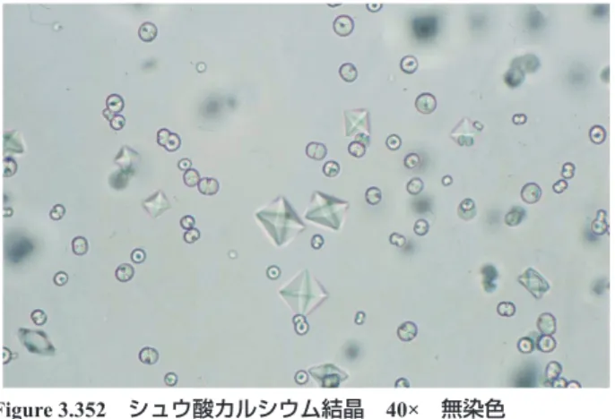
シュウ酸カルシウム結晶 40× 無染色
Calcium oxalate crystals 40× No staining 正八面体の結晶で,弱酸性からアルカリ性まで広い範囲で観察さ れる。酢酸に溶解せず,塩酸で溶解する。pH 6.5
These are regular octahedral crystals that can be observed in a wide range of pH from slightly acidic to alkaline. They do not dissolve in acetic acid but dissolve in hydrochloric acid. pH 6.5
Figure 3.351 シュウ酸カルシウム結晶 40× 無染色
Calcium oxalate crystals 40× No staining 中央は正八面体の結晶で,背景にはビスケット状(円形)の結晶 がみられる。塩酸で溶解する。pH 7.0
In the center, regular octahedral crystals are found, and in the background, biscuit-like (circular) crystals are observed. They dissolve in hydrochloric acid. pH 7.0
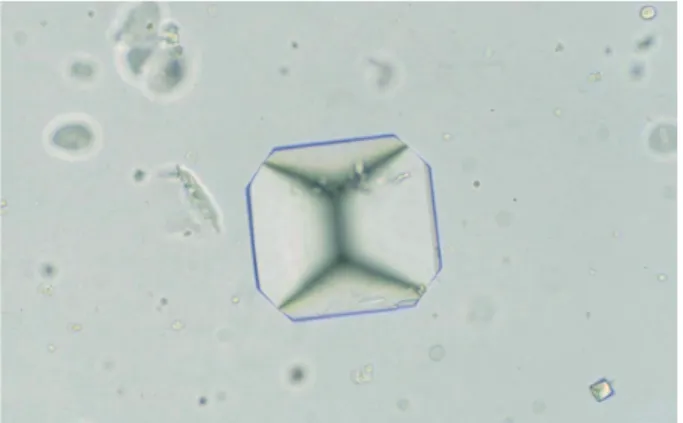
シュウ酸カルシウム結晶 40× 無染色
Calcium oxalate crystals 40× No staining コマ状の結晶で,シュウ酸カルシウム結晶は尿中のシュウ酸濃度 やカルシウム濃度,各種イオン濃度により析出する形状が異なる。 pH 7.0
Spinning top-shaped crystal. Calcium oxalate crystals differ in the shape due to the concentration of oxalic acid, calcium, and various ions in the urine. pH 7.0
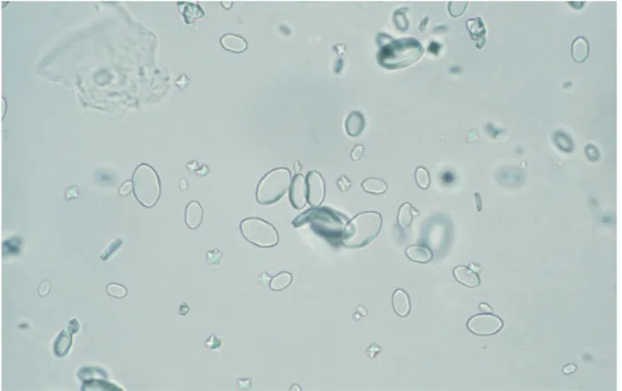
Figure 3.353 シュウ酸カルシウム結晶 40× 無染色
Calcium oxalate crystals 40× No staining
大小不同で楕円形の結晶である。ビスケット状(Figure 3.352)や楕円形の 結晶は赤血球(矢印)と類似する場合がある。しかし,赤血球は淡黄色を 呈するのに対し,この結晶は無色であり,また大小不同や光沢があること などで鑑別できる。pH 6.5
Oval-shaped crystals of different sizes. Biscuit-like (Figure 3.352) and oval-shaped crystals may be similar to red blood cells (arrows). However, red blood cells exhibit a pale yellow color, whereas these crystals are colorless, of different sizes, and glossy. Thus, the crystals can be differentiated from red blood cells. pH 6.5 Figure 3.354*
シュウ酸カルシウム結晶 40× 無染色
Calcium oxalate crystals 40× No staining
楕円形の結晶がみられる。厚みがなく透明感のある結晶である。 pH 7.0
Oval-shaped, transparent and thin crystals. pH 7.0
Figure 3.355 シュウ酸カルシウム結晶 40× 無染色
Calcium oxalate crystals 40× No staining 亜鈴状(鉄アレイ状)の結晶で,炭酸カルシウム結晶も同様の形 状を示す場合がある。しかし,炭酸カルシウム結晶は酢酸で気泡 を産生しながら溶解する。pH 6.5
Dumbbell-shaped crystals. Calcium carbonate crystals may exhibit the same shape in some cases. However, calcium carbonate crystals dissolve while producing bubbles in acetic acid. pH 6.5
Figure 3.356
シュウ酸カルシウム結晶 40× 無染色
Calcium oxalate crystals 40× No staining ビリルビン陽性尿では,円形または楕円形の層状構造を示す結晶 がしばしば観察される。色調はビリルビンに染まり黄褐色を呈す る。pH 7.5
In bilirubin-positive urine, crystals exhibiting a circular- or oval-layered structure are often observed. The color tone is yellowish brown due to the presence of bilirubin. pH 7.5
Figure 3.357 シュウ酸カルシウム結晶 40× Alizarin red 染色 Calcium oxalate crystals 40× Alizarin red staining Figure 3.357の結晶を Calcium 染色(Alizarin red 染色)したもの で,Calcium 陽性の赤色に染色されている。
The crystal shown in Figure 3.357 is stained with calcium staining (alizarin red staining). It stained red and indicates calcium-positive.
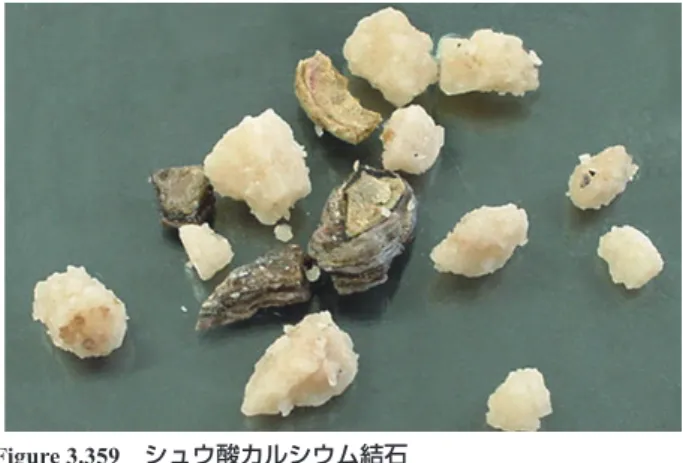
シュウ酸カルシウム結石
Calcium oxalate calculi
体外衝撃波結石破砕術(ESWL)で採取されたシュウ酸カルシウム 結石である。シュウ酸カルシウム結石は,黄褐色や黒褐色で,表 面に凹凸を有する。
Calcium oxalate calculi collected with extracorporeal shock wave lithotripsy (ESWL). Calcium oxalate calculi are yellowish brown or dark brown, with irregular surfaces.
Figure 3.359 尿酸塩 40× 無染色
Urates 40× No staining
酸性尿で析出する褐色の無晶性塩類である。遠心後の尿の外観は レンガ色(紅色)を呈する。析出量が多い場合は,溶解後,観察 するとよい。pH 6.5
Brown amorphous salts deposited from acidic urine. The urine after centrifugation is brick red in color. If a large amount of salt is deposited, it is better to perform observation after dissolving them. pH 6.5
Figure 3.360
尿酸塩 40× 無染色
Urates 40× No staining
酸性尿で析出する褐色の無晶性塩類である。大きくなると 2,8-DHA 結晶と類似するので加温または EDTA 加生理食塩水にて洗浄する など鑑別を要する。pH 6.0
Brown amorphous salts deposited from acidic urine. When the deposition is large, it resembles a 2,8-DHA crystal. For differentiation, it is necessary to heat or wash the specimen with EDTA added physiological saline. pH 6.0
Figure 3.361* 尿酸結晶 40× 無染色
Uric acid crystals 40× No staining
黄褐色で菱形の結晶である。尿酸結晶は酸性尿で析出する。水酸 化カリウムで溶解する。pH 6.0
Yellowish brown rhomboid shape crystals. Uric acid crystals are deposited from acidic urine and dissolve in potassium hydroxide. pH 6.0
Figure 3.362
尿酸結晶 40× 無染色
Uric acid crystal 40× No staining 黄褐色で菊花状の結晶である。pH 5.5
A yellowish brown chrysanthemum blossom-shaped crystal. pH 5.5
Figure 3.363 尿酸結晶 40× 無染色
Uric acid crystal 40× No staining 黄褐色で亜鈴状(鉄アレイ状)の結晶である。pH 6.0 A yellowish brown dumbbell-shaped crystal. pH 6.0
尿酸結晶 40× 無染色
Uric acid crystal 40× No staining 黄褐色で亜鈴状(鉄アレイ状)の結晶である。pH 5.5 A yellowish brown dumbbell-shaped crystal. pH 5.5
Figure 3.365 尿酸結晶 10× 無染色
Uric acid crystals 10× No staining
黄褐色で棒状の結晶である。比較的大型の結晶で肉眼でも観察可 能な場合がある。カバーガラスを載せる際,上手く載せられない ことがしばしばある。pH 5.5
Yellowish brown bar-shaped crystals. These are relatively large crystals and may be observed by the naked eye. It is often difficult to place the coverslip on such specimens. pH 5.5
Figure 3.366
リン酸塩 40× 無染色
Phosphates 40× No staining
無色~灰白色を呈し,多量に析出すると観察の妨げとなる。析出 量が多い場合は,溶解後,観察するとよい。酢酸,塩酸で溶解す る。pH 7.5
Phosphate appears colorless to whitish gray and obstructs observation when the deposition amount is large. In such cases, it is better to perform the observation after dissolving them in acetic or hydrochloric acid. pH 7.5
Figure 3.367 リン酸カルシウム結晶 40× 無染色
Calcium phosphate crystals 40× No staining 無色~灰白色で菊花状の結晶である。背景には板状の結晶がみら れる。酢酸,塩酸で溶解する。pH 8.0
A colorless to whitish gray crystal exhibiting a chrysanthemum blossom shape. There is a plate-shaped crystal in the background. It dissolves in acetic acid and hydrochloric acid. pH 8.0
Figure 3.368
リン酸カルシウム結晶 40× 無染色
Calcium phosphate crystal 40× No staining 無色~灰白色で板状の結晶である。板の表面は顆粒状を呈する。 pH 8.5
A plate-shaped crystal that appears colorless to whitish gray. The surface of the plate is granular. pH 8.5
Figure 3.369 リン酸アンモニウムマグネシウム結晶 40× 無染色
Magnesium ammonium phosphate crystal 40× No staining
無色~淡黄色で封筒状の結晶である。アルカリ性尿で観察される。 ウレアーゼ産生菌による尿路感染症などで出現することがある。 pH 8.5
A colorless to pale yellow envelope-shaped crystal observed in alkaluria. It may appear with a urinary tract infection with urease-producing bacteria. pH 8.5
リン酸アンモニウムマグネシウム結晶 40× 無染色
Magnesium ammonium phosphate crystals
40× No staining
無色~淡黄色で棒状の結晶である。背景には多数の細菌がみられ る。pH 8.5
Colorless to pale yellow bar-shaped crystals. Many bacteria are observed in the background. pH 8.5
Figure 3.371 リン酸アンモニウムマグネシウム結晶 10× 無染色
Magnesium ammonium phosphate crystals
10× No staining
無色~淡黄色で棒状,封筒状の結晶である。比較的大型の結晶で 肉眼でも観察可能な場合がある。pH 8.0
Colorless or pale yellow crystals appearing in a bar or envelope shape. They can be observed with the naked eye due to the relatively large size of the crystals. pH 8.0
Figure 3.372
リン酸アンモニウムマグネシウム結晶 20× 無染色
Magnesium ammonium phosphate crystal
20× No staining
無色~淡黄色で蝶の羽状の結晶である。pH 8.5
A colorless to pale yellow butterfly wing-shaped crystal. pH 8.5
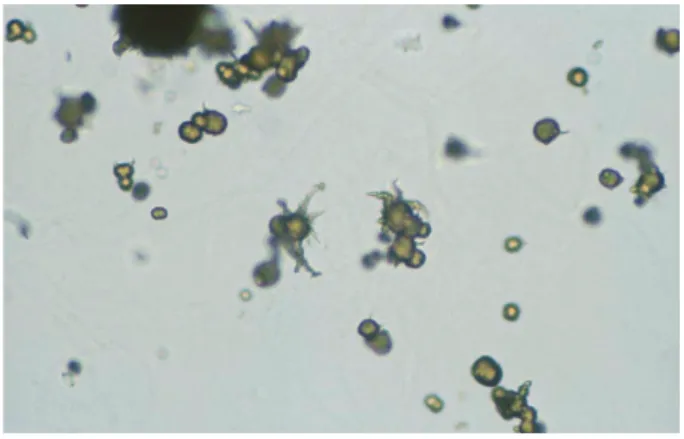
Figure 3.373 尿酸アンモニウム結晶 40× 無染色
Ammonium urate crystals 40× No staining 褐色~淡黄色で棘を有する球状の結晶である。大小不同の棘が特 徴で,一般にアルカリ性尿で観察されるが,しばしば酸性尿でも 観察される。pH 8.5
Brown to pale yellow spherical crystals with thorns. Thorns of various sizes are characteristic. Generally, the crystals are observed in alkaluria; however, they can also be observed in acidic urine. pH 8.5
Figure 3.374
尿酸アンモニウム結晶 40× 無染色
Ammonium urate crystals 40× No staining 褐色~淡黄色で棘を有する球状の結晶である。尿路感染症と関連 がある場合があり,背景に細菌を伴うことがしばしばある。pH 8.0 Brown to pale yellow spherical crystals with thorns that may be associated with urinary tract infections. Bacteria are often observed in the background. pH 8.0
Figure 3.375 酸性尿酸アンモニウム結晶 40× 無染色
Ammonium acid urate crystals 40× No staining 形態的には尿酸アンモニウム結晶と同様である。幼児の感染性胃 腸炎や緩下剤の乱用時に本結石による腎後性急性腎不全の原因と なる。pH 6.0
Ammonium acid urate crystals are morphologically similar to ammonium urate crystals. They may cause postrenal acute renal failure due to the calculi in young children with infectious gastroenteritis or people who abuse laxatives. pH 6.0
炭酸カルシウム結晶 40× 無染色
Calcium carbonate crystals 40× No staining 無色~灰白色で亜鈴状(鉄アレイ状)の結晶である。酢酸,塩酸 で気泡を産生しながら溶解する。pH 8.0
Colorless to whitish gray dumbbell-shaped crystals. They dissolve while producing bubbles with acetic acid and hydrochloric acid. pH 8.0
Figure 3.377 炭酸カルシウム結晶 40× 無染色
Calcium carbonate crystals 40× No staining 無色~灰白色で亜鈴状(鉄アレイ状)の結晶である。シュウ酸カ ルシウム結晶も,しばしば同様の形態を示す場合がある。酢酸や 塩酸添加で気泡を産生する。pH 7.5
Colorless to whitish gray dumbbell-shaped crystals. Calcium oxalate crystals often exhibit a similar morphology. They produce bubbles when acetic acid or hydrochloric acid is added. pH 7.5
Figure 3.378
ビリルビン結晶 40× 無染色
Bilirubin crystals 40× No staining
黄褐色で針状の結晶である。放射状に集合したり,上皮細胞上に 析出している場合がある。背景にみられる上皮細胞なども黄染す る。pH 6.5
Yellowish brown needle-shaped crystals. They may aggregate radially or may be deposited on epithelial cells. Epithelial cells found in the background also turned yellow. pH 6.5
Figure 3.379 ビリルビン結晶 40× 無染色
Bilirubin crystals 40× No staining
上皮細胞上に析出した,黄褐色で針状の結晶である。pH 6.5 Yellowish brown needle-shaped crystals precipitated on an epithelial cell. pH 6.5
Figure 3.380
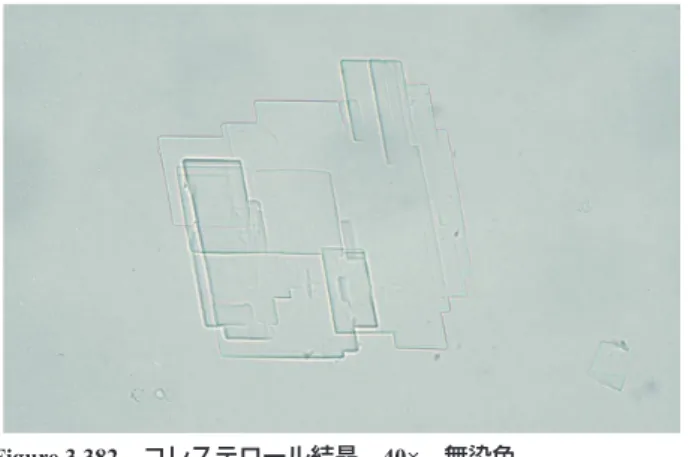
コレステロール結晶 40× 無染色
Cholesterol crystals 40× No staining
無色で歪んだ正方形や長方形の板状の結晶である。重なり合うと, シスチン結晶に類似する場合がある。シスチン結晶は六角形の輪 郭を有する。pH 6.5
Colorless, distorted square or rectangular plate-like crystals. When overlapping, they may look similar to cystine crystals, but cystine crystals have a hexagonal contour. pH 6.5
Figure 3.381 コレステロール結晶 40× 無染色
Cholesterol crystals 40× No staining
無色で歪んだ正方形や長方形の板状の結晶である。重なり合った 結晶である。pH 7.0
Colorless, distorted square or rectangular plate-shaped crystals that are overlapping. pH 7.0
シスチン結晶 40× 無染色
Cystine crystals 40× No staining
無色で六角形の板状結晶である。いく層も重なり合うとコレステ ロール結晶に類似する場合がある。酸性尿でみられる。細菌尿で は直ちに溶解し観察が困難な場合がある。pH 6.0
A colorless, hexagonal plate-shaped crystal. When the crystals overlap in many layers, they may look similar to cholesterol crystals. Cystine crystals are found in acidic urine. In bacterial urine, they may dissolve instantly, making them difficult to observe. pH 6.0
Figure 3.383 シスチン結晶 40× 無染色
Cystine crystals 40× No staining
無色で六角形の板状結晶である。重なり合った結晶である。六角 形の輪郭が残る。結晶の角は 120 度である。pH 6.5
Colorless, hexagonal plate-shaped crystals that are overlapping. A hexagonal outline remains. The angle of the crystal is 120°. pH 6.5
Figure 3.384
2,8-ジヒドロキシアデニン結晶 40× 無染色
2,8-Dihydroxyadenine crystals 40× No staining 褐色で円形の結晶である。先天性プリン代謝異常の APRT 欠損症 でみられる。水酸化カリウムで溶解する。pH 7.0
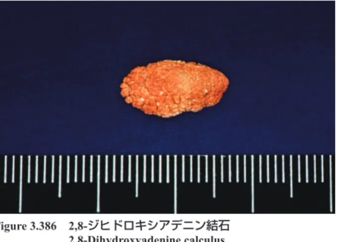
Brown, circular crystals found in cases of APRT deficiency of congenital purine metabolism abnormality. They dissolve in potassium hydroxide. pH 7.0 Figure 3.385 2,8-ジヒドロキシアデニン結石 2,8-Dihydroxyadenine calculus 2,8-ジヒドロキシアデニンは,腎臓より尿中に排泄されると結晶と なり結石を形成する。この結石は X 線透過性で,X 線撮影では結 石像は描出されない。
When 2,8-dihydroxyadenine is excreted in the urine from the kidney, it becomes crystalized and forms a calculus. As X-rays penetrate this stone, no stone image is produced via X-ray photography.
Figure 3.386
薬物結晶 40× 無染色
Drug crystals 40× No staining
投薬薬物によると思われる結晶である。多剤の投薬により薬物の 同定は困難なことが多い。
A crystal that appears to be derived from medication. Causative drug identification is often difficult due to multidrug administration.
Figure 3.387 薬物結晶 40× 無染色
Drug crystal 40× No staining 投薬薬物によると思われる結晶である。 A crystal that appears to be derived from medication.
VII
その他
Others
薬物結晶 40× 無染色
Drug crystal 40× No staining 投薬薬物によると思われる結晶である。 These crystals appear to be derived from medication.
Figure 3.389
ヘモジデリン顆粒 40× 無染色
Hemosiderin granule 40× No staining
無染色で暗褐色調の顆粒成分である。中央部はヘモジデリン顆粒 を含有した尿細管上皮細胞で,背景に散在した黄色の小型顆粒が ヘモジデリン顆粒である。
An unstained dark brownish granule component. In the center, there is a tubular epithelial cell containing hemosiderin granules, and the yellow small granules scattered in the background are hemosiderin granules.
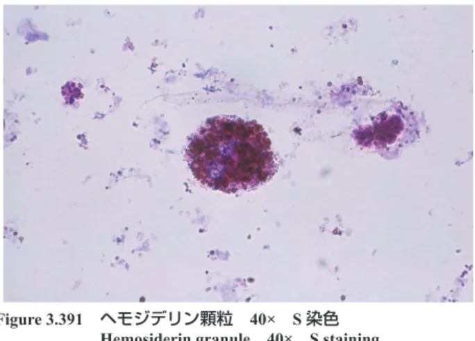
Figure 3.390 ヘモジデリン顆粒 40× S 染色
Hemosiderin granule 40× S staining
ヘモジデリン顆粒は,S 染色では暗赤褐色に染まり,顆粒円柱の 顆粒成分に類似する。このような場合は,無染色での観察や Berlin
blue染色で確認する。
Hemosiderin granules stain dark reddish brown with S staining and are similar to the granular components of granular casts. In such cases, confirmation must be performed by unstained observations or Berlin blue staining.