INTRODUCTION
Accessory spleen is a congenital anomaly char-acterized by ectopic splenic tissue separated from the main body of the spleen. It is a relatively com-mon condition that appears in 10% to 30% of autopsy findings and is usually asymptomatic (1). Torsion is a possible complication and occurs exceptionally. Its clinical presentation is characterized by a nonspecific
acute onset or recurrent abdominal pain. The diag-nosis is difficult, even with the modern imaging techniques (2, 3).
We report a case of acute torsion of an accessory spleen with situs inversus in a three-year-old girl presenting with acute abdomen. The literature is also reviewed
CASE REPORT
A three-year-old girl was admitted to our hospi-tal with an 11-day history of right flank pain with fever. Her medical history revealed an operation of coarctation of the aorta with situs inversus at
CASE REPORT
Torsion of an accessory spleen with situs inversus
in a child
Hiroki Ishibashi
1,2), Takehito Oshio
1), Tomoko Sogami
1), Akira Nii
1),Hiroki Mori
2),
and Mitsuo Shimada
2)1)
Department of Pediatric Surgery, National Kagawa Children’s Hospital, Kagawa, Japan ; and2)
De-partment of Pediatric Surgery and Pediatric Endoscopic Surgery, Tokushima University Hospital, Tokushima, Japan
Abstract : We present an unusual case of acute abdomen caused by torsion of an acces-sory spleen with situs inversus in a child. A three-year-old girl was admitted to our hos-pital with an 11-day history of right flank pain with fever. Her medical history revealed an operation of coarctation of the aorta with situs inversus at one month of age. Physi-cal examination revealed a right flank mass and tenderness. A contrast-enhanced CT scan showed a normally enhanced small spleen in the right upper quadrant and a 7.0 6.0 3.5 cm, hypodense, marginal enhancing mass in the right midabdomen adjacent to the intestine. An emergency laparotomy was decided upon with a preoperative diagno-sis as an acute abdomen. During surgery, a mass was found under the greater omentum and two accessory spleens of 1.5 cm in diameter were found surrounding the main spleen. Several loops of bowel were adherent to the mass. The loops of bowel were dissected away. A pediculated congested mass was observed as an accessory spleen emerging from the greater omentum. The mass was twisted on its vascular pedicle and strangulated. The necrotic mass was removed and the postoperative recovery was uneventful. Though tor-sion of an accessory spleen is extremely rare, it should be considered in the differential diagnosis of acute abdomen in childhood. J. Med. Invest. 59 : 220-223, February, 2012
Keywords : accessory spleen, torsion, situs inversus
Received for publication December 5, 2011 ; accepted Decem-ber 28, 2011.
Address correspondence and reprint requests to Hiroki Ishibashi, M.D., Department of Pediatric Surgery and Pediatric Endo-scopic Surgery, Tokushima University Hospital, Kuramoto - cho, Tokushima 770 - 8503, Japan and Fax : + 81 - 88 - 631 - 9698.
The Journal of Medical Investigation Vol. 59 2012 220
one month of age.
Physical examination revealed a right flank mass and tenderness with voluntary guarding. Laboratory findings disclosed only a white blood cell count of 12,060 /mm3with 64.6% neutrophils and a c-
reac-tive protein level of 3.38 mg/dl.
Plain abdominal X-ray films revealed a stomach bubble in the right upper abdomen without an ileus pattern (Fig. 1). An ultrasound examination was per-formed and revealed a well-defined oval mass with a central hypoechoic region, measuring 7.0 cm in
diameter, anterior to the right kidney (Fig. 2). A contrast-enhanced CT study showed a normally enhanced small spleen in the right upper quadrant and a 7.0
!
6.0!
3.5 cm, hypodense, marginal en-hancing mass in the right midabdomen adjacent to the intestine (Fig. 3).An emergency laparotomy was decided upon with a preoperative diagnosis as an infectious intestinal duplication.
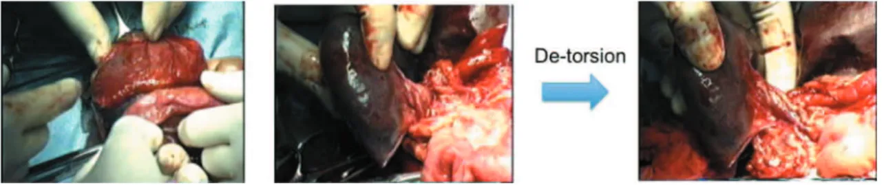
During surgery, a mass was found under the greater omentum and two accessory spleens of 1.5 cm in diameter were found surrounding the main spleen. Several loops of bowel were adherent to the mass. The loops of bowel were dissected away. A pediculated congested mass was observed as an ac-cessory spleen emerging from the greater omen-tum. The mass was twisted anti-clockwise by 360! around its pedicle and strangulated. The necrotic mass was removed (Fig. 4).
Fig. 3 :A contrast- enhanced CT study showed a normally enhanced small spleen in right upper quadrant (S) and a 7.0x6.0x3.5 cm, hypodense, marginal enhancing mass in the right midabdomen adjacent to the intestine (M).
Fig. 1 :The abdominal X- ray shows a stomach bubble in the right upper abdomen and reveals situs inversus.
Fig. 2 :The US shows a well - defined oval mass with a central hypoechoic region.
The patient‘s postoperative course was unevent-ful, and she was discharged in good condition on the 13th postoperative day. Histological examina-tion of the mass was consistent with hemorrhagic and necrotic splenic tissue.
DISCUSSION
Situs inversus is a congenital condition. It can either be partial or total. This entity is considered to have a genetic predisposition that is autosomal recessive with the defect being localised on the long arm of chromosome 14. The incidence of situs inver-sus has been thought to be 1 : 5,000 to 1 : 20,000. This condition may affect thoracic organs, abdomi-nal organs or both. It is associated with a number of other conditions such as Kartagener’s (bronchiec-tasis, sinusitis, situs inversus) and cardiac anomalies. There is no current evidence showing increased in-cidence of accessory spleens in patients with situs inversus.
The spleen arises during the fifth week of gesta-tion from mesenchymal tissue in the dorsal mesogas-trium between the pancreas and stomach. Incom-plete fusion of these mesenchymal buds during embryogenesis can result in the formation of ac-cessory spleens. Because the spleen is formed in the dorsal mesogastrium and then rotates to the left side, accessory spleens are always situated on the left side of the abdomen (4). Therefore, if there is situs inversus as in our case, it reverses left and right. Most are not separated very far and are vas-cularized by branches of the splenic artery.
The most common sites of an accessory spleen are the splenic hilum (75%), pancreatic tail (20%), splenic artery, gastrosplenic and splenocolic liga-ment, and gastrocolic ligament. Other rare locations are mesenterium, splenorenal ligament, greater omentum, jejunal wall, presacral area, adnexal region, scrotum, and mediastinum (1-6). Accessory
spleens are rarely found in more than two locations in one person (7).
Usually, accessory spleens are asymptomatic ; torsion and infarction, rupture with bleeding, and infection with abscess are very rare complications. Torsion of an accessory spleen with resultant in-farction may cause an acute abdomen (2, 6, 8, 9). In our case where the vascular pedicle of an acces-sory spleen was very long, there might be pedicu-lar torsion.
Alexander et al. (10) reported the first adult case with torsion of an accessory spleen in 1914. A review of the literature revealed only 16 cases (including ours) in the pediatric age of torsion of accessory spleen presenting as acute abdomen (1-3, 6, 7, 11-18) (Table 1). The youngest patient in the literature was described by Gardikis et al. (7) in a 14-day-old female patient. There is no case with situs inversus as in our case. All the reported cases went on to
Fig. 4 :A mass adhering to several loops of bowel was found on the greater omentum. A rounded violet mass measuring 7 cm in di-ameter with a 360!torsion around its vascular pedicle was resected.
Table 1 Case reports of children with torsion of an accessory spleen
Author Age Sex Location
Settle, 1940 4y M Gastrosplenic ligament 8y F Gastrocolic ligament Babcock et al., 1974 5y F Not stated
Onuigbo et al., 1978 9y M Jejunal wall 5y F Not stated Nutman et al., 1982 3.5y F Greater omentum Muller, 1988 15mo F Greater omentum Seo et al., 1994 10y M Greater omentum Chateil et al., 1996 15y F Not stated Valls et al., 1998 13y F Pancreatic tail Pèrez Fontàn et al., 2001 13y M Greater omentum Gardikis et al., 2005 14d F Greater omentum Mendi et al., 2006 12y F Splenic hilus Impellizzeri et al., 2009 12y M Mesenterium Yousef et al., 2010 12y M Greater omentum Present case, 2012 3y F Greater omentum
H. Ishibashi, et al. A case report and torsion of an accessory spleen 222
the excision of the strangulated accessory spleen, and in only one case described by Yousef et al. (18), was a laparoscopic excision performed.
Diagnostic imagings (US, CT) can provide abun-dant but aspecific information ; however, radiologi-cal findings are mandatory for preoperative diagno-sis despite not being always available in emergency situations. The correct diagnosis is still quite diffi-cult. In all the previous reported cases with torsion of an accessory spleen, as in our case, the diagno-sis was made in the operating room.
Wacha et al. (19) reported the value of diagnostic laparoscopy in finding an accessory spleen. Grinbaum
et al. (4) reported a laparoscopic excision of the
strangulated accessory spleen in a young adult and found that in the face of the acute presentation and the radiologic findings, laparoscopy was the pre-ferred action for definitive diagnosis and therapy.
In conclusion, torsion of an accessory spleen should be considered in the differential diagnosis of acute abdomen in childhood. The difficulty of distinguishing this extremely rare entity makes surgical intervention a necessity for diagnosis and treatment.
CONFLICT INTEREST
The author and co-authors disclosure that there are no conflicts of interest associated with the pre-sent study.
REFERENCES
1. Valls C, Monés L, Gumà A, López-Calonge E : Torsion of a wandering accessory spleen : CT findings. Abdom Imaging 23 : 194-195, 1998 2. Seo T, Ito T, Watanabe Y, Umeda T : Torsion
of an accessory spleen presenting as an acute abdomen with an inflammatory mass. US, CT, and MRI findings. Pediatr Radiol 24 : 532-534, 1994
3. Pérez Fontán FJ, Soler R, Santos M, Facio I : Accessory spleen torsion : US, CT and MR find-ings. Eur Radiol 11 : 509-512, 2001
4. Grinbaum R, Zamir O, Fields S, Hiller N : Tor-sion of an accessory spleen. Abdom Imaging 31 : 110-112, 2005
5. Pearigen PD : Unusual causes of abdominal pain. Emerg Med Clin North Am 14 : 593-610, 1996
6. Impellizzeri P, Montalto AS, Borruto FA, Antonuccio P, Scalfari G, Arena F, Romeo C : Accessory spleen torsion : rare cause of acute abdomen in children and review of literature. J Pediatr Surg 44 : e15-18, 2009
7. Gardikis S, Pitiakoudis M, Sigalas I, Theocharous E, Simopoulos C : Infarction of an accessory spleen presenting as acute abdomen in a neonate. Eur J Pediatr Surg 15 : 203-205, 2005
8. Habib FA, Kolachalam RB, Swason K : Abscess of an accessory spleen. Am Surg 66 : 215-218, 2000
9. Coote JM, Eyers PS, Walker A, Wells IP : Intra-abdominal bleeding caused by spontaneous rupture of an accessory spleen : the CT find-ings. Clin Radiol 54 : 689-691, 1999
10. Alexander RC, Romanes A : Accessory spleen causing acute attacks of abdominal pain. Lancet 184 : 1089-1091, 1914
11. Settle EB : The surgical importance of acces-sory spleens : with report of two cases. Am J Surg 50 : 22-26, 1940
12. Babcock TL, Coker DD, Haynes JL, Conklin HB : Infarction of an accessory spleen causing an acute abdomen. Am J Surg 127 : 336-337, 1974
13. Onuigbo WI, Ojukwu JO, Eze WC : Infarction of accessory spleen. J Pediatr Surg 13 : 129-130, 1978
14. Nutman J, Mimouni M, Zer M, Grünebaum M, Varsano I : Accessory spleen as a cause of an abdominal mass. Z Kinderchir 37 : 71-72, 1982 15. Müller H, Schneider H, Rückauer K, Greiner P : Accessory spleen torsion. Clinical picture, sonographic diagnosis and differential diagno-sis. Klin Padiatr 200 : 419-421, 1988
16. Chateil JF, Arboucalot F, Perel Y, Roy D, Vergnes P, Diard F : Acute torsion of an acces-sory spleen. J Radiol 77 : 209-211, 1996 17. Mendi R, Abramson LP, Pillai SB, Rigsby CK :
Evolution of the CT imaging findings of acces-sory spleen infarction. Pediatr Radiol 36 : 1319-22, 2006
18. Yousef Y, Cameron BH, Maizlin ZV, Boutross-Tadross O : Laparoscopic excision of infarcted accessory spleen. J Laparoendosc Adv Surg Tech A 20 : 301-303, 2010
19. Wacha M, Danis J, Wayand W : Laparoscopic resection of an accessory spleen in a patient with chronic lower abdominal pain. Surg En-dosc 16 : 1242-3, 2002