Review J Tokyo Wom Med Univ 89(Extra1): E110-E117, 2019 Jul
Role of Microenvironment and Transient Tissue Hypoxia
in Regeneration of the Endometrium
Asuka Yoshii,1
Hisashi Ueta,2
and Shuji Kitahara1
1
Department of Anatomy and Developmental Biology, School of Medicine, Tokyo Women s Medical University, Tokyo, Japan
2
Department of Anatomy (Macro), Dokkyo Medical University, Tochigi, Japan (Accepted October 29, 2018)
During various events in the reproductive period (viz., menstruation and delivery) of a woman, the endometrium displays phenomenal regenerative ability to repeat the cycle of shedding and regeneration. However, the mecha-nisms responsible for complete inner membrane repair and regeneration remain mostly unexplored. Gynecologi-cal diseases, such as placenta accreta and implantation failure, are caused by insufficient repair of the en-dometrium. However, because the mechanism behind these diseases are unclear, there are no known preventive methods. Changes in oxygen concentration in the endometrium are known to be involved in defense against bac-teria and trophoblast invasion for the establishment of pregnancy. Additionally, it is also thought to affect angio-genesis and tissue repair. The difficulty of obtaining human samples, hampers investigation along this line of re-search. Using a postpartum mouse model and mice with sub-involuted uterus, we have reported that changes in intrauterine oxygen concentration, including hypoxia, due to involution of the uterine smooth muscle are respon-sible for scarless endometrial repair. Elucidation of the regenerating mechanism of the endometrium provides possibilities to prevent complications in obstetrics and gynecology, and a strong basis for scarless tissue repair through a new approach of generating microenvironments based on oxygen concentration adjustment in normal tissues. In this review, we discuss the role of microenvironment in the process of healing and regeneration of the endometrium.
Key Words: myometrial contraction, hypoxia, regenerative endometrium, macrophage, scarless wound healing
Introduction
The uterus is a mesoderm-derived tissue composed of the uterine smooth muscle and the endometrium. The uterine smooth muscle, with its inherent con-tractility, can dramatically change its volume dur-ing the reproductive period and constantly changes the intrauterine environment to adjust the oxygen concentration in the tissue.1
The endometrium, on the contrary, has a high regenerative ability, unlike
other tissues, and repeatedly cycles through prolif-eration, differentiation, shedding, and regeneration. The endometrium has drawn considerable atten-tion as the representaatten-tion of scarless tissue, with its ability to repair without forming scars.2,3
However, the mechanisms of regeneration in the en-dometrium are still unknown, and there are several obstetric and gynecological conditions, such as im-plantation failure, malposition of the placenta,
pla-Corresponding Author: Asuka Yoshii and Shuji Kitahara, Department of Anatomy and Developmental Biology, School of Medicine, Tokyo Women s Medical University, 8-1 Kawada-cho, Shinjuku-ku, Tokyo 162-8666, Japan. [email protected] doi: 10.24488/jtwmu.89.Extra1_E110
Copyright Ⓒ 2019 Society of Tokyo Women s Medical University. This is an open access article distributed under the terms of Creative Commons Attribution License (CC BY), which permits unrestricted use, distribution, and reproduction in any medium, provided the original source is properly credited.
centa accreta,4,5
and excessive bleeding from uterine sub-involution, for which no preventive methods are currently available. Basic research to better un-derstand the mechanism underlying these diseases are imperative. However, factors such as the diffi-culty of obtaining human samples, and the limita-tion of animals with similar histology and hormone cycle as humans, hinders progress in this area of re-search.
To date, there have been reports of infection pre-vention and trophoblast infiltration associated with transient hypoxia in the endometrium, particularly during the establishment of pregnancy.6
As a dogma of menstruation mechanism,2
their relation-ship to angiogenesis and restorative regeneration under hypoxia has been discussed; however, there have only been a few basic research reports
in vivo.2,7,8
While focusing on uterine volume change before and after parturition in a postpartum mouse model, and while studying a mouse model with sub-involuted uterus, we found that an intrauterine hy-poxic environment promotes endometrial regenera-tion.9
We also found that a transient hypoxic envi-ronment occurs in the uterus because the area of the uterine cavity is sharply reduced after delivery. Immunohistochemical observations revealed that during the repair process, there is transient infiltra-tion of M 2 macrophages, primarily in the endometrium-deficient area, with peak infiltration on postpartum day (D) 3. When the normal postpar-tum mouse model was compared with the sub-involuted mouse model using theβ-2 adrenergic ceptor agonist, a difference in the time to uterine re-covery and hematoma formation was observed. Furthermore, at the transcript level, there were dif-ferences in the amounts of hypoxia inducible factor 1 a ( HIF 1 a ) , vascular endothelial growth factor ( VEGF ) , and transforming growth factor beta (TGFB). These results show that uterine contrac-tion not only converges the hemorrhage from the vascular stump in endometrial defects, but also in-duces macrophages by adjusting oxygen concentra-tion in the tissues by creating a transient low oxy-gen environment, which creates a
microenviron-ment for inner membrane repair and regeneration. With this review, we aim to clarify the mechanism of endometrial repair and regeneration, describe a research approach to investigate scarless tissue re-pair, and to observe the construction of the mi-croenvironment induced by the regulation of oxy-gen concentration in normal tissues. These insights are expected to contribute to the development of preventive medicine in the future.
Volume Change and Local Hypoxia Affect Myometrial Contraction in Postpartum Uterus
During pregnancy, the uterus changes its size dra-matically through growth and stretching of the uterine smooth muscle. The hemorrhage after de-livery is controlled by contraction of the myometrium.10
Postpartum hemorrhage is one of the major causes of maternal mortality worldwide, and is primarily caused by sub-involution.4
It is also believed that autolysis and phagocytosis during in-volution leads to complete tissue repair. Clinically, medical intervention is required for the prevention and therapy of postpartum hemorrhage.
Because of the decrease in the size of the uterus, but not the number of uterine muscles during invo-lution, the uterus of parous women are slightly dif-ferent from that of non-parous women. Therefore, our study used mice in their first pregnancy to ex-clude these factors. Changes in uterine size and in-trauterine oxygen concentration based on the de-gree of uterine smooth muscle contraction, were compared in normal postpartum mice ( group A ) , and mice given ritodrine, aβ-2 adrenergic receptor agonist, which is used clinically for smooth muscle relaxation (group B). The size of the uterus after de-livery was assessed using the ratio of the total area of the uterine cross section in groups A and B. The time to return to their prepregnant size in group B was found to be 4 days later than in group A. In both groups, the rates of involution increased sig-nificantly until D 2 ( Figure 1 ) . To assess the ischemic states associated with involution in the postpartum uteri, we examined hypoxia using pi-monidazole immunohistochemistry. Pipi-monidazole in the endometrium was strongly detected on D3, and was more abundant in group A than in group B.9
Figure 1 Rates of change in postpartum uterine size.
Group A, sham-operated mice, with a mini osmotic pump for administration of saline after caesarean sec-tion (CS). Group B, mice treated with ritodrine (10 mg/mL) after CS. The total areas of the cross section, including the lumen space, in group A (white square) and group B (black square) mice were compared with those of the non-pregnant uteri (n = 8; the mean value was scored as 1). The rate of change in the volume was significant until day (D) 2 in both groups. In group A, the size of the uterus returned to its prepregnancy volume by D10 and in group B, this was only attained on D14 (n = 5-8 mice in each column). *p<0.05.
Time after operation (day)
Ratio of total area of uterine cross -section non-pr egnan t uter ine (= 1) 0 1 2 3 4 5 6 7 8 9 10 1 2 3 5 7 10 14 * * * * 㻳㼞㼛㼡㼜㻭㻦㻌㼂㼑㼔㼕㼏㼘㼑 㻳㼞㼛㼡㼜㻮㻦㻌㻾㼕㼠㼛㼐㼞㼕㼚㼑
Figure 2 Expression of HIF1a and VEGFa related to postpartum tissue repair.
(a) Double positive cells for HIF1a and VEGFa were seen in the healing endometrium (arrow head). Scale bar, 25 μm. (b-1,2) Expression of Hif1a and Vegfa transcripts are shown as fold increases compared to the control (untreated samples) after normalization to expression of hypoxanthine phosphoribosyltransferase. The Hif1a transcript level peaked on day (D) 1 in group A, and was later for group B. The Vegfa levels peaked on D2 in group A (p<0.05). However, no significant change was seen in the Vegfa levels over a period of 3 days in group B. *p<0.05. Data are presented are mean ± SD.
HIF1a, hypoxia inducible factor 1a; VEGFa, vascular endothelial growth factor a.
HIF1a
VEGFa
a
Quantif icaion o f Hif1a m RNA Ex pr ession Fold incr ease 0 0.5 1 1.5 2 1 2 3 * * *Time aŌer operaƟon (day)
b-1
Q u a n ti fi c a io n o f Vegfa m RNA Ex pression Fold increase 0 1 2 3 4 5 1 2 3 㻳㼞㼛㼡㼜㻭㻦㻌㼂㼑㼔㼕㼏㼘㼑 㻳㼞㼛㼡㼜㻮㻦㻌㻾㼕㼠㼛㼐㼞㼕㼚㼑 *Time aŌer operaƟon (day)
b-2
The pimonidazole immunostaining pattern re-turned to the prepregnant pattern in both groups by D7.
It has been known that hypoxia triggers epithelial-mesenchymal transition ( EMT ) of en-dometrial cells.11,12
Emerging evidence suggests that EMT contributes to wound healing,13,14
tissue regen-eration,15
the invasion of endometrium16,17
and im-plantation of the embryo15
Furthermore, HIF1a is a transcription factor induced by hypoxia.18,19
and is responsible for hypoxic stress response in cells. It is also known to be involved in homeostatic mecha-nisms, such as maintenance of stem cells and con-trol of inflammation, including hypoxic adaptive re-sponses.20,21
Therefore, to observe hypoxic state in the uterine tissue, changes in HIF 1 a during the postpartum process was examined. In group A, the amount of HIF1a peaked at D1 after giving birth and rapidly decreased after D2 (Figure 2b-1) , to-gether with rapid uterine contraction. In addition, secretion of VEGF peaked after D2 postpartum, fol-lowing the secretion peak of HIF1a (Figure 2b-2). It has been reported that hypoxia increases VEGF ex-pression via HIF 1 a in the endometrial epithelial cells and primary human endometrial stromal cells
in vitro.21,22
between HIF1a and VEGF in vivo (Figure 2), sug-gesting that uterine contraction promotes angio-genesis.
HIF1 a is also known to be associated with de-creased TGFB1 production. Some studies have sug-gested that fibrosis can develop in a TGFB 1-independent manner.23
TGFB1 is a pleiotropic factor that is important in stimulating wound contraction and acts as a chemoattractant for monocytes, macrophages, and fibroblasts. Murray and Wynn demonstrated models of idiopathic pulmonary fibro-sis in which the pro-fibrotic function of macro-phages had been attributed to their production and activation of the pro-fibrotic cytokine TGFB1.23
Con-versely, TGFB3 stimulates fibroblast cell migration into healing wounds, resulting in marked improve-ment or absence of scarring.24
Low levels of TGFB1 and high levels of TGFB3 have been described as key factors in scarless wound healing. In our study, the ratio of TGFB 3 to TGFB 1 was significantly higher in the normal postpartum group and the hy-poxic environment owing to uterine contraction played a crucial role in scarless wound healing. In group B, HIF1a transcript levels increased after D2, which suggests that uterine contractions up to D1 after birth, immediately after parturition, is impor-tant for hemostasis and regeneration.
Tissue Repair and Local Microcirculation in the Postpartum Uteri
Ferenczy et al.25
reported that endometrial regen-eration from stromal cells occurs within 48 h after endometrial peeling in humans, whereas in rats it occurs 60 h after endometrial peeling. Regeneration of the stromal tissue begins after the endothelial epithelium is regenerated, which is similar to the process of normal wound healing in other tissues. In addition, Ludwig and Spornitz26
report that the en-dometrial vascular vessels are repaired on day 5 of menstruation.
Furthermore, during the endometrial regenera-tion process, the presence of the estrogen proges-terone receptor in the uterine glandular epithelium was reported, and the stromal cell is known to be hormone-dependent in the endoplasmic regenera-tion following compleregenera-tion of epithelial repair.27,28
In-terestingly, even in cases lacking ovarian hormonal support, hemorrhage from the endometrium stops and endometrial regeneration is completed, sug-gesting that there is another regeneration-promoting factor that is different from the hor-mone.2
Endometriosis is an estrogen-dependent gy-necological disease where endometrium-like tissue grows outside the uterine cavity. The pathogenesis of endometriosis is multifactorial, and factors such as genetics, environment, the immune system, and intrinsic abnormalities in the endometrium play an intrinsic role.29-31
In our study, we performed bilateral salpingo-oophorectomy during delivery and observed regen-eration in the absence of the ovarian hormone.9
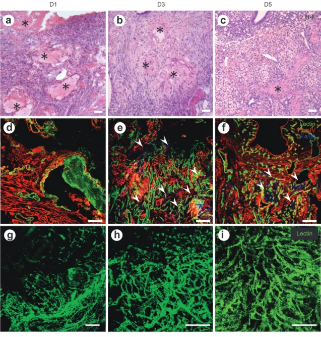
Dur-ing endometrial regeneration after placental abrup-tion, massive hemorrhage and defects in the en-dometrium were seen on D1. The leakage of lectin from the edge of the vessels and the extravasation of lectin gradually reduced and ceased by D3. The reduction of uterine size and hemostasis occurred concurrently and the epithelium was repaired by D 5. Type IV positive continuous epithelium was seen on D5 (Figure 3d-f). Although the speed of recovery and degree of hematoma formation of the postpar-tum uterus are different, the endometrial epithelial regeneration was completed by D5 in both the nor-mal postpartum and sub-involuted mouse models. As previously reported, the endometrium regener-ates without the ovarian hormone, thereby return-ing to its initial point for the next cycle.
Scarless Wound Healing and Role of Inflammatory Cells, Including M2 Macrophages
The wound healing process consists of the four phases, hemostasis, inflammation, proliferation and remodeling, which involves complex interactions between the epithelium, immune cells, and extracel-lular matrix components.32,33
The endometrium is particularly abundant with cells of the myeloid line-age, including neutrophils, eosinophils, macro-phages and monocytes. The changes in the en-dometrium can be regarded as an inflammatory process.2
Macrophages differentiated from mono-cytes play critical roles in regulating a wide range of processes, in all stages of repair. Although early
Figure 3 Morphological time course of postpartum uteri.
(a-c) Hematoxylin and eosin staining. (d-f) Immunohistochemical staining for lectin (green), type IV basement membrane (red), and F4/80 (blue). (g-i) Whole mount observed under a confocal laser microscope for tomato lectin-labeled vascular architecture. Massive hemor-rhage and fibrin deposits (marked with *) were seen on day (D) 1 (a), which gradually di-minished in accordance with uterine size reduction (b, c). The continuous type IV positive basement membrane showed that the epithelium regenerated within D5 of postpartum (d-f). Infiltrated F4/80 positive macrophages were seen in the repairing endometrium and quickly disappeared after D5 (d-f). To characterize microcirculation in postpartum uteri, FITC-labeled tomato lectin was administered. Tomato lectin-labeled vascular architecture can be seen in the uteri and microvascular repair occurs rapidly in the endometrium. The * indicate the location of fibrin. Arrowheads indicate the infiltrated macrophages. Scale bars; 50 μm (a-f), 200 μm (g-i).
㻖
㻖
㻖
b
a
c
Lectin TypeIV F4/80e
d
f
g
h
i
H-E Lectin㻖
㻖
㻖
㻖
㻖
D3 D1 D5research focused on the role of macrophages as scavenger cells in wound healing, the complex roles of monocytes and macrophages in tissue repair, and mechanisms of fibrosis and tissue regeneration are
being investigated.34
If macrophages are depleted in early stages of injury, the inflammatory response is often diminished.35
Their removal can also result in decreased wound debridement and lead to delayed
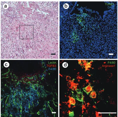
Figure 4 Microscopic changes in the uterine tissue on postpartum day 3.
(a) Hematoxylin and eosin staining. (b) TUNEL (green) and Dapi (blue) staining. (c) Immuno-histochemical staining for lectin (green), TGFB3 (red), and F4/80 (blue). (d) Enlarged hema-toxylin and eosin staining of (a). F4/80 (green) and Arginase 1 (red). Many F4/80 positive macrophages are seen beneath the epithelial defect. Scale bar, 50 µm (a-d).
b
c
a
TUNELDAPI Lectin TGFB3 F4/80 F4/80 Arginase1d
rates of structural and functional recovery.36
Recent research suggested two phenotypes of macro-phages, M1 and M2, based on their gene expression profile in response to specific stimuli.23,37
M1 cells are classically activated macrophages, comprised of im-mune effector cells with an acute inflammatory phenotype for host defense, and M2 cells are alter-natively activated macrophages that regulate the maintenance of tolerance and tissue repair. Hesse et al.38
and Briken and Mosser39
demonstrated that al-ternatively activated macrophages play important roles in the clearance of helminthic parasites. M 2 macrophages synthesize arginase, an enzyme that inhibits nitric oxide production, which kills cellular pathogens and allows them to produce ornithine, a precursor of hydroxyproline and polyamines. In our previous study, we showed that M2 macrophages increased in ischemic sites in early phase of healing, with a rapid response to hypoxia, which involved
al-teration in expression of a wide array of genes9
(Fig-ure 4). We suggest that the balance of M1 and M2
macrophages is controlled by transient hypoxia with uterine contraction, which plays a predomi-nant role in scarless endometrial healing.
Conclusion
We propose that uterine contractility promotes scarless endometrial regeneration, with infiltration of inflammatory cells under transient hypoxia. Un-derstanding the normal endometrial healing proc-ess provides greater insight into several obstetrical and gynecological complications resulting from en-dometrial defects caused by its inadequate repair. Furthermore, these results are useful in the preven-tion and management of various healing disorders involving other tissue.
Acknowledgments
Anatomy (Macro), Dokkyo Medical University, Tochigi, Japan for the patient guidance, encouragement and ad-vice he has provided. We are also thankful to Dr. Hide-hiko Nakanishi, Kitasato University Hospital, Tokyo, Ja-pan for clinical advice he has provided, research team members from the Department of Anatomy (Macro ) , Dokkyo Medical University, Tochigi, Japan and the De-partment of Anatomy of Developmental Biology, Tokyo Women s Medical University, Tokyo, Japan.
Part of our study was supported by the Global Center of Excellence program, Multidisciplinary Education and Research Center for Regenerative Medicine, from the Ministry of Education, Culture, Sports Science, and Technology, Japan.
Conflicts of Interest: The authors declare no con-flicts of interest associated with this manuscripts.
References
1.Lee YH, Bae YS: Phospholipase D2 downregulation induces cellular senescence through a reactive oxy-gen species-p53-p21 Cip1 / WAF 1 pathway. FEBS Lett 588: 3251―3258, 2014
2.Salamonsen LA: Tissue injury and repair in the fe-male human reproductive tract. Reproduction 125: 301―311, 2003
3.Cousins FL, Kirkwood PM, Saunders PT et al: Evi-dence for a dynamic role for mononuclear phago-cytes during endometrial repair and remodelling. Sci Rep 6: 36748, 2016
4.Bohlmann MK, Rath W : Medical prevention and treatment of postpartum hemorrhage : a compari-son of different guidelines. Arch Gynecol Obstet 289: 555―567, 2014
5.Rosen T: Placenta accreta and cesarean scar preg-nancy: overlooked costs of the rising cesarean sec-tion rate. Clin Perinatol 35: 519―529, 2008
6.Daikoku T, Matsumoto H, Gupta RA et al: Expres-sion of hypoxia-inducible factors in the peri-implantation mouse uterus is regulated in a cell-specific and ovarian steroid hormone-dependent manner. Evidence for differential function of HIFs during early pregnancy. J Biol Chem 278 : 7683 ― 7691, 2003
7.Tsuzuki T, Okada H, Cho H et al: Hypoxic stress si-multaneously stimulates vascular endothelial growth factor via hypoxia-inducible factor-1 alpha and inhibits stromal cell-derived factor-1 in human endometrial stromal cells. Hum Reprod 27: 523―530, 2012
8.Tranquilli AL, Landi B: The origin of pre-eclampsia: from decidual hyperoxia to late hypoxia. Med Hy-potheses 75: 38―46, 2010
9.Yoshii A, Kitahara S, Ueta H et al: Role of uterine contraction in regeneration of the murine postpar-tum endometrium. Biol Reprod 91: 32, 2014
10.Cunningham FG, Leveno KJ, Bloom SL et al: Wil-liams Obstetrics, (25th edition) . Mc Graw-Hill Edu-cation, New York (2018)
11.Xiong Y, Liu Y, Xiong W et al: Hypoxia-inducible factor 1alpha-induced epithelial-mesenchymal tran-sition of endometrial epithelial cells may contribute to the development of endometriosis. Hum Reprod 31: 1327―1338, 2016
12.Liu H, Du Y, Zhang Z et al: Autophagy contributes to hypoxia-induced epithelial to mesenchymal tran-sition of endometrial epithelial cells in endometrio-sis. Biol Reprod 2018, doi: 10.1093/biolre/ioy128 13.Kim YS, Yi BR, Kim NH et al: Role of the
epithelial-mesenchymal transition and its effects on embry-onic stem cells. Exp Mol Med 46: e108, 2014 14.Ahmed N, Maines-Bandiera S, Quinn MA et al:
Mo-lecular pathways regulating EGF-induced epithelio-mesenchymal transition in human ovarian surface epithelium. Am J Physiol Cell Physiol 290: C1532―C 1542, 2006
15.Kalluri R, Weinberg RA : The basics of epithelial-mesenchymal transition. J Clin Invest 119 : 1420 ― 1428, 2009
16.Bartley J, Jülicher A, Hotz B et al: Epithelial to mes-enchymal transition (EMT) seems to be regulated differently in endometriosis and the endometrium. Arch Gynecol Obstet 289: 871―881, 2014
17.Proestling K, Birner P, Gamperl S et al: Enhanced epithelial to mesenchymal transition ( EMT ) and upregulated MYC in ectopic lesions contribute in-dependently to endometriosis. Reprod Biol Endocri-nol 13: 75, 2015
18.Semenza GL, Wang GL: A nuclear factor induced by hypoxia via de novo protein synthesis binds to the human erythropoietin gene enhancer at a site required for transcriptional activation. Mol Cell Biol 12: 5447―5454, 1992
19.Weidemann A, Johnson RS: Biology of HIF-1alpha. Cell Death Differ 15: 621―627, 2008
20.Majmundar AJ, Wong WJ, Simon MC : Hypoxia-inducible factors and the response to hypoxic stress. Mol Cell 40: 294―309, 2010
21.Schödel J, Oikonomopoulos S, Ragoussis J et al: High-resolution genome-wide mapping of HIF-binding sites by ChIP-seq. Blood 117 : e 207 ― e 217, 2011
22.Critchley HO, Osei J, Henderson TA et al: Hypoxia-inducible factor-1 alpha expression in human en-dometrium and its regulation by prostaglandin E-series prostanoid receptor 2 (EP2). Endocrinology 147: 744―753, 2006
23.Murray PJ, Wynn TA: Obstacles and opportunities for understanding macrophage polarization. J Leu-koc Biol 89: 557―563, 2011
24.O Kane S, Ferguson MW : Transforming growth factor beta s and wound healing. Int J Biochem Cell Biol 29: 63―78, 1997
25.Ferenczy A: Studies on the cytodynamics of human endometrial regeneration. II. Transmission electron microscopy and histochemistry. Am J Obstet
Gyne-col 124: 582―595, 1976
26.Ludwig H, Spornitz UM: Microarchitecture of the human endometrium by scanning electron micros-copy : menstrual desquamation and remodeling. Ann N Y Acad Sci 622: 28―46, 1991
27.Okulicz WC, Scarrell R : Estrogen receptor alpha and progesterone receptor in the rhesus en-dometrium during the late secretory phase and menses. Proc Soc Exp Bio Med 218: 316―321, 1998 28.Bigsby RM: Control of growth and differentiation of
the endometrium : the role of tissue interactions. Ann N Y Acad Sci 955: 110―117, 2002
29.Giudice LC, Kao LC : Endometriosis. Lancet 364 : 1789―1799, 2004
30.Fung JN, Mortlock S, Girling JE et al: Genetic regu-lation of disease risk and endometrial gene expres-sion highlights potential target genes for en-dometriosis and polycystic ovarian syndrome. Sci Rep 8: 11424, 2018
31.Xiong W, Zhang L, Yu L et al: Estradiol promotes cells invasion by activating beta-catenin signaling pathway in endometriosis. Reproduction 150: 507― 516, 2015
32.Singer AJ, Clark RA: Cutaneous wound healing. N Engl J Med 341: 738―746, 1999
33.Mahdavian Delavary B, van der Veer WM, van Eg-mond M et al: Macrophages in skin injury and re-pair. Immunobiology 216: 753―762, 2011
34.Wynn TA, Chawla A, Pollard JW: Macrophage biol-ogy in development, homeostasis and disease. Na-ture 496: 445―455, 2013
35.Duffield JS, Forbes SJ, Constandinou CM et al: Se-lective depletion of macrophages reveals distinct, opposing roles during liver injury and repair. J Clin Invest 115: 56―65, 2005
36.Zhang MZ, Yao B, Yang S et al: CSF-1 signaling me-diates recovery from acute kidney injury. J Clin In-vest 122: 4519―4532, 2012
37.Wynn TA, Vannella KM : Macrophages in Tissue Repair, Regeneration, and Fibrosis. Immunity 44 : 450―462, 2016
38.Hesse M, Modolell M, La Flamme AC et al: Differ-ential regulation of nitric oxide synthase-2 and arginase-1 by type 1 / type 2 cytokines in vivo : granulomatous pathology is shaped by the pattern of L-arginine metabolism. J Immunol 167 : 6533 ― 6544, 2001
39.Briken V, Mosser DM: Editorial: switching on ar-ginase in M2 macrophages. J Leukoc Biol 90: 839― 841, 2011 組織内酸素濃度変化を背景とした子宮内膜再生における微小環境変化 1東京女子医科大学医学部解剖学・発生生物学講座 2獨協医科大学解剖学(マクロ) ヨ シ イ ア ス カ ウ エ タ ヒ サ シ キタハラ シュウジ 吉井明日香1 ・上田 祐司2 ・北原 秀治1 子宮内膜は月経や分 といった内膜の脱落と再生を繰り返す驚異的再生能力を持ち,瘢痕を残さず再生を遂げ るため,非瘢痕性組織の代表として注目されている.また,自動収縮能を持つ子宮平滑筋はその組織内酸素濃度の 調整を自ら行い,子宮内の環境を絶えず変化させている.このような高い再生能力と特殊な再生環境を背景とした 子宮内膜再生のメカニズムについては未だ不明な点が多く,再生不全に起因する癒着胎盤や着床不全などの産婦 人科合併症も近年増加傾向であるにも関わらず,明確な予防法がないのも現状である. 子宮内膜における組織内酸素濃度変化は,特に妊娠成立時の感染予防やトロホブラスト浸潤に関わることが既 に知られており,血管新生や修復再生との関わりも論じられている.しかしながら in vivo での研究報告が未だ少 ない.疾患のメカニズム解明には基礎研究によるアプローチが重要であるが,本テーマの研究遂行には,ヒト検体 の収集が困難であること,また,ヒトと同じ性周期を持ち,かつ組織学的にもヒトに類似するような実験動物が限 られている点も,基礎研究が進まない理由と考えられる.われわれはこれまでに,子宮容積変化に着目した産褥マ ウスモデルおよび子宮復古不全マウスモデルを独自に作製し,子宮内膜修復に関する研究を行ってきた.その中 で,子宮平滑筋収縮が誘引する組織内低酸素環境が子宮特有の再生環境背景として存在し,非瘢痕性修復を担うメ ディエーターが産生され,子宮内膜組織修復を促進させる事象について報告した.これら研究結果を含め,子宮内 膜治癒および再生における微小環境について論じる.