緒 言
海綿静脈洞部硬膜動静脈瘻(cavernous sinus dural arteriovenous fistula;CS dAVF)に対する血管内治療 は,コイルを用いた経静脈的塞栓術が第一選択の治療法 とされ,治癒率は80~100%と報告されている5).しかし,
シャント部位が複雑なCS dAVFでは経静脈的にCSに到 達できたとしてもcoil packingで完全閉塞が得られない ことがあり,また過度のcoil packingは脳神経麻痺を来 たす恐れがある10).我々は,このような症例にn-butyl cyanoacrylate(n-BCA,Histoacryl; B.Braun, Melsungen, Germany)を用いた経静脈的塞栓術を行ったので,手技
上の工夫,留意点,有効性について報告する.
対象と方法
当院および関連施設で治療した64例のCS dAVFのう ち,n-BCAを用いた経静脈的塞栓術を施行した3例を 対象とした.塞栓術は全例,全身麻酔下に,経大腿静脈 的に下錐体静脈洞(inferior petrosal sinus;IPS)経由 で行った.ガイディングカテーテルは4Fr/6Frコアキ シャルシステム(4FrセルリアンG, Medikit, Tokyo, Japan/6Fr Envoy XB, Cordis Neurovascular, Miami Lakes, FL, USA)を用い,4FrインナーカテーテルをIPSに 誘導した.マイクロカテーテルは,Renegade(Boston
海綿静脈洞部硬膜動静脈瘻に対するn-Butyl Cyanoacrylateを用いた経静脈的塞栓術
宮本直子1) 内藤 功1) 高玉 真1)
岩井丈幸1) 清水立矢2)
Transvenous Embolization Using n-Butyl Cyanoacrylate for Cavernous Sinus Dural Arteriovenous Fistulas
Naoko MIYAMOTO1) Isao NAITO1) Shin TAKATAMA1) Tomoyuki IWAI1) Tatsuya SHIMIZU2)
1) Department of Neurosurgery, Geriatrics Research Institute and Hospital 2) Department of Neurosurgery, Gunma University Graduate School of Medicine
●Abstract●
Objective: Transvenous embolization using coils is one of the standard treatment options for cavernous sinus dural arteriovenous fistulas (CS dAVFs). However, complete obliteration is not always obtained by transvenous coil embolization alone. We report here three patients treated by transvenous infusion of n-butyl cyanoacrylate (n-BCA), and discuss the technical aspects, safety and efficacy of this technique.
Method: Two patients presented with ocular symptoms and one with intracerebral hemorrhage. Affected CS was accessed via transfemoral- inferior petrosal sinus route in all patients. One patient was treated with transvenous n-BCA infusion alone and two patients with transvenous n-BCA infusion after coil embolization.
Result: Complete obliteration was achieved in two patients, and reduction of the shunt with obliteration of a dangerous drainage in one patient.
No complications associated with the procedures were observed in any of the patients.
Conclusion: Transvenous n-BCA infusion is an effective alternative in CS dAVFs refractory to transvenous coil embolization.
●Key Words●
cavernous sinus, dural arteriovenous fistula, n-butyl cyanoacrylate, transvenous embolization
(Received March 6, 2010:Accepted March 31, 2010)
1)老年病研究所附属病院 脳神経外科 2)群馬大学大学院医学系研究科 脳神経外科
<連絡先:宮本直子 〒371-0847 群馬県前橋市大友町3-26-8 E-mail:[email protected]>
3例中2例で完全閉塞が得られた.1例でシャントが 残存したが頭蓋内静脈への逆流は消失しその目的が達成 された.手技に伴う合併症はなかった.
症例呈示
1.症例1
患者:71歳,女性.
複視,右眼充血,拍動性耳鳴で発症し,6ヵ月後に意 識障害,構音障害,左半身麻痺が出現した.CTで右前 頭葉,側頭葉に著明な浮腫を伴う皮質下出血を認め,
MRIで右CS dAVFが疑われ当院に紹介された.第1回 経静脈的塞栓術を施行した.血管造影では両側内頚動脈
(internal carotid artery;ICA)からmeningohypophyseal trunk(MHT),眼動脈を介しrecurrent meningeal artery,
外頚動脈(external carotid artery;ECA)からinternal maxillar artery(IMA),middle meningeal artery(MMA),
ascending pharyngeal artery(APhA)などが流入動脈と なり,右CS内側後方からintercavernous sinusにシャン トし,両側上眼静脈(superior orbital vein;SOV),両 側IPS,右superficial sylvian vein(SSV)に導出して いた.右IPS経由にてintercavernous sinus,右CS内側 をコイルで塞栓した.僅かにシャントが残存したが SSVへの逆流が消失したため終了した.follow-up MRI で,右SSVの描出を認め再開通が疑われたため,初回 治療から3ヵ月後,第2回経静脈的塞栓術を施行した.
右CS内側にシャントが残存し,前回留置したコイルの 隙間をぬけ,右SSV,SOVに導出していた(Fig. 1A).
右IPS経由にて右SSVにマイクロカテーテルを挿入し,
コイルで塞栓した.右SSVへの逆流は消失したが,右 SOV, 右 deep sylvian vein, 右 superior petrosal sinus
(SPS)などの導出静脈が顕著となった.右SPSへの流 出部をコイルで塞栓した後に,CS中央で全ての導出静 脈が描出される部位にマイクロカテーテルを進め(Fig.
1B),33% n-BCAを注入した(Fig. 1C).その際,右
ICAか らMHT, 右ECAか らIMA,MMA,APhA, 右 ECAからIMA,MMAなどが流入動脈となり,左CS前下 方と右CS内後方,右sphenoparietal sinusにシャントし,
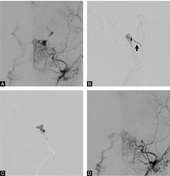
両側SOV,右SSV,右petrosal vein(PV)に導出して いた.右IPS経由にて,左CS前下方と右CS内後方のシ ャント部をコイルで塞栓したが,シャントの完全閉塞が 得られず,第2回経静脈的塞栓術を施行した(Fig. 2A).
右IPS経由にて右sphenoparietal sinusのシャント部,右 SSV,右SOV,右PVをコイルで塞栓した.次いで,左 IPS経由にて左SOVをコイルで塞栓した.左PVへの流 出が残存したが,ここへのマイクロカテーテル挿入は困 難であったため(Fig. 2B),50% n-BCAを注入した.
CSのPV流出部にn-BCA castが形成され(Fig. 2C),
PVへの流出は消失し,右IPSへの導出のみ残存した(Fig.
2D).術後,神経脱落症状の出現なく退院した.
3.症例 3
患者:75歳,女性.
右眼充血と痛み,眼球突出,複視で発症した.血管撮 影で右CS dAVFと診断され当院に紹介された.入院時,
右眼瞼浮腫,結膜浮腫,右外転神経麻痺を認めた.経静 脈的塞栓術を施行した.血管造影では流入動脈は両側 ICA,ECAで,右MHT,MMAが右CS内側後方に(Fig.
3A),左medial clival artery,MMAがintercavernous sinus にシャントを形成(Fig. 3B),右SOVに導出し末梢で停 滞していた.IPSは両側閉塞していた.右IPS経由にて,
マイクロガイドワイヤーを進め,難渋ながら内側のシャ ント近傍にマイクロカテーテルを挿入した.同部位でコ イルを留置すると,マイクロカテーテルが押し戻される 危険があり,また右CS内側後方のシャントが残存する 可能性があるため,n-BCAで塞栓する方針とした.マ イクロカテーテルより造影剤を圧入すると左medial clival arteryに逆流した(Fig. 3C).n-BCA注入時に左 medial clival arteryへの逆流は前後像で確認できるが,
右MHTを介するICAへの逆流は確認できないため,右
ICAのMHT分岐部をHyperForm 7mm×7mm(Micro Therapeutics, Irvine, CA, USA)にて一時的に閉塞しな がら40% n-BCAを注入した(Fig. 3D).シャント部近 傍から右CS,近位SOVにn-BCA castが形成され,シャ ントの完全消失が得られた(Fig. 3E,F).右外転神経麻 痺は,術後約2ヵ月で改善した.
考 察
CS dAVFの血管内治療は,コイルを用いた経静脈的 塞栓術が第一選択の治療法とされ,種々のアプローチル
ートが報告されている2-6,9,13).しかし,経静脈的にCSに 到達できたとしても,シャント部位が複雑な場合,その 同定は必ずしも容易ではなく,またCS内の隔壁のため シャント部位にカテーテルを誘導しコイルを留置するこ とが困難なことがある.また,再発例では先に留置した コイルのため,シャント部の同定やカテーテルの誘導は さらに困難となる.また,過度のcoil packingによる脳 神経麻痺も問題となる10).
このような場合,液体塞栓物質を用いた経動脈的塞栓 術が考えられるが,その報告は少ない7,8,11).経動脈的塞 Fig. 1 Right internal carotid angiogram (lateral view) before 2nd transvenous embolization (A)
showing a recurrence of dural AVF draining into the right superficial sylvian vein (SSV) and superior orbital vein (SOV). After coil embolization of the right SSV via right IPS approach, drainage into the right SOV, deep sylvian vein and superior petrosal sinus became prominent. A microcatheter was placed in the CS (arrow indicating a tip of the microcatheter, B), and 33% n-BCA was injected while the medial angle of the right orbit was manually compressed to prevent n-BCA droplets from migrating into the right SOV (C). After 2nd embolization, dural AVF completely disappeared (D).
A B
C D
栓術で根治を得るにはn-BCAをシャント部に十分到達 させる必要があり,そのためマイクロカテーテルを流入 動 脈(MMA,accesory meningeal artery,APhAな ど ) にwedgeさせ,15~25%の低濃度n-BCAを注入する方法 が用いられているが,CSの解剖学的特徴から,流入動 脈が脳神経の栄養血管になっている場合が多く,また ICAから短いdirect feederが流入し,n-BCAの迷入の危 険がある.
一 方, コ イ ル を 用 い た 塞 栓 術 で 治 癒 し に く いCS dAVFに対し,n-BCAを用いた経静脈的塞栓術が有効で
あったとの報告がされている14).実際,我々が報告した 3例のうち2例はシャント部位が複雑であり(症例 1,2),初回治療ではコイルを用いてシャント部のみの 選択的塞栓術を行ったが,1例は再発し,1例は閉塞が 得られなかたためn-BCAを用いた再治療が行われた.
経静脈的アプローチによるn-BCA塞栓術は,必ずし もカテーテルをシャント部位に誘導できなくても,症例 3のように1回の注入でn-BCAをCSから導出静脈まで 到達させ完全閉塞が得られる可能性がある.しかし,ア プローチルートに逆流すると再治療が困難となるため,
Fig. 2 Left external carotid angiogram (anteroposterior view) before 2nd transvenous embolization (A) showing a residual shunt in the left cavernous sinus draining into the bilateral SOV, SSV and right IPS. The draining veins (bilateral SOV, SSV and right petrosal vein) were occluded using coils. Angiogram (lateral view)injected from the microcatheter placed in the left cavernous sinus showing drainage into the left basal vein of Rosenthal via the petrosal vein (arrow) (B). After 50% n-BCA injection (C, lateral view), drainage into the left petrosal vein disappeared. After 2nd embolization, a shunt draining into the right IPS remained patent (D, anteroposterior view).
A B
C D
Fig. 3 Right middle meningeal artery (MMA) angiogram (anteroposterior view) showing a dural AVF at the posteromedial portion of the right CS (arrow, A) draining into the right SOV. The left internal carotid angiogram (anteroposterior view) showing a shunt at the intercavernous sinus (white arrow, B), which is located medial to the shunt from the right middle meningeal artery. Right meningohypophyseal trunk (MHT) and left MMA also feed the dural AVF (not shown). Venogram of forced injection from the microcatheter navigated into the intercavernous sinus through the occluded right IPS (arrowhead indicating a tip of the microcatheter) shows a reflux into the left medial clival artery (black asterisk) of the left ICA (white asterisk, C, anteroposterior view). 40% n-BCA was slowly injected while the right ICA was temporarily occuluded with a balloon catheter at the origin of MHT to prevent n-BCA droplets from migrating into the right ICA (D, anteroposterior view). Left (E) and right (F) common carotid angiograms (anteroposterior views) after embolization showing complete obliteration of the fistula.
B A
C D
E F
dAVFの導出静脈としては,sphenoparietal sinusを介する SSV,uncal veinを 介 す るdeep sylvian veinとbasal vein of Rosenthal,SPSを介するPVなどがあり,high flowの場合 にはCSや導出静脈に予めコイルを留置し血流を落とす か,低濃度n-BCAの使用を避ける必要がある(症例 1,2).症例3はSOVが唯一の導出静脈で停滞してい たためn-BCAを単独で用いた.
次の問題点は流入動脈への逆流である.ECAの流入 動脈に逆流した場合は脳神経麻痺が起こる可能性があ り,ICAの流入動脈に逆流した場合は脳梗塞のリスクを 伴う.安全に手技を行うためには血管構築の詳細な把握 が重要であり,ICAとの吻合を評価するにはCSに誘導 したマイクロカテーテルから強い圧をかけた静脈造影が 有用である(Fig. 3C).n-BCAの注入は,流入動脈に逆 流しないように強い圧をかけないこと,場合によっては バルーンでICAからの流入動脈起始部を一時的に閉塞す ることによりICAへの迷入を予防するなどの手技上の工 夫を行うことが重要である(Fig. 3D)11).
Wakhlooら14)は,同様の方法で治療を行った14例を報 告している.6例はn-BCA単独,7例はコイルとの併用,
1例はpolyvinyl alcohol particleを用いた経動脈的塞栓 術後に経静脈的にn-BCAを注入し,全例で完全閉塞が 得られたと報告している.合併症として,1例で無症候 性であったが,ICAの流入動脈へn-BCAが逆流し,中大 脳動脈へ迷入したと報告している.また,1例で治療数 日後に一過性外転神経麻痺が生じており,これは遅発性 に起こっていることから脳神経の栄養血管の閉塞ではな く,CSに注入したn-BCAの炎症反応に起因するもので はないかと考察している.
近 年, 非 接 着 性 液 体 塞 栓 物 質 で あ るOnyx(Micro Therapeutics, Irvine, CA, USA)を用いたCS dAVFの 経静脈的塞栓術が報告されている1,12).非接着性である ため,n-BCAに比べ時間をかけてゆっくり注入でき,
制御しやすく,目的部位に十分到達させることができる.
や再発例で追加治療が必要な症例に限定し,安易な適応 は避けるべきと思われる.
結 論
コイルを用いた経静脈的塞栓術で根治しにくい複雑な CS dAVFや再発例に対し,n-BCAを用いた経静脈的塞 栓術は有用な治療法になりうる.
文 献
1) Arat A, Cekirge S, Saatci I, et al: Transvenous injection of Onyx for casting of the cavernous sinus for the treatment of a carotid-cavernous fistula. Neuroradiology 46:1012-1015, 2004.
2) Benndorf G, Bender A, Lehmann R, et al: Transvenous occlusion of dural cavernous sinus fistulas through the thrombosed inferior petrosal sinus: report of four cases and review of the literature. Surg Neurol 54:42-54. 2000.
3) Benndorf G, Bender A, Campi A, et al: Treatment of a cavernous sinus dural arteriovenous fistula by deep orbital puncture of the superior ophthalmic vein.
Neuroradiology 43:499-502, 2001.
4) Goldberg RA, Goldey SH, Duckwiler G, et al: Management of cavernous sinus-dural fistulas: indications and techniques for primary embolization via the superior ophthalmic vein. Arch Ophthalmol 114:707-714, 1996.
5) Klisch J, Huppertz HJ, Spetzger U, et al: Transvenous treatment of carotid cavernous and dural arteriovenous fistulae: results for 31 patients and review of the literature. Neurosurgery 53:836-857, 2003.
6) Kuwayama N, Endo S, Kitabayashi M, et al: Surgical transvenous embolization of a cortically draining carotid cavernous fistula via a vein of the sylvian fissure. AJNR 19:1329-1332, 1998.
7) Li MH, Tan HQ, Fang C, et al: Trans-arterial embolisation therapy of dural carotid-cavernous fistulae using low concentration n-butyl-cyanoacrylate. Acta Neurochir (Wien) 150:1149-1156, 2008.
8) Liu HM, Huang YC, Wang YH, et al: Transarterial embolisation of complex cavernous sinus dural arteriovenous
fistulae with low-concentration cyanoacrylate. Neuroradiology 42:766-770, 2000.
9) Miller NR, Monsein LH, Debrun GM, et al: Treatment of carotid-cavernous sinus fistulas using a superior ophthalmic vein approach. J Neurosurg 83:838-842, 1995.
10) Nishino K, Ito Y, Hasegawa H, et al: Cranial nerve palsy following transvenous embolization for a cavernous sinus dural arteriovenous fistula: association with the volume and location of detachable coils. J Neurosurg 109:208-214, 2008.
11) Niimi Y, Song JK, Berenstein A: Cavernous sinus dural arteriovenous fistula completely occluded by transarterial NBCA embolization with balloon assisted technique.
JNET 2:29-33, 2008.
12) Suzuki S, Lee DW, Jahan R, et al: Transvenous treatment of spontaneous dural carotid-cavernous fistulas using a combination of detachable coils and Onyx. AJNR 27:1346 -1349, 2006.
13) Venturi C, Bracco S, Cerase A, et al: Endovascular treatment of a cavernous sinus dural arteriovenous fistula by transvenous embolisation through the superior ophthalmic vein via cannulation of a frontal vein. Neuroradiology 45:574-578, 2003.
14) Wakhloo AK, Perlow A, Linfante I, et al: Transvenous n-butyl-cyanoacrylate Infusion for complex dural carotid cavernous fistulas: technical considerations and clinical outcome. AJNR 26:1888-1897, 2005.
JNET 4:9-15, 2010
要 旨
【目的】海綿静脈洞部硬膜動静脈瘻(CS dAVF)に対する血管内治療は,コイルを用いた経静脈的塞栓術が第一選択の治 療法とされているが,coil packingで完全閉塞が得られないことがある.このような症例に,n-butyl cyanoacrylate(n-BCA)
を用いた経静脈的塞栓術を行った3例を報告し,手技上の工夫,留意点,有効性について検討した.【方法】全例,下錐
体静脈洞経由で塞栓術を行った.1例はn-BCA単独で,2例はコイル塞栓術後の再発や不完全閉塞に対しn-BCAを注入 した.【結果】3例中2例で完全閉塞が得られ,1例でシャントが残存したが頭蓋内静脈への逆流は消失した.手技に伴う
合併症はなかった.【結論】コイルを用いた経静脈的塞栓術で根治しにくいCS dAVFや再発例に対し,n-BCAを用いた経
静脈的塞栓術は有用な治療法になりうる.