緒 言
上行咽頭動脈と内頚静脈との間の動静脈瘻(ascending pharyngeal-internal jugular arteriovenous fistula:AP-IJ AVF)は稀な病態で,これまでわずか4例の症例報 告しかない1,3,4,11).これらの報告例の中では,2例が特 発性であり,1例は頚部手術,1例は頭部外傷に合併し た症例であった.4例すべてが頚静脈孔で上行咽頭動脈 から直接内頚静脈に流入するsingle-hole AVFであった. 今回,拍動性耳鳴および聴力低下が約5年の経過で進行 し,経動脈的塞栓術を行ったAP-IJ AVFの1例を経験 したので,文献的考察を加え報告する.
症例呈示
症例は32歳女性で,約5年前より右側の拍動性耳鳴が 出現し,徐々に増強してきた.約1年半前には右眼球結 膜充血が出現したが2,3ヵ月で軽快した.約半年前か らは右聴力低下とめまいを来し,低音障害型感音性難聴 と診断された.その後,めまいが頻回に起こるようにな ったためMRIが施行された.MRAでは右頚静脈球が明 瞭に描出されていたため,硬膜動静脈瘻が疑われ当科に 紹介入院となった(Fig. 1).入院時,右耳介後部に血 管雑音が聴取され,聴力検査では右低音が65dBまで低 下していた.その他に神経学的な異常は認められなかっ た.外傷や頭頚部手術の既往はなかった.CTでは右頚 静脈球の上端は内耳道下縁レベルにあり,いわゆる高位 頚静脈球(high jugular bulb)を呈しており,鼓室壁と の間の骨欠損も認められた(Fig. 2).MRA元画像では 淡い右頚静脈球の信号の内側に伴走する右上行咽頭動脈 の高信号が認められ,右頚静脈球内へのAVFのジェッ トも認められた(Fig. 3).血管撮影では,右後頭動脈 から分岐する右上行咽頭動脈のjugular branchが栄養動 脈で,右頚静脈球へのsingle-hole AVFが認められた(Fig. 4).右外頚動脈撮影にて他の硬膜動脈の流入は認めら当麻直樹
1)佐藤 裕
1)山道 茜
2)朝倉文夫
1)阪井田博司
2)松島 聡
1)滝 和郎
1,2)Ascending pharyngeal-internal jugular arteriovenous fistula: case report
Naoki TOMA
1)Yu SATO
1)Akane YAMAMICHI
2)Fumio ASAKURA
1)Hiroshi SAKAIDA
2)Satoshi MATSUSHIMA
1)Waro TAKI
1,2)1) Department of Neurosurgery, Mie University Graduate School of Medicine 2) Department of Innovative Neuro-Intervention Radiology, Mie University Graduate School of Medicine
●Abstract●
Objective: We report a rare case of an ascending pharyngeal-internal jugular arteriovenous fistula (AP-IJ AVF).
Case presentation: A 32-year-old woman was admitted with a 5-year-history of pulsatile tinnitus. Left external carotid angiography demonstrated a single-hole AVF between the ascending pharyngeal artery and the superior bulb of the internal jugular vein. The fistula was completely occluded by transarterial embolization using detachable coils. The patient showed immediate improvement after embolization. Conclusion: AP-IJ AVF is considered a subgroup of parachordal AVFs. Transarterial embolization is an effective treatment for this condition.
●Key Words●
arteriovenous fistula, ascending pharyngeal artery, embolization, internal jugular vein
(Received July 9, 2009:Accepted August 7, 2009) 1)三重大学大学院医学系研究科 脳神経外科
2)三重大学大学院医学系研究科 先進的脳血管内治療学
A B C D
Fig. 2 Axial (A, B, C) and sagittal views (D) of contrast-enhanced CT scan demonstrating an enlarged high jugular bulb on the right side (asterisks). The top of the jugular bulb (black arrows) extends up to the level of the internal auditory canal (black arrowheads). Note the lack of bony separation between the jugular bulb and the tympanic cavity (white arrowhead). The anterior condylar canal can be identified (white arrow).
れなかった.右下錐体静脈洞への逆流は認められず,流 出路は右内頚静脈のみで静脈還流障害は認められなかっ た. 局所麻酔下で右大腿動脈から経動脈的塞栓術を施行し た.6Frガイディングカテーテル(Envoy,Cordis, Miami,FL,USA)を右後頭動脈に留置し,マイクロ カ テ ー テ ル(Echelon10,ev3,Irvine,CA,USA) を 右後頭動脈から分岐する上行咽頭動脈のjugular branch に誘導した.Jugular branchは拡張しておりマイクロカ テーテルは全く抵抗なく瘻孔を通過して頚静脈球内に入 った.そこで,瘻孔を確実に閉鎖するため,マイクロカ テーテルの先端を頚静脈球内から引き戻しつつ,1本の コイルで静脈側から動脈側へと瘻孔を跨いで塞栓するこ とを試みた.しかし,離脱型コイル(GDC 18 soft 2mm/ 8cm,Boston Scientific,Natick,MA,USA) を 挿 入 していくと,マイクロカテーテルを引き戻してきてもコ イルはすべて静脈側に流れていくほど瘻孔は大きかっ た.そこで,コイルは静脈内に少し巻いた状態で,もう 1本のマイクロカテーテル(Excelsior SL-10,Boston Scientific,Natick,MA,USA)を瘻孔の遠位まで誘導 し,そこからもう1本の離脱型コイル(GDC 18 soft 2mm/8cm)で瘻孔の近位までpackingしてから,1本 目のコイルの残りを挿入していくと,静脈側に抜けずに 塞栓できた.この時点で耳鳴は消失したが,まだわずか にシャントが描出されたため,さらに離脱型コイル (EDC 10 ES 1.5mm/3cm,Kaneka Medix,Osaka)3 本でtight packingしシャントの完全消失を確認した(Fig. 5).手技に関連した合併症はなく,術後は耳鳴とめま いはともに消失した.聴力も正常となり,感音性難聴は 雑音によるものであったと考えられた.術後1年の経過 で再発は認められていない.
Fig. 1 Anterior (A) and lateral views (B) of initial MR angiography demonstrating an abnormal signal of the right jugular bulb (arrowheads).
Fig. 3 Serial MR angiography source images demonstrating flow signals of the ascending pharyngeal artery (arrowheads) adjacent to the jugular bulb (asterisks). Note the jet of the fistula between the ascending pharyngeal artery and the jugular bulb (black arrows). The anterior condylar canal can be identified (white arrow).
A B
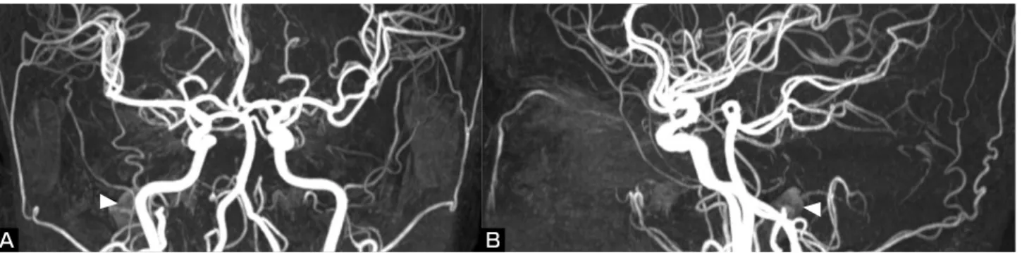
Fig. 4 Anteroposterior (A) and lateral views (B) of the right external carotid angiogram demonstrating the fistula (arrowheads) between the jugular branch (arrows) of the ascending pharyngeal artery branching off the occipital artery (large arrows) and the superior bulb of the internal jugular vein (asterisks). The pharyngeal branch of the ascending pharyngeal artery (white arrows) arising from the external carotid artery can be identified.
考 察
動静脈瘻(arteriovenous fistula:AVF)は動脈と静 脈が近接する部位に起こる病態であるが,頚静脈孔内で 近接している上行咽頭動脈と内頚静脈との間のAP-IJ AVFの報告はこれまでに4例と稀である1,3,4,11)(Table 1). 1978年のFoxら,1988年のGuglielmiらによる最初の2例 は特発性で,glomus tumorとの鑑別が重要であると報告 されている3,4).その後,1992年にChaloupkaらは頚部手 術の2年後に眼球突出,眼球結膜充血で発症した症例を1), 2004年にLiliangらは頭部外傷の5ヵ月後より拍動性耳 鳴が出現した症例を報告している.頚部手術後の症例で は内頚静脈が切除されているため下錐体静脈洞から海綿 静脈洞への逆流があり,頚動脈海綿静脈洞瘻と同様の眼 症状を呈した1)が,それ以外は本症例を含めすべて拍動 性耳鳴で発症しており,内頚静脈へ順行性に流出し,逆 流は認められず,耳鳴以外の症状はなかった. AP-IJ AVFの形態の特徴として上行咽頭動脈のみか ら直接内頚静脈に流入するsingle-hole AVFであるため, 保存的に加療された1例を除き,経動脈的に上行咽頭動 脈を塞栓することで根治している.塞栓物質としては, 下位脳神経を栄養するjugular branchを塞栓すること, 瘻孔が大きいhigh-flow AVFであるためコントロールが 困難であることから,液体塞栓物質は適していないと考 えられる.これまでの症例においては,最初の2例の報 告ではGelfoamやIvalon,頭部外傷後の症例ではプラチ ナファイバーコイルで塞栓されている.本症例では,塞 栓物質には離脱型プラチナコイルを選択した.当初は硬 膜動静脈瘻である可能性も考慮していたため,確実に瘻 孔を閉鎖すべくdouble catheter techniqueを用いて瘻孔 の静脈側から動脈側へと離脱型コイルで塞栓したが,結 局この疾患はsingle-hole AVFであったため動脈側を確 実に塞栓するだけで根治できたと考えられた.上行咽頭動脈は多くは外頚動脈の近位部後外側面から
A B C D
Fig. 5 Anteroposterior views of the fluoroscopic images showing the first (black arrow) and the second microcatheters (white arrow) (A), and configuration of the coils deposited in the ascending pharyngeal artery (B). Note the first coil passing through the fistula into the jugular bulb. Anteroposterior (C) and lateral views (D) of the right external carotid angiogram obtained immediately after embolization demonstrating disappearance of the fistula between the ascending pharyngeal artery and the jugular bulb.
Author Age/Sex Side Symptom/Duration Etiology Treatment Outcome
Fox 1) 54/M right bruit, dizziness / 5 months spontaneous embolization recovery
Guglielmi 2) 62/F left bruit, tinnitus, vertigo / 5 years spontaneous embolization recovery
Chaloupka 3) 64/M left eye pain, proptosis / 6 weeks neck surgery acetazolamide blurry vision
Liliang 4) 31/M left bruit, tinnitus / 5 months head trauma embolization recovery
Present case 32/F right bruit, tinnitus / 5 years spontaneous embolization recovery Table 1 Summary of reported cases of ascending pharyngeal-internal jugular arteriovenous fistula
branchとmedial branchに分岐する.lateral branchはS状 静脈洞の硬膜壁に沿って走行し後頭動脈の硬膜枝と吻合 する.medial branchは下錐体静脈洞の硬膜に沿って走行 し内頚動脈からのlateral clival arteryと吻合する6,7,10).
本症例ではこれらの硬膜枝は栄養動脈となっていなかっ た.
CTやMRIから本症例では頚静脈球の内側の下錐体静 脈洞の合流部近くに瘻孔が存在していることが分かる. この部位は解剖学的に頚静脈球と下錐体静脈洞および anterior condylar veinと が 合 流 す るanterior condylar confluent15,16)に非常に近く,硬膜動静脈瘻の好発部位で ある2).しかし,同様にその近傍に発生するAP-IJ AVF は頭蓋外動静脈瘻で,その病態は硬膜動静脈瘻とは全く 異なるものである. 内頚静脈のsuperior bulb,すなわち頚静脈球は胎生期 には形成されず,通常は2歳頃になって認められるよう になる.頚静脈球は骨性組織に囲まれているため拡張し にくいが,立位になってから右心房からの上行性の陰圧 波により骨構造を拡張し頚静脈窩が形成されるとされて いる14).本症例では頚静脈球は拡張して上方に突出した いわゆる高位頚静脈球を呈していた.高位頚静脈球では 突出した頚静脈球と鼓室壁との間の骨欠損を伴うことも あり,それ自体でも拍動性耳鳴,感音性難聴,めまいの 原因となることがある17).また,高位頚静脈球の組織学 的検討によると,頚静脈球の壁の厚さは高位であるほど 薄いとされる5). これまでのAP-IJ AVFの症例報告の中では,特発性 の1例で同様に高位頚静脈球が報告されている3).頚部 手術後および外傷後に発症した2例では頚静脈球の高さ についての記述はないものの,近接した上行咽頭動脈か らのAVF発生に頚静脈球の壁構造の脆弱性が関与した 可能性はあると推測される.また,特発性の場合でも軽 微な外傷の影響は否定できず,この病態に高位頚静脈球 の存在が関与する可能性はあると考えられる.今後,報 告症例数が増えることにより,この点が明らかにされる ことが期待される. Lasjauniasらは頭蓋外の鰓弓動静脈瘻(branchial AVF) 経孔に動静脈瘻をもつ.上行咽頭動脈,後頭動脈はそれ ぞれhypoglossal arteryやC1,C2の体節動脈であり, 椎骨動脈もC3からC8の体節動脈が体軸方向に吻合し て形成された体節構造としてみなされるため,AP-IJ AVFや椎骨動脈から椎骨静脈叢へのvertebro-vertebral AVF(VVAVF)にもこの疾患概念が適用される.実際, Lasjauniasらのシリーズの中でも,詳細な症例報告では ないがAP-IJ AVFの若年成人例が1例記載されている 8,9).
Parachordal AVF は high-flow single-hole AVF であ り,多くは小児期に雑音により偶然発見されるが,心不 全により致命的となることや,逆に認識されないことも ある.同じくparachordal AVFに属する傍脊椎動静脈瘻 の5例の報告では,全例2,3歳で持続性の雑音により 発見されている13).また,新生児症例で心不全にて発症 したVVAVFの報告もある12).一方,症例数は少ないも ののこれまでのAP-IJ AVFの症例はすべて成人例で小 児例の報告はないが,他のparachordal AVFの症例と比 較してこの疾患が高年齢で発症する理由は明らかではな い.今後症例数が増えることにより,さらにこの病態が 正確に解明されることが期待される.
結 論
拍動性耳鳴で発症したAP-IJ AVFの1例を経験した. この疾患はanterior condylar confluent近傍に生じるが, 硬膜動静脈瘻とは異なる頭蓋外動静脈瘻であり,特発性 のAP-IJ AVFは発生学的にはparachordal AVFに属す るものと考えられた.この疾患はsingle-hole AVFであ るため,離脱型コイルを用いて経動脈的に上行咽頭動脈 からシャント部を塞栓することにより根治が得られた.文 献
1) Chaloupka JC, Kibble MB, Hoffman JC: Ascending pharyngeal artery-internal jugular vein fistula complicating radical neck dissection. Neuroradiology 34:524-525, 1992. 2) Ernst R, Bulas R, Tomsick T, et al. Three cases of dural
the hypoglossal canal. AJNR Am 20:2016-2020, 1999. 3) Fox AJ, Allcock JM: Successful embolization of a fistula
between the ascending pharyngeal artery and internal jugular vein. Neuroradiology 15:149-152, 1978.
4) Guglielmi G, Guidetti G, Mori S, et al: Therapeutic embolization of an ascending pharyngeal artery-internal jugular vein fistula. Case report. J Neurosurg 69:132-133, 1988.
5) Kawano H, Tono T, Schachern PA, et al: Petrous high jugular bulb: a histological study. Am J Otolaryngol 21:161-168, 2000.
6) Lasjaunias P, Berenstein A: Surgical Neuroangiography, Vol 1. Functional anatomy of craniofacial arteries. Berlin. Springer-Verlag, 1987, 123-153.
7) Lasjaunias P, Moret J: The ascending pharyngeal artery: normal and pathological radioanatomy. Neuroradiology 11:77-82, 1976.
8) Lasjaunias P, TerBrugge K, Berenstein A: Surgical Neuroangiography, Vol 3. Berlin. Springer-Verlag. 2006, 687-720.
9) Lasjaunias P, TerBrugge K: Vascular diseases in neonates, infants and children: Interventional neuroradiology management. Berlin. Springer-Verlag. 1997, 501-530. 10) Lasjaunias P, Theron J, Moret J: The occipital artery.
Anatomy: normal arteriographic aspects: embryological significance. Neuroradiology 15:31-37, 1978.
11) Liliang P-C, Hsu S-W, Kuo Y-L, et al: Trauma-induced ascending pharyngeal artery-internal jugular vein fistula: case report. Surg Neurol 61:89-91, 2004.
12) Nakano S, Agid R, Klurfan P, et al: Limitations and technical considerations of endovascular treatment in neonates with high-flow arteriovenous shunts presenting with congestive heart failure: report of two cases. Childs Nerv Syst 22:13-17, 2006.
13) Niimi Y, Berenstein A, Fernandez PM, et al: Pediatric nonvertebral paraspinal arteriovenous fistulas along the segmental nerve: clinical, imaging, and therapeutic considerations. J Neurosurg (Pediatrics 2) 103:156-162, 2005.
14) Okudera T, Huang YP, Ohta T, et al: Development of posterior fossa dural sinuses, emissary veins, and jugular bulb: morphological and radiologic study. AJNR 15:1871-1883, 1994.
15) San Millan Ruiz D, Gailloud P, Rufenacht DA, et al. The craniocervical venous system in relation to cerebral venous drainage. AJNR 23:1500-1508, 2002.
16) Takahashi S, Sakuma I, Omachi K, et al. Craniocervical junction venous anatomy around the suboccipital cavernous sinus: evaluation by MR imaging. Eur Radiol 15:1694-1700, 2005.
17) Yetiser S, Kertmen M, Yildirim A: An analysis of correlation between the unusual location of the jugular bulb and audiovestibular symptoms. Auris Nasus Larynx 31:369-377, 2004.
JNET 3:100-105, 2009
要 旨
【目的】稀なascending pharyngeal-internal jugular arteriovenous fistula(AP-IJ AVF)の1例を報告する.【症例】32歳女性. 約5年の経過で右側の拍動性耳鳴および聴力低下が進行し,MRAにて頚静脈球の異常信号が認められた.血管撮影で右後 頭動脈から分岐した上行咽頭動脈のjugular branchが栄養する頚静脈球への動静脈瘻を認め,離脱型コイルによる経動脈的 塞栓術で根治した.【結論】AP-IJ AVFの瘻孔はanterior condylar confluent(ACC)の近傍に存在するが,硬膜動静脈瘻 とは異なる頭蓋外動静脈瘻であり,発生学的には傍脊索動静脈瘻(parachordal AVF)に属し,形態としてはsingle-hole AVFであるため,経動脈的塞栓術が有効であった.