光干渉断層映像装置(OCT)の計測精度と計測に影響を与える
因子についての検討
緒 言 近年,経皮的冠動脈形成術(percutaneous coronary intervention:PCI)は手技の向上やデバイスの進歩に より慢性完全閉塞病変や左冠動脈主幹部病変,分岐 部病変などの適応症例が増えてきている1∼3).また,薬剤溶出性ステント(drug eluting stent:DES)の出現
により以前から使用していたベアメタルステント(bare metal stent:BMS)の問題点であった再狭窄が減少し た4).さらに,冠動脈の画像診断やステント留置後の 評価として,血管内超音波法(intravascular ultrasound: IVUS)やCT,MRIなどの画像診断装置が数多くの施 設で使用されるようになってきている.特にIVUSは 血管径や形態などの冠動脈内の評価に優れており, PCI時には一般的に使用されている5, 6).しかしIVUS の解像度は100∼150emであり,冠動脈内腔や血管内 径の把握は可能であるが,解像度の限界があり,ス テント圧着不良やDESの新生内膜被覆の状態の観察 が困難である. 1991年,IVUSの10∼20倍の高解像度を有する光干 渉断層映像(optical coherence tomography:OCT)が
発明され7),数年前より,冠動脈内の微細な構造の観 察などに,臨床使用されるようになった8).それに 神戸大学医学部附属病院放射線部 1)神戸大学医学部附属病院循環器内科 論文受付 2009年 1 月27日 論文受理 2009年 6 月 5 日 Code No. 890
根冝典行・古東正宜・志手淳也
1)・澤田隆弘
1)・川光秀昭
Examination of Accuracy and Factors Influencing Optical Coherence
Tomography(OCT)Measurements
Noriyuki Negi, Masanobu Koto, Junya Shite,1) Takahiro Sawada,1)and Hideaki Kawamitsu
Department of Radiology, Kobe University Hospital 1)Department of Cardiology, Kobe University Hospital
Received January 27, 2009; Revision accepted June 5, 2009; Code No. 890
Summary
Introduction: Optical coherence tomography(OCT)is a new imaging modality with increasing clinical application. In the present study, we examined the accuracy and factors influencing the OCT measurements (luminal diameter, area, and stent strut thickness). Methods: We evaluated several luminal sizes of phantom
model by OCT with several situations(frame-rate: 8.2 or 15.6 F/s; pullback-speed: 1.0 or 2.0 mm/s; position of Image Wire: in-center; off-center)and compared them with the actual value and the intravascular ultrasound measurements. We also evaluated the accuracy of the stent strut thickness(5 bare metal stents and 2 drug eluting stents)in stents that were implanted to phantom models. Results: The accuracy of OCT measurements was affected by frame-rate and the position of OCT Image Wire. The distortion and the change in brightness of the OCT image were detected when the Image Wire was positioned off-center, especially at low frame-rate. In this condition, OCT measured the luminal diameter and area larger than the actual size. As for the strut thickness, when we unified the measurement points of the stent strut surface, the precision of the OCT measurements was satisfactory. Conclusion: We revealed that the precision of the OCT measurements was satisfactory. However, we should note that the OCT measurements were affected by frame-rate and position of the Image Wire.
Key words: optical coherence tomography (OCT), accuracy of measurement, stent strut
別刷資料請求先:〒650-0017 兵庫県神戸市中央区楠町7-5-2
神戸大学医学部附属病院放射線部 根冝典行 宛
よって,IVUSなどの画像診断装置では不可能であっ たDES留置後の新生内膜の厚さやステント圧着不良 の観察を10emまで評価することができる9, 10).当院で は2004年よりOCTを使用しているが,2008年10月より 保険償還の適用を受け,今後PCI時などに,より普及 すると考えられる.今回,われわれは模擬血管ファン トムとステントを用い,OCTの計測精度や計測値に影 響を与える因子について検討した. 1.方 法 1-1 使用機器 ・OCT Image Wire;0.016インチ Light Lab社製 ・IVUS Revolusion;3.2Fr 周波数;45MHz 機械走査式 Volcano社製 ・模擬血管ファントム ゴムチューブ(3mmφ) アクリルパイプ(正円形;1.8・3.3・4.9・6.0mmφ/正 四角形;1.8mm) ・ステント;3mmφ ExpressTM(Boston社製) TunamiTM(TERUMO社製) DuraflexTM(Goodman社製) DriverTM(Medtronic社製)
VelocityTM(Johnson & Johnson社製)
CypherTM(Johnson & Johnson社製)
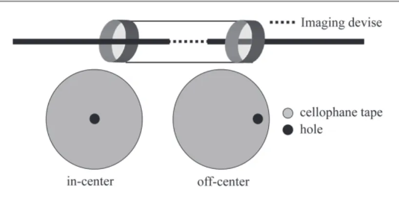
TaxusTM(Boston社製) 1-2 OCTによる血管内腔径,内腔面積の計測精度 1-2-1 計測方法 模擬血管ファントムとして正円形のアクリルパイプ (1.8・3.3・4.9・6.0mmφ)を使 用し,OCTはフレーム レ ート(8.2・15.6F/s), プ ル バ ック ス ピ ード(1.0・ 2.0mm/s)をそれぞれ変化させることにより,次の 4 種類の組み合わせ(8.2F/s-1.0mm/s・8.2F/s-2.0mm/s・ 15.6F/s-1.0mm/s・15.6F/s-2.0mm/s)に関して 計 測し た.IVUSはフレームレート30F/s,プルバックスピー ド0.5mm/sにて計測を行った.OCTのImage Wireはア クリルパイプの中心(以下in-center) と中心外(以下off-center)を走行するように,IVUSのRevolusionカテー テルはアクリルパイプの中心を走行するよう固定し, 37˚Cに調整した乳酸リンゲル液内にて計測を行った (Fig. 1). 1-2-2 検討方法 得られた画 像 からOCTでは10フレームごとに, IVUSでは0.5mmごとに内腔径,内腔面積を計測し た.また,画像のゆがみの指標として短径と長径の 比率も計算した.そしてin-centerとoff-centerのOCT 画像,IVUS画像の内腔径,内腔面積,短径長径比, さらには実際の値との比較を行った.OCTの計測値 の観察者間における計測誤差についてはBland and Altman plotにて検討した. 1-3 OCT画像の再現性 1-3-1 計測方法 正円形(1.8mmφ)と正四角形(1.8mm)のアクリルパ イプを使用し,OCTのフレームレート,プルバックス ピードを 1-2-1 と同様に変化させて,4 種類の組み合 わせにて行った. 1-3-2 検討方法 断面形の再現性は得られた動画像,静止画像より 視覚的に画像のゆがみや輝度の変化,モーション アーチファクトについて検討した.
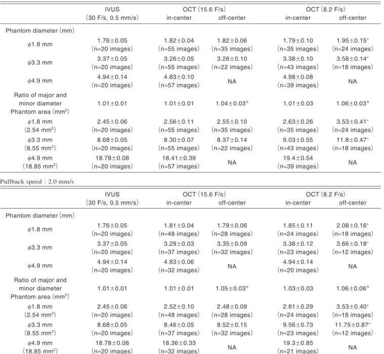
1-4 ステントストラット厚の計測精度 1-4-1 計測方法 ゴムチューブに 7 種類(5 種類のBMS,2 種類の DES)の 3mmφのステントをそれぞれの最大耐用圧に て拡張留置し,ゴムチューブに密着させ,その後, OCT(フレームレート15.6F/s,プ ル バックスピード 1.0mm/s)を行った. 1-4-2 検討方法 得られた画像からステントストラットの厚さの計測 を行った.計測点は描出されているストラット中央か らゴムチューブ内層までの距離にて行った11)(Fig. 2). またVelocityにおいては,観察者間における計測誤 差 をBland and Altman plotを 用 い て 評 価を 行 い, Image Wireの走行位置の違い(in-center,off-center) による計測誤差についても検討した.計測点は10フ レームごとに行った. 2.結 果 2-1 OCTの計測精度 OCTとIVUSの計測値はTable 1に示した.OCTでは 6.0mmφのファントム以外はImage Wireがin-centerに 位置していれば 全景観察可能であった(4.9mmφの ファントムではImage Wireがoff-centerに位置してい る場合は全景観察が不可能であった).フレームレー トを15.6F/sに設定すればImage Wireの走行に影響な くIVUSの計測値とほぼ同等であった.しかしフレー ムレートを8.2F/sに設定した場合,Image Wireの走行 がoff-centerになるとIVUSより過大評価された.また 短 径と長 径の比率はImage Wireの走行位置がoff-centerの場合大きくなった(Table 1).
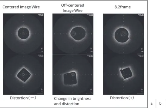
Bland and Altman plotによる計測値の観察者間誤 差は認容可能な範囲であった(内腔径;mean differ-ence:0.018w0.045mm,内腔面積;mean difference: 0.143w0.11mm2(Fig. 3).) 2-2 OCT画像の特性 OCT画像の断面形の再現性はImage Wireがin-center を走行し,フレームレートが15.6F/sの場合,良好で あった.一方,Image Wireがoff-centerの場合,ゆが みが現れ,Image Wireから離れたファントム壁の輝 度も低下していった.また,フレームレートを低下さ せることにより,モーションアーチファクトが現れる 頻 度 が 高くなる傾向が みられ た(8.2F/s;15.5% vs 15.6F/s;8.1%)(Fig. 4). 2-3 ステントストラット厚の計測精度 OCTによるステントストラット厚の計測値は,7 種 類のステントすべてにおいてメーカ公称値に近い計 測値が得られた(Table 2).
Bland and Altman plotによるVelocityでのストラッ ト厚の観察者間の計測値の差も同様に,認容可能な 範囲であった(mean difference:1.09w11.4mm).Image Wireの走行位置の違いによる計測誤差については, Image Wireとストラットの距離が遠くなるほど誤差が 大きくなった(Fig. 5). 3.考 察 今回のわれわれが行った検討の結果,OCTによる 内腔径および内腔面積,ストラット厚の計測値は実 際の値に非常に近いものであったが,フレームレート やImage Wireの走行位置により影響を受けることが 判明した.off-centerでの誤差原因は短径と長径の比 率でも現れていたが,この現象は主にImage Wireと ファントムの長軸が同軸でないために起こる斜位断面 によるものと思われる.これは視覚的にも断面形のゆ がみとして確認された.この視覚的なゆがみはフレー ムレートを低下させたときに特に顕著に現れ,Image Wireの回転ムラや画像収集時の時間分解能不足によ る影 響も考えられる.また,ファントムの辺 縁 が Image Wireから遠くなるほど輝度が低下したが,これ は赤外線光の光量不足と思われた.一方,フレーム レートを15.6F/sに設 定 すれば,ゆが みが 減 少し, IVUSと比較しても計測誤差が少なく,視覚的に観察 しても再現性が高いことが判明し,臨床的にも使用 Fig. 2 Method of measuring stent strut thickness.
φ3.3 mm
(n=20 images) (n=55 images) (n=22 images) (n=43 images) (n=18 images)
φ4.9 mm 4.94w0.14 4.83w0.10
NA 4.98w0.08 NA
(n=20 images) (n=57 images) (n=39 images) Ratio of major and
minor diameter 1.01w0.01 1.01w0.01 1.04w0.03* 1.01w0.03 1.06w0.03* Phantom area(mm2)
φ1.8 mm 2.45w0.06 2.56w0.11 2.55w0.10 2.63w0.26 3.53w0.41+
(2.54 mm2) (n=20 images) (n=55 images) (n=35 images) (n=35 images) (n=24 images)
φ3.3 mm 8.68w0.05 8.30w0.07 8.37w0.14 9.03w0.55 11.8w0.47+
(8.55 mm2) (n=20 images) (n=55 images) (n=22 images) (n=43 images) (n=18 images)
φ4.9 mm 18.78w0.08 18.41w0.39
NA 19.4w0.54 NA
(18.85 mm2) (n=20 images) (n=57 images) (n=39 images) Pullback speed;2.0 mm/s
IVUS OCT(15.6 F/s) OCT(8.2 F/s)
(30 F/s, 0.5 mm/s) in-center off-center in-center off-center Phantom diameter(mm)
φ1.8 mm 1.76w0.05 1.81w0.04 1.79w0.06 1.85w0.11 2.08w0.16+
(n=20 images) (n=48 images) (n=28 images) (n=24 images) (n=18 images)
φ3.3 mm 3.37w0.05 3.29w0.03 3.35w0.09 3.38w0.12 3.66w0.18+
(n=20 images) (n=37 images) (n=32 images) (n=23 images) (n=12 images)
φ4.9 mm 4.94w0.14 4.83w0.06
NA 4.94w0.14 NA
(n=20 images) (n=32 images) (n=20 images) Ratio of major and
minor diameter 1.01w0.01 1.01w0.01 1.05w0.03* 1.03w0.03 1.06w0.06* Phantom area(mm2)
φ1.8 mm 2.45w0.06 2.52w0.10 2.48w0.09 2.81w0.29 3.53w0.40+
(2.54 mm2) (n=20 images) (n=48 images) (n=28 images) (n=24 images) (n=18 images) φ3.3 mm 8.68w0.05 8.46w0.05 8.52w0.15 9.56w0.73 11.75w0.87+
(8.55 mm2) (n=20 images) (n=37 images) (n=32 images) (n=23 images) (n=12 images)
φ4.9 mm 18.78w0.08 18.36w0.33
NA 19.3w0.85 NA
(18.85 mm2) (n=20 images) (n=32 images) (n=21 images) *P<0.0001 vs IVUS, P<0.0001 vs OCT in-center, + P<0.001 vs IVUS
NA: not applicable
Fig. 4 Reproducibility of the OCT image.
Stent Manufacture’s specification OCT measurements Stent strut thickness
VelocityTM 140 em(0.0055 inch) 134.7w13.9 em (232 struts) (difference, 5.4w13.8) ExpressTM 132 em(0.0052 inch) 127.3w11.0 em (166 struts) (difference, 4.7w11.0) DuraflexTM 114 em(0.0045 inch) 111.0w12.1 em (158 struts) (difference, 3.0w12.1) DriverTM 91 em(0.0036 inch) 91.5w8.5 em (118 struts) (difference, 0.5w8.5) TunamiTM 79 em(0.0031 inch) 79.0w11.4 em (212 struts) (difference, 0.04w11.4) CypherTM 150 em 142.5w11.9 em
(149 struts) (0.0055 inch+polymer) (difference, 7.5w11.9)
TaxusTM 148 em 146.8w10.9 em
(177 struts) (0.0052 inch+polymer) (difference, 2.3w12.6) Table 2 Comparison between stent strut thickness measurements with OCT
and the manufacturer’s specification of stent strut thickness
Fig. 5 Variability in the OCT mea-surements of stent strut thick-ness owing to the difference in Image Wire distance from the phantom surface.
績であった.これは計測対象が比較的大きいもので あったからと思われる.計測対象が小さいもの,例え ばDES内の新生内膜や菲薄化線維性被膜の場合, IVUSでは解像度の限界のためほぼ不可能であり明ら かにOCTの方が優ると考えられる12, 13). 今回,ステントストラット厚の計測精度についての 計測点はストラットからの光反射を考慮し,ストラッ ト中央からゴムチューブ内層までとした11).今回の検 討結果より,この計測点に統一すれば,どのステント でも精度の高いストラット厚の計測が可能と考えられ た.また観察者間においての計測誤差は少なかった が,Image Wireとストラットの距離が離れたり近づい たりすることにより誤差が大きくなることは注意が必 要と考える.これらのことより計測点を統一すれば, OCTによりステント圧着状態の評価やステント留置後 の定量的な新生内膜の評価ができ,PCI前後の検討 撮像条件によって,血管内腔径,内腔面積の計測精 度はIVUSとほぼ同等であり,またIVUSなどの画像診 断装置では評価できなかったステントストラット厚の 計測やステント留置後の微細な変化の定量的な評価 ができると考えられる.しかし,今回の基礎的評価に より画像収集時の条件によってはサイズの過大評価 や画像のゆがみが生じ,定量的評価を行う際には注 意が必要である. 謝 辞 本研究を行うにあたり,ご協力いただいた神戸大 学医学部附属病院循環器内科カテーテルグループの 医師および放射線部の技師諸兄に感謝いたします. なお,本論文の要旨は第17回日本心血管インター ベンション学会学術集会において発表した. 参考文献
1) Sirnes PA, Golf S, Myreng Y, et al. Stenting in Chronic Coronary Occlusion(SICCO): a randomized, controlled trial of adding stent implantation after successful angio-plasty. J Am Coll Cardiol 1996; 28(6): 1444-1451.
2) Yamashita T, Nishida T, Adamian MG, et al. Bifurcation lesions: two stents versus one stent−immediate and f ollow-up results. J Am Coll Cardiol 2000; 35(5): 1145-1151.
3) Park SJ, Hong MK, Lee CW, et al. Elective stenting of unprotected left main coronary artery stenosis: effect of debulking before stenting and intravascular ultrasound guidance. J Am Coll Cardiol 2001; 38(4): 1054-1060. 4) Morice MC, Serruys PW, Sousa JE, et al; RAVEL Study
Group. Randomized Study with the Sirolimus-Coated Bx Velocity Balloon-Expandable Stent in the Treatment of Patients with de Novo Native Coronary Artery Lesions. A randomized comparison of a sirolimus-eluting stent with a standard stent for coronary revascularization. N Engl J Med 2002; 346(23): 1773-1780.
5) Fitzgerald PJ, St Goar FG, Connolly AJ, et al. Intravascu-lar ultrasound imaging of coronary arteries. Is three layers the norm? Circulation 1992; 86(1): 154-158.
6) Görge G, Ge J, Haude M, et al. Intravascular ultrasound: a guide for management of complications during interven-tion? Eur Heart J 1995; 16(Suppl L): 86-92.
7) Huang D, Swanson EA, Lin CP, et al. Optical coherence tomography. Science 1991; 254(5035): 1178-1181.
8) Fujimoto JG, Boppart SA, Tearney GJ, et al. High resolu-tion in vivo intra-arterial imaging with optical coherence tomography. Heart 1999; 82(2): 128-133.
9) Sawada T, Shite J, Shinke T, et al. Persistent malapposition after implantation of sirolimus-eluting stent into intramu-ral coronary hematoma: optical coherence tomography observations. Circ J 2006; 70(11): 1515-1519.
10) Matsumoto D, Shite J, Shinke T, et al. Neointimal cover-age of sirolimus-eluting stents at 6-month follow-up: evaluated by optical coherence tomography. Eur Heart J 2007; 28(8): 961-967.
11) 寺島充康.OCTとintervention.光干渉断 層法(Optical Coherence Tomography) 新しい冠動脈イメージング. 南江堂,東京,2008:45-58.
12) Sawada T, Shite J, Garcia-Garcia HM, et al. Feasibility of combined use of intravascular ultrasound radiofrequency data analysis and optical coherence tomography for detecting thin-cap fibroatheroma. Eur Heart J 2008; 29 (9): 1136-1146.
13) Sonoda S, Morino Y, Ako J, et al. Impact of final stent dimensions on long-term results following sirolimus-eluting stent implantation: Serial intravascular ultrasound analysis from the sirius trial. J Am Coll Cardiol 2004; 43(11): 1959-1963.
14) Shite J, Matsumoto D, Yokoyama M. Sirolimus-eluting stent fracture with thrombus, visualization by optical coherence tomography. Eur Heart J 2006; 27(12): 1389.
Fig. 1 Image Wireの固定方法
Phantomの両断面にセロハンテープを張り,そこに穴を開けカテーテルを挿入し,固定した. Fig. 2 ステントストラット厚の計測方法
描出されているステントストラットの反射点の中央からゴムチューブ内層までの距離を実際のストラット厚とした. Fig. 3 OCT計測値の観察者間の計測誤差
Bland Altman plotによるOCT計測値の観察者間誤差は認容可能な範囲であった. Fig. 4 OCT画像の再現性
(a)in-center 15.6F/sのOCT画像.円形のphantomは正円形に,四角形のphantomは正四角形に描出されている.
(b)off-center 8.2F/sのOCT画像.OCT画像はゆがみ,Image Wireから遠ざかっているphantomの表面の輝度は低下してい る.
(c)モーションアーチファクトのOCT画像.
Fig. 5 Image Wireとステントストラットの距離の違いによる計測誤差 Image Wireとストラットの距離が遠くなるほど誤差が大きくなった. Table 1 OCT計測値とIVUS計測値との比較 OCTによる内腔径および内腔面積,ストラット厚の計測値はIVUS計測値と同様に実際の値に非常に近いものであったが, フレームレートやImage Wireの走行位置により影響を受けた. Table 2 OCT計測値とメーカ公称値との比較 数種類のステントでもOCT計測値はメーカ公称値に近かった.