学位論文
Complete loss of ciliary motility in Ktu-knockout mice;
Insights into tissue-specific mechanisms
for rotational polarity
マウス
Ktu
変異体を用いた繊毛の配向形成における
繊毛の運動性の役割の解析
平成 24 年 12 月博士(理学)申請
東京大学大学院理学系研究科
生物科学専攻
松尾 萌
(
)
Contents
Abbreviations ... 3
Abstract ... 4
General introduction ... 5
Chapter 1: Characteristics of mouse Kintoun (Ktu) ... 9
Introduction
... 10Materials and Methods
... 12Results
... 16Discussion
... 19Chapter 2: Phenotypic analysis of Ktu-knockout mice ... 20
Introduction
... 21Material and Methods
... 23Results
... 27Discussion
... 30Chapter 3: Contribution of ciliary motility to ... 31
rotational polarity ... 31
Introduction
... 32Material and Methods
... 34Results
... 36Discussion
... 38General Discussion ... 41
Figures ... 43
Reference ... 62
Acknowledgements ... 66
Abbreviations
Ca.: Circa
CCDC: Coiled-coil domain containing
CP complex: Central pair
CSF: Cerebral spinal flow
DAPI: 4',6-diamidino-2-phenylindole
DNAAF: axonemal dynein assembly factor
Hsp: Heat shock protein
IFT: Intra flagellar transport
LRRC: Leucine-rich-repeat
MTEC: Mouse tracheal epithelial cell culture
PCD: Primary ciliary dyskinesia
PCP: Planar cellular polarity
Abstract
Coordinated uni-directional beating is an essential feature of multi cilia in various
organs to guarantee proper fluid transport. Recent works have suggested that ciliary
motility and planar polarity are required during the process of ciliary alignment to
achieve coordinated beating. However, the extent to which ciliary motility is involved in
this process in mammals has not yet been fully clarified. Here, in order to address the
role of ciliary motility in the process of coordinated ciliary alignment, I analyzed
Kintoun knockout-mice (Ktu-/-). Ktu-/- exhibited typical phenotypes of PCD with complete loss of ciliary motility in the trachea and brain ependyma.
Immunohistochemistry using antibodies against axonemal dyneins confirmed the loss of
multiple axonemal dynein components in mutant cilia. Observation of ciliary orientation
based on the basal foot direction revealed that ciliary motility was not required for the
alignment of airway cilia, whereas a strong requirement was observed in brain
ependymal cells. Thus I conclude that the involvement of ciliary motility in the
establishment of coordinated ciliary alignment varies among tissues. I propose that the
balance of power between fluid flow and planar polarity in establishment of coordinated
orientation of motile cilia differ among tissues according to the difference in the
General introduction
Cilia are hair-like cell organelles that project from the surface of almost all eukaryotic
cells and consist of microtubule-based axoneme (9+0 or 9+2 structure) surrounded by a
specialized ciliary membrane. The ciliary axoneme is build on the basal body, a
derivative of the centriole that is positioned under the cell membrane. This ciliary
architecture is highly conserved among species, ranging from protists to mammals. Cilia
are generally assembled in differentiated or quiescent cells that exit mitosis (Quarmby
and Parker, 2005). Cilia possess receptors for Hedgehog, platelet-derived growth factor
(PDGF), Wnt and other signaling molecules on its ciliary membrane. Thus cilia are
thought to function as sensors for mechanical and chemical cues from the extra-cellular
environment and fine tune the cellular differentiation (Plotnikova et al., 2009). In
addition to the above sensory cilia, specialized cilia exist in several organs. Cilia that
contain dynein arms are motile and involved in cell motility and extracellular fluid
transport (Satir and Christensen, 2007) (Fig. 1B). In mammals, fluid flow driven by
motile cilia is important in many organ systems, such as the respiratory tract, the
oviduct to the ventricles of the brain. In trachea, hundreds of motile cilia projecting
from the apical surface of the epithelial cells beat in the direction from lung to the oral
cavity, in order to clear contaminants (mucociliary clearance) (Wanner et al., 1996).
Mucociliary clearance plays a pivotal role in the prevention of respiratory disease. Multi
ciliated cells lining the walls of ventricles (ependymal cells) generate directed
cerebrospinal fluid (CSF) flow that helps CSF homeostasis by transporting secreted
guidance factors (Sawamoto, 2006) and nutrients. In addition, this flow guides the
long-range migration of neuroblasts along the lateral walls to the olfactory bulb
sperm requires flagella motility to swim toward the egg and ciliated cells in the female
reproductive tract transport eggs from the fallopian tube to uterus. Furthermore in early
developmental stages, a leftward flow created by motile cilia in the node determines the
left-right body axis. In mice, motile cilia generally grow in a patch–like style, where
hundreds of cilia protrude from a single cell. One exception is the node cilia which is
solitary (mono cilia). The node cilium is also different in movement, as it shows
rotational movement while others move in a waving form. As motile multi cilia is the
majority in the mammalian body, we mostly discuss motile multi cilia in this doctoral
thesis.
Given its critical and diverse functions, defects in motile cilia in human cause primary
ciliary dyskinesia (PCD) which affects one in 16000 people, known as Kartagener
syndrome (Fliegauf et al., 2007) (Fig. 1A). Defects in airway cilia cause recurrent
respiratory infection such as bronchiectasis and chronic sinusitis. Defective nodal cilia
lead to organ mislocation (situs inversus), and flagellar and ciliary motility defect in
reproductive organs cause infertility (Fliegauf et al., 2007). Stagnation of the CSF flow
raises the CSF pressure within the brain ventricles and cause hydrocephalus. In addition,
one of the overlooked issues of PCD is that the coordinated uni-direcional alignment of
cilia are disturbed in some of the PCD patients (Rautiainen et al., 1990). Thus, it is
assumed that ciliary disorientation is a byproduct of abnormal ciliary motility.
In previous reports, a question as to how PCD occurs by motile ciliary defects has
been addressed using various knockout mice, including ciliary motility defect mutants.
However, they have several problems. First, cilia retained residual motility in most of
ciliary mutants used. Indeed, previous reports mentioned that weakend beating of
effect of the residual motility. As is suggested in recent papers, the role of ciliary
motility is not only production of the extra-cellular flow but also enhancement of the
ciliary sensory function (Mitchell et al., 2007). Thus defective beating could have some
roles. Furthermore, some of the studies used knockout mice with ciliogenesis defects in
addition to motility defects, which made the analyses complicated. Finally, as described
above, ciliary disorientation was interpreted as a result of ciliary motility defects in
PCD patients. In general, ciliary motility was simply regarded as a flow generator and
was thought to have less effect on ciliary development. However recent reports
suggested that ciliary motility may play a role in ciliary development (alignment).
However, previous studies of mice mutants with ciliary motility defects did not
examined this possibility. Taking all these problems into account, a knockout mouse that
shows complete loss of ciliary motility in multiple organs is required to understand the
role of ciliary motility in development more precisely.
For this purpose, I focused on a cytoplasmic protein Kintoun (Ktu) in this thesis. In the
ktu/pf13 medaka mutant, outer and a part of inner dynein arms are missing, leading to a
complete loss of motility. As Ktu is required for axonemal dynein pre-assembly,
depletion of Ktu was expected to disrupt the assembly of multiple dyneins, leading to
greater reduction of ciliary motility than previously reported for mouse models (Fig.
1C). Thus I generated and analyzed Ktu-knockout mice (Ktu-/-). In the present thesis, I
first characterized murine Ktu in vivo and in vitro using immunohistochemistry (chapter
1). In the second chapter, detailed phenotypic analysis of Ktu-/- mice was performed. As
expected, the mutant mice exhibited a complete loss of ciliary motility in trachea and
brain ventricles, providing an ideal model of PCD (chapter 2). In the last chapter, using
rotational polarity of the cilium/basal body in brain ependyma and trachea epithelia. My
results demonstrate that ciliary motility is required for the unidirectional alignment of
cilia in the brain ependyma, but not in the tracheal ciliated cells (chapter 3). Thus I
conclude that the involvement of ciliary motility in their orientation differs dramatically
Introduction
Motile cilia develop specifically in cells that express specific transcription factors that
regulate ciliogenesis such as Foxj1 and Rfx3 (Thomas et al., 2010). In general,
ciliogenesis begins with the generation of a basal structure called “basal body”. Unlike a
cell producing solitary cilia (mono cilia) in which a single basal body is generated only
from the duplication of mother centrioles, massive numbers of additional basal bodies
are produced from non-microtubule-based structure called “deuterosome” in the
cytoplasm of a multi-ciliated cell (Dirksen, 1991; Hagiwara et al., 2004). Basal bodies
dock to the cell surface and protrude cilia. Intraflagellar transport (IFT) proteins
transport ciliary proteins (membrane components, receptors, dynein components,
accessory proteins and so on) between the cytoplasm and ciliary compartments. As cilia
lack translational machineries, ciliary components are produced in the cytoplasm and
transported to cilia via IFT.
Ciliary motility is generated by two types of axonemal dyneins, inner arm dynein and
outer arm dynein. Axonemal dyneins are protein complexes that consist of three types
of proteins, light chains, intermediate chains and heavy chains (Fig. 1B). The core
ciliary components for motility (the central pair complex, the radial spoke) are also
important for proper beating of cilia. Like other ciliary proteins, axonemal dynein
components are produced in the cytoplasm and transported to cilia during ciliogenesis.
It was reported that the axonemal dynein complex is assembled in the cytoplasm and
transported to the ciliary compartment (Fowkes and Mitchell, 1998). Thus, proteins that
are involved in dynein assembly are also essential for ciliary motility. Today, several
cytoplasmic proteins in these groups have been identified including Leucine-rich-repeat
DNAAF3 (Mitchison et al., 2012). Kintoun (Ktu) is one of the earliest known proteins
that function in cytoplasmic dynein pre- assembly (Omran et al., 2008).
The ktu gene was first identified in a medaka mutant showing a defect in left-right
patterning, and found to be the causative gene for some PCD human patients and the
pf13 mutant of Chlamydomonas. Subsequent genetic and biochemical analyses revealed
that Ktu is required for preassembly of axonemal dyneins in the cytoplasm (Omran et al.,
2008). Further analysis showed the interaction of Ktu with dynein intermediate chain
DNAI2 and heatshock protein Hsp70. Thus Ktu is thought to form a pre-assemble
dynein complex in the cytoplasm with Hsp70 (Fig. 1C). The previous study explored
the function of Ktu in medaka, mouse, human and Chlamydomonas (Omran et al., 2008),
but the expression of Ktu in tissues and cells was not examined in detail except for in
the medaka renal tubule. To analyze whether the expression of Ktu is similar in
Materials and Methods
Section immunohistochemistry (IHC)
Mice were killed and tissues were isolated in ice-cold PBS. Tissues were mounted in
Tissue-Tek O.C.T. compound (Sakura finetek) and frozen in liquid nitrogen. Frozen
samples were sectioned by cryostat into 12-16 µm thick sections (12 µm for trachea, 16 µm for brain). Sections were dried and fixed with 4% PFA/PBS for 10 min and permeabilized with 0.5% or 10% triton X-100 for 10 min. When staining with anti-Tuj1
or anti-S100β brain samples were dissected and fixed in 4% PFA overnight at 4ºC. After cryoprotection by 20% sucrose/PBS, brains were mounted in O.C.T. compound and
sectioned as described above. After washing sections with PBS or PBDT, sections were
blocked with 5% BSA or 5% skim milk diluted in PBS. Primary antibodies were diluted
in PBS or 1% skim milk and incubated for 2 h at room temperature (or overnight at 4ºC).
After incubation, sections were washed in PBS or PBDT for 10 min, three times.
Secondary antibodies were diluted in PBS or 1% skim milk and incubated for 2 h at
room temperature and washed by PBS or PBDT for 10 min, three times. For some
samples, double or triple staining with DAPI or rhodamine phalloidin (Invitrogen,
R415) was performed. Sections were mounted with 50% glycerol and analyzed by
confocal microscopy (LSM 710, Zeiss). Primary antibodies used in IHC are as follows:
Mouse anti-Foxj1 mAb (1:250; eBioscience, 14-9965-80), mouse anti-acetylated
α-tubulin mAb (1:500; Sigma, T7451), mouse anti-S100β mAb (1:300; Sigma, S2532), mouse anti-TUJ1 mAb (1:300; Covance, MMS-435P), rabbit anti-Ktu pAb (1:200;
Omran et al., 2008). Secondary antibodies are donkey anti-mouse Alexa 488/555/647
(Invitrogen), donkey anti-rabbit Alexa 488/555 (Invitrogen), donkey anti-goat Alexa
Western Blotting
Tissues were dissected in ice-cold PBS and homogenized in HMEK buffer containing
NP-40 (10 mM HEPES, 5 mM MgSO4, 5 mM EDTA, 25 mM KCl, complete protease
inhibitor cocktail (Roche), 0.25% NP-40) as protein samples. Protein samples were
boiled with 2x SDS buffer (0.1 M Tris-HCl (pH 6.8), 20% Glycerol, 4% SDS, 10%
2-mercaptoethanol, 0.01% BPB) and separated by 5%, 7%, 10% polyacrylamide gels.
Gels were transferred onto PVDF membranes (Millipore) and blocked with 5% skim
milk/PBS (Morinaga) for 1h at room temperature. Primary antibodies were diluted in
PBS or 1% skim milk and incubated for 2 h at room temperature or overnight at 4ºC.
After serial washing with PBST, membranes were incubated with secondary antibodies.
Protein bands were visualized using ECL prime kit (GE) or Chemilumi one kit
(Funakoshi) and detected by ImageQuant (GE Healthware).
Antibodies used in western blotting are as follows: rabbit anti-Ktu pAb (1:1500)
(Omran et al., 2008), goat anti-actin pAb (1:3000; Santa Cruz, sc-1616), anti-rabbit
HRP (1:3000; Sigma, A0545), anti-goat HRP (1:3000; Chemicon, AP106P).
In vitro mouse trachea epithelial cell culture (MTEC)
MTEC was produced using methods described previously (You et al., 2002). For
MTEC production, mice, 3-5 week of age were used. After cervical dislocation, mice
were briefly soaked to 70% ethanol. Trachea (from larynx to near bronchial main
branches) was dissected from the surrounding tissues. Trachea was trimmed of excess
muscle and connective tissues in ice-cold Ham’s F-12 (Ham’s F-12 containing 100 U/ml
were collected by pronase digestion (Ham’s F-12 containing 1.5 mg/ml pronase) at 4ºC
overnight (16-18 h). In general, 200 µl/ trachea of pronase solution was used. Samples were gently shaked several times during the incubation. Sample tubes were put on ice
and FBS was added (final 10%). Sample tubes were inverted for 12 times and tracheas
were transferred to another tube with Ham’s F-12 with 10% FBS and inverted. Tracheas
were transferred to another tube with Ham’s F-12 with 10% FBS and inverted to further
release cells. The collected cells were centrifuged at 400 G for 10 min at 4ºC.
Supernatants were discarded and cell pellets were resuspended in Ham’s F-12
containting DnaseI (Ham’s F-12 containing 100 U/ml penicillin, 100 µg/ml streptomycin, 10 mM HEPES pH7.4, 0.5 mg/ml pancreatic DnaseI, 10 mg/ml BSA).
Samples were incubated on ice for 5 min. After centrifugation (400 G for 5 min at 4ºC),
cell pellets were resuspended in MTEC basic media FBS (DMEM-Ham’s F-12 (1:1
vol/vol), 15 mM HEPES, 100 U/ml penicillin, 100 µg/ml streptomycin, 0.25 µg/ml fungizone) with 10% FBS and incubated in tissue culture dish for 3-4 h in 5% CO2 at
37ºC to eliminate fibroblasts. After incubation, non-adherent cells in the medium were
collected by centrifugation (400 G for 5 min at 4ºC) and resuspended to MTEC/PLUS
(MTEC basic media supplemented with 10 µg/ml insulin, 5 µg/ml transferrin, 0.1 µg/ml cholera toxin, 25 ng/ml epidermal growth factor, 30 µg/ml bovine pituitary extract, 5% FBS, 0.01 µM retinoic acid) seeded on polyester membrane dishes (Transwell). Polyester membrane dishes were coated by 50 µg/ml type I rat tail collagen diluted in 0.02 N acetic acid. Cells were cultured in 5% CO2 at 37ºC to reach confluent. Media
filling upper and lower chambers were changed every 2 days. 2 days after confluent,
media in the upper chamber was removed to establish air-liquid interface (ALI), while
supplemented with 2% NuSerum (BD) and freshly added 0.01 µM retinoic acid). After several days of incubation, cultured cells were fixed with 0.5% PFA/PBS for 5 min
followed by –20ºC MetOH for 7 min. Cells were permeabilized with 0.5% Triton-X 100
for 3 min. Samples were blocked wih 5% BSA for 30 min and incubated in primary
antibodies for 2 h at room temperature. After incubation, samples were washed with
PBS (10 min x3) and incubated with secondary antibodies for 30 min. Samples were
washed and mounted with 50% glycerol/PBS between slide glass and coverslip.
Dilution of primary and secondary antibodies was the same as the protocol for section
Results
Ktu is expressed in multiciliated tissues.
I started the analysis of murine Ktu with western blots to various tissues from P14
mice using a specific antibody to murine Ktu. Ktu protein was detected as a band
around 120 kDa as previously reported (Omran et al., 2008). In mouse, Ktu is expressed
in all tissue that harbor motile cilia or flagella, high in testis and moderate to low in
oviduct, trachea and brain. In contrast, Ktu expression was undetectable in other
non-ciliated organs such as spleen and heart (Fig. 2A). We then examined the
subcellular localization of Ktu in multiciliated mouse tissues by immunohistochemistry.
Tracheal tissues taken from new born mice at P14 were immunolabeled with anti-Ktu,
acetylated α-tubulin (a marker for cilia), IFT88 (a component of the intraflagellar transport machinery present in cilia) and Foxj1 antibodies. Foxj1 transcription factor is
a master regulator of the motile ciliogenic program (Yu et al., 2008) and exclusively
expressed in cells that develop motile cilia (Blatt et al., 1999; Hackett, 1995; Jacquet et
al., 2009). Thus Foxj1 demarcates ciliated cells in the tracheal epithelium that consists
of multiciliated cells and non-ciliated cells including goblet cells. In the tracheal
epithelium, Ktu is expressed only in Foxj1-positive cells and is present broadly in the
cytoplasm (Fig. 2C). There was no signal detected in the ciliary compartment (Fig. 2B).
The walls of the lateral ventricles of the brain at P7 are mostly lined by ependymal
cells as shown by the localization of S100β (calcium binding protein, a marker for ependymal cells) (Jacquet et al., 2009) (Fig. 2E), whereas neurons positive for Tuj1
(Neuronal class III beta-tubulin) are present underneath the ependymal layer (Fig. 2F).
In lateral ventricles, Ktu is exclusively detected in ependymal cells and again, its
epithelium, however, Ktu was reported to be localized in the apical portion of the cells
(Omran et al., 2008). This apparent discrepancy in subcellular localization between
mouse and fish could be due to the timing of observation; as the observation was
performed against adult tissues with mature cilia in the medaka kidney whereas the
observation in mice may have been done in much earlier stages. Ktu may change
distribution depending on the step of ciliogenesis.
Ktu expression in cultured tracheal cells
To monitor Ktu distribution at all steps of ciliogenesis, I applied mouse tracheal
epithelial cell (MTEC) culture system, which is known to faithfully model ciliogenesis
in the tracheal epithelium in vivo (You et al., 2002) (Fig. 3A). The culture is started by
seeding tracheal cells isolated from P21-35 mice onto a porous filter suspended in
medium. During plating, most ciliated cells are unable to attach to the filter so that
ciliogenesis will start de novo in this system. Under submerged conditions, cells
proliferate into a confluent state that resembles a polarized epithelium. Ciliogenesis is
then induced by placing cells in an air–liquid interface. A certain proportion of cells
begin ciliogenesis 2 - 3 days after induction and differentiate into a maximally ciliated
epithelium at ∼14 days (fig. 3B). Similar to in vivo situations, Ktu is expressed
exclusively in Foxj1-positive cells in MTECs (Fig. 3C). Ciliogenesis did not proceed
synchronously among ciliated cells in vitro under our experimental conditions. Some
cells (ca.10%) began to express Ktu in the cytoplasm 2 days after induction. Ktu is
broadly and evenly distributed throughout the cytoplasm, at time points at which cilia
are not yet visible on the cell surface (Fig. 4A, B). I noticed that a few cells express
Foxj1. At subsequent stages, localization of Ktu did not change significantly (Fig. 4A,
B). Axonemal dynein heavy chain DNAH5 was already localized to young short cilia
(Fig. 4C) in cells at the early ciliogenesis stage (as shown in cell 2 in Fig. 4B),
suggesting that Ktu becomes functional in the cytoplasm to assemble axonemal dyneins
Discussion
We have investigated the expression and subcellular localization of murine Ktu using
the specific antibody. In adult mice, Ktu is highly to moderately expressed in organs that
develop motile cilia/flagella, and in these organs only ciliated cells express Ktu (Fig.
2A). Broad and uniform distribution of Ktu was frequently observed in the cytoplasm of
differentiated ciliated cells (Fig. 2B). An in vitro culture system (MTEC) further
demonstrated that tracheal cells start to express Ktu just before the onset of ciliogenesis
and maintain expression until cilia are fully developed (Fig. 4A, B). Upon induction in
MTECs, a certain proportion of tracheal cells develop multi-cilia de novo and Foxj1 is
expected to regulate the process of ciliogenesis as a master regulator (You et al., 2004).
Ktu is likely to function downstream of Foxj1 because at an early step of differentiation,
some cells express only Foxj1 but not Ktu, while Ktu is expressed exclusively in
Foxj1-positive cells (Fig. 3C). MTEC also demonstrates that subcellular localization of
Ktu does not significantly change during the entire process of ciliogenesis. This
observation appears to conflict with our previous finding in the medaka renal epithelium
in which Ktu mainly localizes to the apical cytoplasm (Omran et al., 2008). This may
reflect a difference in cell type; medaka renal cells have a single motile cilium, while
tracheal cells generate and maintain hundreds of cilia during differentiation and growth,
requiring a large amount of ciliary components including axonemal dyneins. In
conclusion, murine Ktu expresses exclusively in motile ciliated cells which is consistent
with previous report suggesting a conserved function of Ktu in dynein pre-assembly in
Introduction
Ciliary motility is controlled by the coordinated interaction between microtubule
doublets and axonemal dyneins. The axonemal dynein on A-tubules produce ciliary
motility through ATP-dependent sliding on neighbouring B-microtubules. The radial
spoke and the central pairs are involved in beat regulation. Coordinated
activation/inactivation of inner and outer arm dynein arms generate beating of cilia
(Salathe, 2007). Inner-arm dynein is responsible for the bending form of the cilia. On
the other hand, outer-arm dynein regulate the beat frequency (Chilvers et al., 2003; de
Iongh and Rutland, 1995). Mutations of these dyneins greatly affect the ciliary motility
and/or beat frequency. Indeed reduction of ciliary motility was observed in PCD
patients lacking axonemal dynein components such as DNAI1, DNAI2, DNAH5 (IC1,
IC2, γ-HC respectively in Chlamydomonas) (Loges et al., 2008; Olbrich et al., 2002; Pennarun et al., 1999). However, due to the complementary functions among
components, knockout mice with a deletion in any of dynein components were reported
to possess residual ciliary motility. For example, the knockout mouse of DNAH5, a
component of outer arm dynein heavy chain retained residual motility in brain
ependyma (Ibanez-Tallon, 2004). To make matters complicated, tissue-specific
phenotype was observed, as complete loss of motility in the trachea. As Ktu is required
for dynein complex formation, we expected that Ktu-knockout mice would exhibit
severe ciliary motility defect than any dynein mutant mice. Thus I expected that Ktu
-/-would provide an ideal model that exhibit complete loss of ciliary motility in multiple
organs.
Here, I show that Ktu-/- exhibits typical phenotypes of PCD. Cilia of Ktu-/- were
analysis showed reduction in expression of axonemal dynein components. Thus I
conclude that Ktu-/- is a novel PCD model suitable for the investigation of
Material and Methods
Generation of knock-out mouse
I generated a targeting vector with a floxed neo cassette to replace the 1st exon of Ktu,
which would thus remove a translational initiation codon to produce a null allele. The
linearized vector (25 μg) was electroporated into TT2 ES cells (Yagi et al., 1993). G418-resistant cell clones were further selected by PCR. Correct homologous
recombination was confirmed by Southern blotting analysis and targeted cell clones
were aggregated with MCH (a closed ICR colony established at CLEA Japan Inc.,
Tokyo, Japan) 8-cells and transferred to pseudopregnant female recipients. The resulting
chimeric mice were bred with MCH females. Germline transmission of the targeted
allele was confirmed by PCR. The floxed neomycin cassette was later removed by
breeding with a CAG-Cre transgenic mouse (Sakai and Miyazaki, 1997). The
established Ktu+/- line was intercossed to produce homozygous Ktu-/- mice (Fig. 5A).
Genotype of the mice was determined by PCR polymorphism. Amplification by primer
set of 1 and 2 (1&2) generate DNA fragment of 600bp, whereas amplification by primer
set of 1 and 3 (1&3) generate DNA fragment of 300bp. (primer1:
5’-GGAGACATCTGATGGACAGTTGATG-3’, primer2: 5’-TACCACTGAGCCA
CACACCCAGG-3’, primer3: 5’-GGC AGCGAGAGCCGCAGCCGGTAGTC-3’).
Ktu+/+: 1&2 (+) 1&3 (-), Ktu+/-: 1&2 (+) 1&3 (+), Ktu-/-: 1&2 (-) 1&3 (+). Ktu-/- were also identified visually by enlarged head and abnormal location of milk spot. All
experiments were performed with the approval of the animal experiment ethics
committee at the University of Tokyo according to the University of Tokyo guidelines
Histology
Mice were killed and tissues were dissected in ice-cold PBS. Brains were fixed in 4%
PFA/PBS overnight at 4ºC. After cryoprotection by 20% sucrose/PBS, brains were
mounted in Tissue-Tek O.C.T. compound (Sakura finetek) and sectioned into 20 µm slices in -20 ºC. Sections were immersed in 70% Ethanol for 4 h and dehydrated. They
were then rinsed with water and stained with hematoxylin and eosin. After being
substituted with Clear plus (Farma), sections were mounted with Entellan new (Merck).
Production of antibody
Rabbit polyclonal antibodies against mouse DNAH5 and DNAH9 were produced as
follows. To produce antigen, mDNAH5 cDNA fragment (nt 2752-3621 of NM_133365)
and mDNAH9 cDNA fragment (nt 3094-4143 of NM_001099633) was obtained by
PCR using set of primers (DNAH5, primer 1&2; DNAH9, primer 3&4) and subcloned
into pET24a plasmid. His-tag fused protein was produced in Escherichia coli strain,
BL21. The fusion protein was purified with Ni-NTA agarose beads (Millipore) and
immunized to rabbits for polyclonal antibody production. Antiserum was purified with
CNBr-Sepharose beads (GE) and used for western blotting and immunohistochemistry.
The primer sequences are in the followings (underlined letters represent restriction
enzyme sites). Primer1: GTCGACAAGACCCCCTGACTTGTGA AGAA, Primer2:
GCGGCCGCGTCCATGGATGTGATAACTTC, Primer3: GTCGAC AATATAGAA
GAAATACCCTCAGCCAG, Primer4: GCGGCCGCTGCATCCCAGGACCTGAAC
TC. The amino acid sequences for the antigen are as follows.
Section immunohistochemistry (IHC)
Mice were killed and tissues were isolated in ice-cold PBS. Tissues were mounted in
Tissue-Tek O.C.T. compound (Sakura finetek) and frozen in liquid nitrogen. Frozen
samples were sectioned by cryostat into 12-16 µm thick sections (12 µm for trachea, 16 µm for brain). Sections were dried and fixed with 4% PFA/PBS for 10 min and permeabilized with 0.5% or 10% triton X-100 for 10 min. After washing sections with
PBS or PBDT, sections were blocked with 5% BSA or 5% skim milk diluted in PBS.
Primary antibodies were diluted in PBS or 1% skim milk and incubated for 2 h at room
temperature (or overnight at 4ºC). After incubation, sections were washed in PBS or
PBDT for 10 min, three times. Secondary antibodies were diluted in PBS or 1% skim
milk and incubated for 2 h at room temperature and washed by PBS or PBDT for 10
min, three times. For some samples, double or triple staining with DAPI or rhodamine
phalloidin (Invitrogen, R415) was performed. Sections were mounted with 50%
glycerol and analyzed by confocal microscopy (LSM 710, Zeiss). Primary antibodies
used in IHC are as follows: mouse anti-DNAI2 mAb (1:250; Zymed), rabbit
anti-DNAI1 pAb (1:250; Sigma, HPA021649), mouse anti-acetylated α-tubulin mAb (1:500; Sigma, T7451), rabbit anti-α-tubulin pAb (1:300, Abcam), goat anti-IFT88 pAb (1:300; Everest, EB07088 ). Secondary antibodies are donkey anti-mouse Alexa
488/555/647 (Invitrogen), donkey anti-rabbit Alexa 488/555 (Invitrogen), donkey
anti-goat Alexa 647. Secondary antibodies were used with a dilution of 1:250.
Video microscopy of cilia motility
Animals were killed and trachea and brain were isolated in PBS. Trachea were
Tissues were sectioned into thin strips using a microsurgery knife (MANI) (~0.5 mm).
Sections were placed on slide glasses with holed electrical tape and mounted with
pre-warmed sHBSS (HBSS+25 mM Hepes). Beating cilia were captured at 26 frames
per second by ARTCAM 130MI-BW (Artray) camera connected to BX61 (Olympus)
Results
Generation of Ktu
-/-Since ktu/pf13 mutation results in complete loss of ciliary motility in Chlamydomonas
and medaka Kuppfer’s vesicle, an organ equivalent to the mouse node, I expected the
same phenotype in Ktu-/- mice, which allowed us to examine the direct relationship
between ciliary motility and orientation. I thus generated Ktu-/- mice, in which exon1
had been deleted through a Cre-recombinase based technique (Fig. 5A). Mice were
genotyped using specific primers (Fig. 5B). A western blot analysis showed the absence
of Ktu protein at 120kDa in Ktu-deficient mice (Fig. 5C). Furthermore, cytoplasmic
localization of Ktu was lost in trachea epithelial cells (Fig. 5D). These results confirmed
that no functional Ktu is produced from the mutated allele.
Ktu-knockout mice exhibit primary ciliary dyskinesia.
Following normal Mendelian inheritance, approximately 25% of the pups born from
Ktu+/- intercrosses were homozygous Ktu-/- mutants. Some of the Ktu-/- mice died during the initial 1-2 days after birth. Most of the pups that survived the neonatal period
showed a reduced rate of weight gain, resulting in small body size (Fig. 6A). More than
90% of the Ktu-/- mice died before weaning. Translocation of circular (heart) and
abdominal organs (stomach and spleen) was frequently reversed in Ktu-/- pups (17 out of
43). This randomized positioning is a typical phenotype of PCD and referred to as situs
inversus totalis (Fig. 6B). The ratio of situs inversus occurrences was 40% in Ktu-/- (n= 43) (Fig. 6C). Ktu-/- also exhibited another typical phenotype of PCD, hydrocephalus
(Fig. 6D-F). Coronal sections of P14 brains of Ktu-/- showed enlarged fluid-filled brain
lateral ventricles were accompanied by thinning and rarefaction of the cerebral
cortex.Due to the accumulation of CSF, cerebrum is enlarged and the olfactory bulb and
cerebellums is compressed (Fig. 6E). We further examined the development of
hydrocephalus by making serial sections of mutant brains of different stages. At
embryonic day 18.5 (E18.5), brains from Ktu-/- mice were indistinguishable from those
of controls (heterozygous and wild-type littermates) and did not show any indication of
hydrocephalus (data not shown). At P0.5, lateral ventricles of the Ktu-/- started to
enlarge and gradually became enlarged during the course of development (N=6/6) (Fig.
7). Hydrocephalus was not observed in human KTU/pf13 patients (Omran et al., 2008).
This may be due to the difference in the size of ventricles suggesting that narrower
canals are sensitive to CSF flow retardation caused by ciliary motility defect. Although
renal cysts were observed in medaka ktu, the overall morphology of Ktu-/- kidneys were
normal and devoid of cysts. In general, fish kidneys possess motile cilia whereas mouse
kidneys are devoid of motile cilia. We suspect that this morphological difference is the
cause of this difference in phenotypes.
As described above, the survival rate of homozygous mutants decreased as embryonic
and postnatal development proceeded, and most mutants died by around stage P21. This
poor viability of Ktu-/- mice prevented investigation of later developing organs such as
the oviduct, one of the well-studied ciliated tissues. I thus focused on the tracheal
epithelium and ventricles of the brain in the following experiments.
As components of the axonemal dynein were reduced in patients with KTU mutation,
I investigated the expression and distribution of axonemal dyneins in Ktu-/- mutant cells
by immunohistochemistry. In brain ependymal cells of P7 mice, outer arm dynein heavy
cilia in Ktu+/+ (Fig. 8). However, positive signals were completely lost in mutant cilia
(Fig. 8). The same was true for tracheal epithelial cellsexcept for DNAH9 (Fig. 9A, C,
D). In trachea epithelial cells, there was a residual signal of DNAH9 in the ciliary tip
(Fig. 9B). These results suggest that incorporation of axonemal dynein components to
cilia is severely affected in Ktu-/- mice. The loss of DNAH9 from ciliary axoneme is
consistent with the result of trachea epitheilial cells of human KTU/pf13 patients
(Omran et al., 2008). However DNAH5 and DNAI2 is lost from the distal tip of cilia
but retain its localization in the proximal axonemes in human patients (Omran et al.,
2008). This difference may be due to the difference in the requirement of Ktu for dynein
pre-assembly among species.
To further confirm the above results, I performed transmission electron microscopy
(TEM). TEM analysis revealed that both outer and inner dynein arms attached to the
doublet of microtubules were lost in Ktu-/- cilia of tracheal epithelial cells (Fig. 9E) and
ependymal cells (not shown). Finally, I examined the motility of cilia in Ktu-/- mice at
P7 by video microscopy. In wild-type mice, multi cilia of both trachea epithelial cells
and brain ependymal cells beat vigorously, but mutant cilia were paralyzed and
completely lost their motility. Taken together, Ktu-/- mice completely lack cilia motility
due to failure in dynein arm formation, despite normal ciliogenesis, thus providing an
Discussion
The Ktu-/- mice exhibited a complete loss of ciliary motility in tracheal and ventricular
epithelia. This is consistent with the phenotype of respiratory cells taken from
KTU-defective human PCD patients caused by loss-of-function KTU mutations (Omran
et al., 2008). Among ciliary motility mutants reported thus far, the Mdnah5-knockout
mouse has been well characterized and served as a model of human PCD (Ibanez-Tallon
et al., 2002). Mdnah5 encodes a heavy chain of outer dynein arms and a mutation of this
gene was found in families of human PCD patients (Olbrich et al., 2002). In
Mdnah5-mutant mice, the beat frequency of ependymal cilia is severely reduced,
irregular and asynchronous, and hydrocephalus begins to emerge at P3-5. In Ktu-/- mice,
hydrocephalus was detectable at P0, much earlier than in Mdnah5 mutants. This
difference in pathogenic timing could be explained by the severity of motility defects.
While cilia are completely paralyzed in Ktu-/- mice, dynein mutants often retain residual
ciliary motility although coherent CSF is not generated in brain ventricles (Olbrich et al.,
2002). Since the Ktu mutation assures the complete loss of ciliary motility without
disruption of ciliogenesis at least in the trachea and brain ventricle, the Ktu-/- mouse
provides an ideal model to study the pathogenesis of human PCD and the assessment of
Chapter 3: Contribution of ciliary motility to
rotational polarity
Introduction
To create a coherent directional fluid flow in multicilated tissues, ciliated cells should
be controlled by at least two types of planar polarity, termed rotational polarity and
tissue-level polarity during their differentiation process (Fig. 10B, C). The former refers
to the ciliary alignment within a cell and is manifested by the position of the basal foot,
a structure on the basal body at the base of each cilium that points in the direction of
effective stroke (Fig. 10A). The latter is an inter-cellular polarity which results from the
coordination of the rotational polarity in all the multi-ciliated cells within a tissue. Since
cilia disorientation was frequently reported in human PCD, understanding the
relationship between ciliary motility and these polarities has been a subject of recent
studies (De Iongh and Rutland, 1989; Rautiainen et al., 1990; Wallingford, 2010).
Previous reports have shown the complex relationship between ciliary motility and
the establishment of ciliary orientation. First, in murine tracheal cilia, basal foots are
oriented by the planar cell polarity (PCP) pathway by the time they are formed, before
the onset of ciliary beating (Vladar et al., 2012). During subsequent development,
ciliary orientation is refined. The involvement of cilia motility was not examined in
these studies, raising the question as to what extent cilia motility contributes to the
establishment of coordinated alignment of cilia in these tissues. Elaborate work with
Xenopus skin and mouse brain ependymal cells has revealed a slightly different
mechanism in the establishment of rotational polarity in these tissues. As blockage of
ciliogenesis and downregulation of PCP components both result in disroientation of cilia,
basal bodies are assumed to re-orient in one direction through coupling of ciliary-driven
hydrodynamic forces and PCP-mediated planar polarity (Guirao et al., 2010; Mitchell et
key determinants is different in the establishment of rotational polarity in these two
examples.
Ciliary motility and the cilia-driven hydrodynamic force was suggested to have a
supportive role in Xenopus skin ciliary alignment, while in mouse ependymal cells, it
plays a dominant role in the decision of ciliary direction. Although these studies were
well performed, they were carried out using IFT-mutant mice in which ciliogenesis was
genetically blocked, leading to no protruding cilium. Because of this, the effect of
ciliary loss and motility loss cannot be clearly separated. Furthermore, as discussed
before, mouse models with genetically disrupted ciliary motility reported so far still
retained residual ciliary motility. In addition, ciliary alignment has not been assessed in
dynein mutant mice. Thus, the extent to which cilia motility is involved in this process
in mammals has not yet been fully clarified. Here, in order to address the role of ciliary
motility in the process of coordinated cilia alignment (rotational polarity), I analyzed the
Material and Methods
Whole mount immunohistochemistry
Mice were killed and brains were collected in ice-cold PBS. The lateral ventricle wall
of the brain was dissected and immersed in 4% PFA at 4ºC overnight. Isolated trachea
were opened longitudinally and fixed in 4% PFA overnight at 4ºC. When staining with
-tubulin, TritonX-100 was added to a final concentration of 0.1% or 0.5% during fixation. After serial washing with PBS (20 min x 3), samples were blocked with normal
goat serum. Primary antibodies were diluted in PBS and incubated for 4ºC overnight.
After serial washing with PBDT (30 min x 3), samples were incubated with secondary
antibodies and rhodamine phalloidin for 4ºC overnight. Samples were trimmed and
mounted with 50% glycerol/PBS after serial washing with PBDT. Confocal images
were obtained using a LSM710 (Zeiss) microscope. Primary antibodies were used in
following concentrations. Rabbit anti-Pericentrin pAb (1:300; Covance, PRB-432C),
rabbit anti-Vangl1 pAb (1:300; Sigma, HPA025235).
Electron microscopy
Tissues were isolated in ice-cold PBS and fixed in pre-fixation solution (2.5 %
glutaraldehyde, 4% PFA, 0.1M phosphate buffer (pH7.4)) overnight at 4ºC and trimmed.
After a series of washing in 0.1M cacodylate buffer (pH7.4), samples were post-fixed in
1% OsO4/ 0.1M cacodylate buffer for 2 h and dehydrated with a graded ethanol series.
After replacing with methyl oxirane. Samples were embedded in epoxy resin (Nissin
EM) and hardened at 60ºC. Samples were sectioned into 80-95nm thick. Ultrathin
sections were coated with iridium and contrasted with lead citrate (TAAB). After
microscopy (JEOL).
Quantitative analysis of basal foot orientation
To quantify the alignment of cilia within each cell, the directionality of the basal foot
was measured by standard protocols (Guirao et al., 2010; Hirota et al., 2010) with
modifications. A basal line was drawn for each picture (Fig. 11B). For each basal foot, a
vector connecting the center of the basal body and the protrusion of basal feet was
drawn. The angle between this vector and the basal line was measured manually using
ImageJ software (Fig. 11C). In brief, 7-14 basal feet were measured per cell and 30-40
cells from 2-3 mice were used for each analysis. Mean angle was calculated for each
cell using Oriana 4.0 software. Mean angle was defined as mean ciliary direction
(shown as 0º in each circular plot graph). Deviation from the mean angle was measured
for all of the basal feet analyzed (Fig. 11D). Deviation angles of the basal feet were
pooled and plotted on a circular graph using Oriana 4.0 software (on average, 300 basal
Results
Translational, but not rotational polarity, is normally established in mutant mice.
In addition to rotational polaritiy and tissue-level polarity, there is a third polarity in
brain ependymal cells (Fig. 10C). Unlike most multi-ciliated cells where cilia (or basal
bodies) cover the entire apical surface, the surface of ependymal cells in the brain
ventricle are only partially covered by clusters of cilia and the position of these clusters
are normally polarized to one end, which is referred to as translational polarity. I first
examined whether translational polarity is established normally in ventricles of Ktu-/- as
well as wild-type littermates. In mutant ventricles, basal bodies stained by
anti-pericentrin antibody migrated to the apical cell border by P10 as in the wild-type
tissue (Fig. 12A). These results suggest that translational polarity is normally
established in the absence of Ktu. However, the migration pattern of basal bodies in
mutants was different from that in wild-type in the following two aspects. First,
probably due to general growth retardation, the migration is slower than that of wild
type. Second, in wild-type ventricles, basal bodies tend to be tightly packed forming a
crescent shape during migration, but they migrate in a loosely packed form in mutant
cells (data not shown). Nonetheless, consistent with polarized distribution of basal
bodies, we observed the asymmetric localization of Vangl1, one of the core PCP
proteins, in mutant ependymal cells. In both wild-type and Ktu-/- ependymal cells,
Vangl1 localizes asymmetrically to the posterior cell cortex, forming a pattern
commonly termed ‘‘crescent’’ (Fig 12B), indicating the normal establishment of
translational polarity in the absence of ciliary motility.
We then examined the rotational polarity in brain ependymal cells. For this, we
sections. The mean angle of the basal foot projections was calculated for each cell.
Deviation against the mean angle was then calculated for all basal feet analyzed and
pooled onto a circular plot (for details, see M & M). As shown in Fig. 12C, basal feet of
Ktu-/- mice pointed in random directions when compared to the basal feet of Ktu+/+ mice that are aligned parallel to each other (Fig. 12C). Deviation angles of basal feet varied in
mutant mice suggesting a rotational orientation defect (Fig. 12D). These results indicate
that cilia motility is required for normal cilium orientation in brain ependymal cells.
Cilium orientation is not determined by ciliary motility in trachea epithelial cells.
I then examined the rotational polarity in trachea epithelial cells of mutant mice. A
recent paper reported that the establishment of ciliary orientation is largely determined
by the PCP pathway in the trachea but involvement of ciliary motility in this process has
not yet been determined (Vladar et al., 2012). In the mouse tracheal epithelium,
ciliogenesis initiates at E16 in the trachea (Toskala, 2005) and PCP-protein asymmetry
emerges prior to the onset of ciliogenesis (Vladar et al., 2012). I first examined the
localization of Vangl1 at P10 (Kunimoto et al., 2012; Vladar et al., 2012) and found that
Vangl1 showed asymmetric distribution within the cell surface (cortex) in both
wild-type and Ktu-/- mice (Fig. 13A), indicating that planar polarity is established
normally in the absence of Ktu. I then examined the orientation of basal feet in tracheal
epithelial cells by the method described above. Surprisingly, the orientation of basal feet
in mutant cells at P10 was almost unidirectional similar to those in control littermates
(Fig. 13B), indicating that the motility of cilia does not contribute to cilium orientation
Discussion
It was recently reported that the rotational orientation of tracheal cilia is largely
determined by the PCP pathway (Vladar et al., 2012). The tracheal epithelium exhibits a
clear anterior-posterior polarity in which PCP components such as Vangl1 are
asymmetrically localized in each cell. This tissue-level polarity is established by E14.5,
and ciliogenesis and the formation of rotational polarity takes place in these molecularly
polarized cells at E16.5 and onward. The timing in appearance of cellular and ciliary
polarities supports the major contribution of the PCP pathway to initial cilium
orientation. Once roughly oriented based on tissue-level polarity, cilium orientation is
progressively refined at late embryonic and early neonatal (E17.5-P5) stages (Vladar et
al., 2012). Like in other multiciliated epithelia (discussed below), this refinement was
also thought to require ciliary motility-driven fluid flow. However, in Ktu-/-, the
refinement process appeared to normally take place, and at P10 the cilium orientation in
mutant cells was indistinguishable from that in wild-type cells, suggesting that both
initial orientation and the subsequent refinement process in tracheal cilia are
independent of ciliary motility. If so, what mechanism drives the refinement process of
the immotile cilia in Ktu-/-? As Ktu-/- cells showed proper alignment, I suspect that multi
cilia are able to sense extracellular cues albeit the loss of motility. Indeed there is
emerging evidence that like primary cilia, motile cilia in the respiratory and
reproductive tracts of humans and mice can also function as sensors to external cues
such as mechanical and chemical ones (Guirao et al., 2010; Kamura et al., 2011; Shah et
al., 2009). Interestingly, it has been reported that bi-directional fluid flow is produced in
the trachea through fetal breathing movements during late embryonic development, a
Ktu-/- cells would sense this environmental flow. Further studies will be necessary to elucidate the mechanism responsible for the motility-independent refinement process in
trachea.
In ependymal cells, the tissue-level and translational polarity was normally
established in the absence of Ktu as indicated by the asymmetric distribution of Vangl1
and clustered cilia. However, rotational polarity was severely affected. Thus in contrast
to trachea, alignment of multi cilia in ependymal cells depend on ciliary motility. These
results are largely consistent with studies with IFT-mutant mice (Guirao et al., 2010;
Mirzadeh et al., 2010). However, the ciliary orientation in Ktu-/- cells is not totally
random but exhibits some bias (Fig. 12D). It was previously shown that IFT-mutant
ependymal cells exhibit no bias in the orientation of basal body docking (Guirao et al.,
2010). This discrepancy, again, could be explained by the sensing ability of motile cilia;
cilia in Ktu-/- cells, albeit lack of motility, would sense the initial CSF flow which is
generated by CSF secretion in the choroid plexus and absorption in the subarachnoid
cisterns (Redzic et al., 2005). Taking together, my results from trachea and ependymal
cells strengthen the idea that multi cilia are not merely a fluid generator but a perception
hub for environmental cues.
Ktu-/- mice provide concrete evidence that the dependency of motility for the establishment of ciliary orientation varies among tissues. What is the biological
significance of this variety? The generation of flow requires a collection of ciliated cells
working in unison, and feedback between flow and refinement would direct reorienting
cilia in response to changes that occur during and after ciliogenesis. In the case of brain
ventricles and the Xenopus epidermis, the surface of tissues continuously and
continuous feedback loop through active beating needs to be functional to assure
directional liquid flow on the surface. In the case of the trachea, however, one
directional rostro-caudal axis is genetically determined for the future clearance of
mucus and this does not change for life, thus trachea cilia may not need the
motility-driven feedback loop. Possibly due to this characteristic feature of organ
development, the dependency of ciliary motility may have differentiated. Hence the
power balance between fluid flow and planar polarity in establishment of coordinated
orientation of motile cilia may be determined by the stability of the tissue morphology
General Discussion
In my Ph.D. study, I characterized the murine Ktu in vivo and in vitro and showed that
Ktu is specifically expressed in cells that have motile cilia. This is consistent with the
previous report (Omran et al., 2008) (Chapter 1). Furthermore, the in vitro study showed
continuous expression of Ktu, prior and post ciliogenesis. Then I produced Ktu
knockout mouse which exhibited complete ciliary motility loss thus proved to be an
ideal PCD model. Ktu-/- was found to exhibit completely immotile cilia leading to
typical PCD phenotypes such as hydrocephalus and situs inversus (Chapter 2). Through
the analysis of these knockout mice, I was able to address the relationship between the
ciliary motility and the establishment of rotational polarity. In the brain ependymal cells,
the rotational polarity is largely dependent on ciliary motility. On the contrary, ciliary
motility plays a minor role in the trachea epithelial cells. These results reveal the
tissue-specific dependency of ciliary motility for coordinated ciliary alignment (Chapter
3, Fig. 14). Thus my study is the first to directly address the effect of ciliary motility in
the establishment of rotational polarity. Based on these findings, I propose that the
balance of power between ciliary motility and planar polarity is determined by the
stability of the tissue morphology. This notion is consistent with the previous studies of
quail oviduct and mouse node cilia in which planar polarity plays a major role in the
establishment of ciliary(Boisvieux-Ulrich et al., 1991).
Finally, I would like to describe my prospect in the future studies of this research field.
The molecular mechanism of how fluid flow and ciliary motility re-orient cilia needs to
be elucidated. The cytoskeletons (actin and microtubules) that are attached to the basal
bodies are suggested to regulate ciliary re-orientation (Werner et al., 2011). Interestingly.
opposite end, striated ciliary rootlet (Kunimoto et al., 2012; Vladar et al., 2012).
Furthermore, it is known that Dvl2, a cytoplasmic effector of PCP pathway regulates
rotational polarity in the Xenopus skin and mouse brain ependymal cilia (Hirota et al.,
2010; Park et al., 2008). Thus it is assumed that Dvl2 transmits the extra-cellular cues
into the cell and control cytoskeltons. It is reported that Dvl binds to Rho GTPase, a
major regulator in actin dynamics (Park et al., 2008). In ciliated cell, Dvl2 is localized
to the ciliary rootlet (Hirota et al., 2010) (Fig. 15). In Ktu-/-, the localization of Dvl2 did
not change significantly, suggesting that ciliary motility did not affect the localization of
Dvl2 (data not shown). Further investigation should be necessary to examine the
activation of Rho family downstream of ciliary motility and reorganization of the actin
filaments. The involvement of microtubules in this process has not been examined so
far.
In conclusion, my thesis study show that the generation of the uni-directional
alignment of cilia within a cell is dependent on the ciliary motility to different levels in
different tissues.
Figures
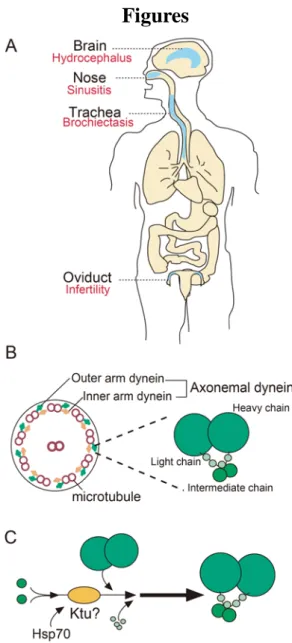
Fig. 1. Schematic picture of motile cilia structure
(A) Schematic picture of multiciliated tissues in human body. Red letters show PCD phenotypes in respective tissues. (B) Left: Transverse section of motile cilia representing 9+2 structure of microtubule circular orientation. Right: Detailed structure of microtubule associated dynein arms. (C) Hypothetical function of Ktu in the process of ourter arm dynein complex formation.
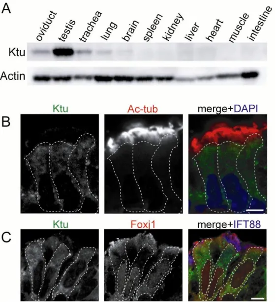
Fig. 2. Ktu is expressed in multiciliated tissues.
(A) Immunofluorescence of P14 mouse organs by anti-Ktu pAb. Expression of Ktu was detected in multiciliated/ flagellated tissues (oviduct, testis, trachea, brain). (B) Anti-Ktu pAb was double-immunolabeled with anti-acetylated α-tubulin mAb (Ac-tub, a cilia marker, B), (C) withanti-Foxj1 mAb (multiciliated cell marker) in trachea epithelial cells,.
Fig. 2. (Continued)
(D-F) Expression of Ktu in mouse ependymal cells in lateral brain ventricles was analyzed by immunofluorescence using anti-Ktu pAb (green) and anti-Foxj1 mAb (red, multiciliated cell marker, D), S100β (red, ependymal cell marker, E) , and Tuj1 (red, neuronal marker, F). Ktu was detected in Foxj1-postive cells, indicating that it is expressed in multiciliated cells. Ktu also co-localized with S100β but did not co-localize with neuronal marker Tuj1 in the lateral ventricles (yellow arrowheads). DAPI is the marker for nuclei (blue). Dotted lines represent the walls of the ventricle lumen. Scale bars: 5 µm in B, C; 10 µm in D, E, F.
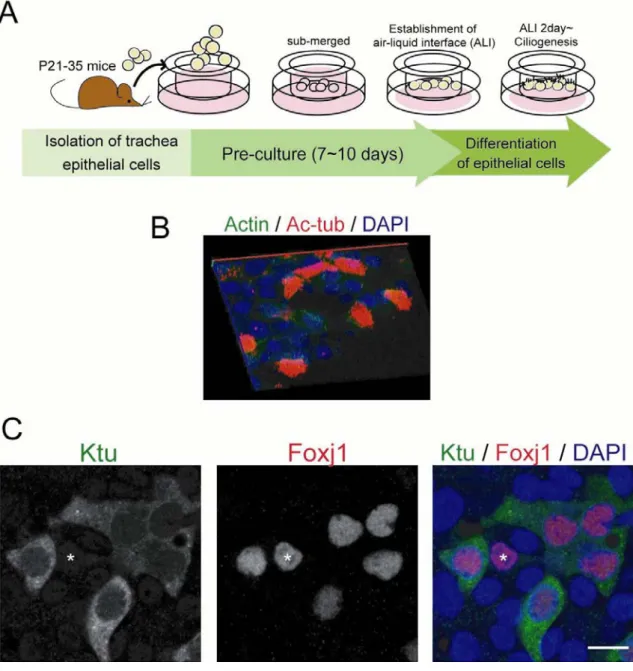
Fig. 3. In vitro studies of ciliogenesis using MTEC
(A) Schematic illustration of the production of MTEC. (B) A 3D reconstruction of confocal microscopy images showing normal ciliogenesis in the MTEC culture system. MTEC are immunolabeled with actin (green, cell border) and acetylated α-tubulin (Ac-tub, red) and DAPI (blue, nucleus). (C) Ktu (green) is expressed in the
Foxj1-positive cells (red). The asterisk shows a Foxj1 (+) Ktu (-) cell. Scale bars: 10 µm in B.
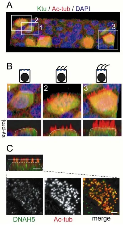
Fig. 4. Localization of Ktu during ciliogenesis
(A) A 3D reconstruction of the MTEC immunolabeled with Ktu (green) and acetylated α-tubulin (Ac-tub, red). (B) Cells at different ciliogenesis stages boxed in A. 1: pre-ciliogenesis period, 2: early-ciliogenesis period, 3: cilia elongation period. Serial confocal images projected in the x-z plane are shown in the lower panels. Ktu is expressed widely within the cytoplasm before and during ciliogenesis. (C) DNAH5 (axonemal dynein protein) localized to cilia at an early ciliogenesis stage. Scale bars: 2 µm in C.
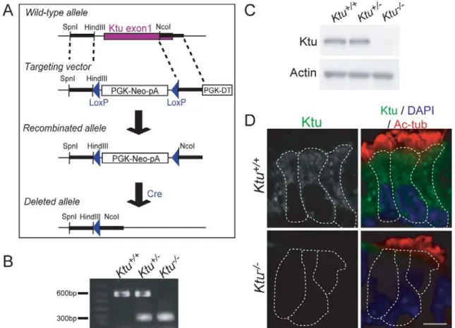
Fig. 5. Generation of Ktu knockout mouse.
(A) Construction of the wild-type allele, targeting vector, recombinated allele, and deleted allele of the mouse Ktu gene. The 1st exon was recombinated with the targeting vector. The recombinated allele was then deleted through Cre recombinase. (B) Genotype of Ktu-/- littermates by PCR. (C) Western blot analysis of Ktu from mouse testis. Ktu protein (120 kDa) is depleted in the homozygous knockout mouse. (D) Indirect immunofluorescence of trachea epithelial cells from P7 mice using anti-Ktu pAb and anti-acetylated α-tubulin mAb. Cytoplasmic expression of Ktu (green) was lost in the Ktu-/-. White dotted lines demarcate individual cells. Scale bar: 5 µm.
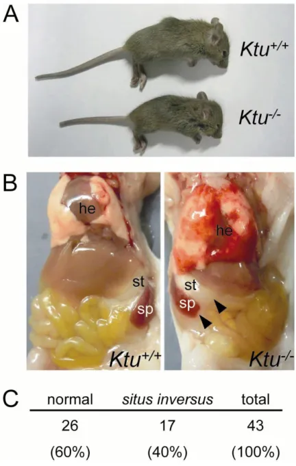
Fig. 6. Ktu knockout mice show primary ciliary dyskinesia
(A) Ktu+/+and Ktu-/- littermates (P14). Ktu-/- shows growth defect. (B) Reversal of organ translocation (situs inversus totalis) in Ktu-/- (P14). Arrowheads indicate reversed localization of stomach and spleen. he, heart; st, stomach; sp, spleen. (C) Ratio of situs inversus totalis within Ktu-/- mice observed at P3.
Fig. 6. (Continued)
(D) The Ktu-/- develops hydrocephalus and exhibits a dome-shaped head at P14 ( arrow). (E) The olfactory bulb is diminished (white arrowhead) and the cerebellum is pressed in
Ktu-/- (white arrow). (F) Coronal section of P14 brains stained with Haematoxylin and Eosin (H&E) staining. The lateral ventricle is enlarged in Ktu-/- (yellow arrow).
Fig. 7. Hydrocephalus initiates between E18-P0
(A)Schematic picture of the section position of the P0.5 brain shown in B. (B) Coronal sections of P0.5 Ktu+/+ (left) and the Ktu-/- (right) brains. In both rostral (1) and caudal (2) positions, lateral ventricles are enlarged (yellow arrows). Haematoxylin and eosin (H&E) staining. The lateral ventricle is enlarged in Ktu-/- (yellow arrows).
Fig. 8. Absence of axonemal dynein components in the Ktu-/- in brain ependyma (A) Ciliary localization of axonemal dynein was visualized by immunofluorescence of ependymal cells in P7 lateral ventricles with anti-DNAH5 pAb and anti-acetylated α-tubulin (Ac-tub, cilia marker) mAb. (B) Double-immunolabeling with anti-DNAH9 pAb and anti-acetylated α-tubulin mAb (Ac-tub).
Fig. 8. (Continued)
(C) Indirect immunofluorescence using anti-DNAI1 pAb and anti-acetylated α-tubulin mAb (Ac-tub). (D) Immunohistochemistry using anti-DNAI2 mAb and anti-α-tubulin pAb. In Ktu+/+, DNAH5 (green, A), DNAH9 (green, B), DNAI1 (green, C) and DNAI2 (green, D) is expressed along the ciliary axoneme (red), but in Ktu-/-, none of them is detected (yellow arrowheads). The white dotted lines indicate the surface of the cells.
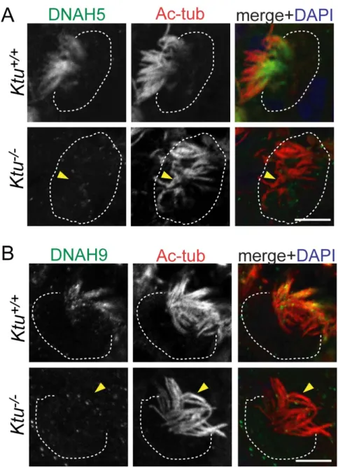
Fig. 9. Absence of axonemal dynein components in the Ktu-/- multicilia of trachea epithelial cells
(A) Ciliary localization of dynein components was visualized by immunofluorescence of P7 trachea epithelial cells with anti-DNAH5 pAb and anti-acetylated α-tubulin (Ac-tub, cilia marker) mAb. (B) Immunohistochemistry using anti-DNAH9 pAb and anti-acetylated α-tubulin In Ktu+/+
, DNAH5 (green, A), DNAH9 (green, B) is expressed along the ciliary axoneme (red), but in Ktu-/-, DNAH5 was absent from the axoneme (yellow arrowheads, A). Similarly, signal of DNAH9 was lost from the ciliary base yellow arrowheads, B). The white dotted lines indicate the surface of the cells.
Fig. 9. (Continued)
Immunohistochemistry of trachea epithelial cells with (C) anti-DNAI1 pAb and anti- Ac-tub mAb. (D) anti-DNAI2 mAb and anti-α-tubulin pAb. In Ktu+/+
, DNAI1 (green, C) and DNAI2 (green, D) is expressed along the ciliary axoneme (red), but in Ktu-/-, none of them are detected (yellow arrowheads) in the axoneme (E) Cross-section of trachea ciliary axoneme visualized by transmission electron microscope. Cilia of Ktu
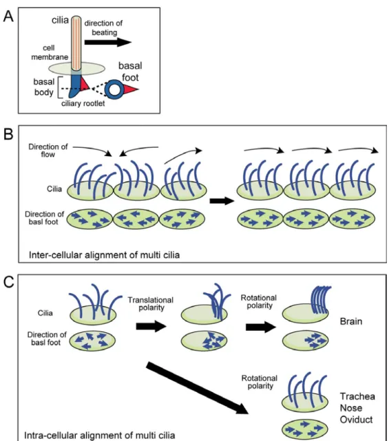
Fig. 10. Schematic view of coordinated ciliary alignment within the cell and among cells
(A) Schematic illustration of the motile cilium and its basal structure. The directionality of ciliary beat corresponds to the direction of the basal foot. Ciliated cells align their cilia to beat in a uni-directional manner in the tissue. (B) The coordinated alignment of cilia between cells (inter-cellular alignment) is called as the tissue-level polarity. (C) Coordinated alignment of cilia also exists within the cell (rotational polarity). Basal foot aligns in the same direction during/ after ciliogenesis in each cell. In brain ependyma, cilia patches migrate toward the one side of the cell (translational polarity) before the establishment of rotational polarity. Other ciliated cells harbor cilia in a uniform manner within the cell surface.
Fig. 11. Quantitive analysis of basal foot orientation
To quantify the alignment of cilia in each cell, the directionality of the basal foot was measured. (A) TEM section of trachea epithelial cells cut in the basal position of cilia. The basal foot are shown in dense arrow-like structure. (B) A basal line was drawn for each picture (yellow line). For each basal foot, a vector connecting the center of the basal body and the protrusion of basal feet was drawn (pink line). (C) Schematic illustration of basal foot that are boxed in B. A basal line that is parallel to the base of the picture was drawn for each picture taken. The angle between each vector and the basal line was measured manually using ImageJ software. The mean angle was calculated for each cell using Oriana 4.0 software. (D) The mean angle was defined as the mean ciliary direction (0º).
Fig. 12. Loss of cilia motility affects alignment of basal bodies in brain ependymal cells.
(A) Double-–label immunohistochemistry of P10 lateral ventricles with anti-Pericentrin pAb (green, basal body marker) and anti-Actin pAb (red, cell border marker). Basal bodies (arrows) migrated toward the anterior side of ependymal cells (yellow arrow) in
Ktu+/+ (left). Migration of basal bodies was unaffected in Ktu-/- (right). (B) Asymmetrical localization of Vangl1 (green) immunolabeled with actin (red) in the brain ependymal cells (P10). Asymmetrical localization of Vangl1 was retained in Ktu -/-(yellow arrowheads). Scale bars: 10 µm in A, B.
Fig. 12. (Continued)
(C) TEM sections of P10 brain ependymal cells. Unidirectional alignment of basal feet was observed in Ktu+/+ (rotational polarity). Rotational polarity was disrupted in Ktu-/-. Directions of basal foot are shown by yellow arrows. (D) Circular plots of deviation angles of the basal feet. Mean angles are pointed to 0. (Ktu+/+, 284 basal feet in 38 cells from 3 mice; Ktu-/-, 350 basal feet in 39 cells from 2 mice).
Fig. 13. Alignment of basal feet in trachea epithelial cells is unaffected in a Ktu knockout mouse
(A) Trachea epithelial cells from P10 mice were immnunolabeled with anti-Vangl1 pAb (green) and anti- actin pAb (red). Posterior-specific localization of Vangl1 was established in Ktu-/- as well as Ktu+/+ (yellow arrowheads). Scale bar: 10 µm. (B) TEM sections of P10 trachea epithelial cells of Ktu+/+ (left) and Ktu-/- (right). Directionality of the basal feet is established in the Ktu-/- (yellow arrows). Circular plots of deviation angles of the basal feet (bottom). Mean angles are pointed at 0. (Ktu+/+, 319 basal feet in 31 cells from 2 mice; Ktu-/-, 341 basal feet in 29 cells from 3 mice.
Fig. 14. Schematic picture of the establishment of rotional polarity in different tissues
Basal foot are aligned within the cell (rotational polarity). The major determinant of rotational polarity is the ciliary motility in the brain ependyma and planar polarity in trachea epithelial cells.
Fig. 15. Hypothetical model of ciliary re-orientation through ciliary motility
Actin filaments and microtubules are attached to striated ciliary rootlet and basal foot respectively. The Dvl-Rho complex is known to regulate actin remodeling. Ciliary motility may activate Dvl-Rho dependent actin remodeling pathway and reorient cilia.