Antitumor Activity of the Novel Oral Heat
Shock Protein 90 Inhibitor in Mono Therapy and
Combination Therapy
year
2020
その他のタイトル
新規経口Hsp90 阻害剤の創製と単剤及び併用による
抗腫瘍効果に関する研究
学位授与大学
筑波大学 (University of Tsukuba)
学位授与年度
2019
報告番号
12102甲第9453号
URL
http://doi.org/10.15068/00160401
Antitumor Activity of the Novel Oral Heat Shock Protein 90
Inhibitor in Mono Therapy and Combination Therapy
January 2020
Antitumor Activity of the Novel Oral Heat Shock Protein 90
Inhibitor in Mono Therapy and Combination Therapy
A Dissertation Submitted to
the Graduate School of Life and Environmental Sciences,
the University of Tsukuba
in Partial Fulfillment of the Requirements
for the Degree of Doctor of Philosophy in Biological Science
(Doctor Program in Biological Sciences)
Table of contents
Contents
Pages
Abstract ... 1
Abbreviations ... 3
General introduction ... 4
Chapter 1 ... 6
Preclinical antitumor activity of the novel heat shock protein 90 inhibitor CH5164840,
against human epidermal growth factor receptor 2 (HER2)-overexpressing cancers ... 6
1. Introduction ... 6
2. Materials and Methods ... 7
3. Results ... 10
4. Discussion ... 12
5. Tables ... 15
6. Figures ... 18
Chapter 2 ... 29
Enhanced antitumor activity of erlotinib in combination with the HSP90 inhibitor
CH5164840, against non-small-cell lung cancer ... 29
1. Introduction ... 29
2. Materials and Methods ... 30
3. Results ... 31 4. Discussion ... 33 5. Tables ... 36 6. Figures ... 37
General discussion ... 43
Acknowledgements ... 45
References ... 46
List of Publications ... 53
1
Abstract
Heat shock protein 90 (HSP90) is a molecular chaperone that plays a significant role in the stability and maturation of client proteins, including oncogenic targets for cell transformation, proliferation, and survival. It is an attractive target for cancer therapy. Recently, a novel HSP90 inhibitor (CH5164840) was identified, and its induction of oncogenic client protein degradation, anti-proliferative activity, and apoptosis investigated using the NCI-N87 gastric and BT-474 breast cancer cell lines. Interestingly, CH5164840 demonstrated tumor selectivity both in vitro and in vivo, binding to tumor HSP90, which forms active super chaperone complexes, rather than the HSP90 of normal cell, which mainly exist as non-complexes with co-chaperons in vitro. It is extensively distributed in mouse model tumors. These facts support the CH5164840-induced phosphorylated AKT level decrease observed in tumor tissues, but not in normal tissues. In addition to being well tolerated, orally administered CH5164840 exerts potent antitumor efficacy, leading to regression in the NCI-N87 and BT-474 tumor xenograft models. Additionally, CH5164840 significantly enhanced antitumor efficacy against the gastric and breast cancer models when co-administered with the human epidermal growth factor receptor 2 (HER2)-targeted agents trastuzumab and lapatinib. These data demonstrate the potential antitumor efficacy of monotherapy with CH5164840, and the significant synergistic efficacy of its co-administration with trastuzumab or lapatinib, validifying clinical development of CH5164840 as an HSP90 inhibitor for combination therapy with targeted agents against HER2-overexpressing tumors.
Epidermal growth factor receptor (EGFR) is one of the most potent oncogenic client proteins of HSP90. Targeted inhibition of EGFR has shown clinical efficacy in the treatment non-small-cell lung cancer (NSCLC) patients. However, primary and acquired resistance to existing EGFR inhibitors is a major clinical problem. In the present study, the antitumor activity of a combination of erlotinib and CH5164840 was investigated. The NSCLC cell lines and xenograft models were treated with CH5164840 and erlotinib to examine their mechanisms of action and cell growth inhibition. CH5164840 showed remarkable antitumor activity against the NSCLC cell lines and xenograft models. Combination therapy with CH5164840 enhanced the antitumor activity of erlotinib against NCI-H292 EGFR-overexpressing xenograft models. In vitro treatment of NCI-H292 cells with erlotinib resulted in increased STAT3 phosphorylation. Next, the role of the STAT3 signal in the mechanism of action of the erlotinib and CH5164840 combination was evaluated, as it is an important tumor growth-related signal. STAT3 phosphorylation increase in erlotinib-treated NCI-H292 cells was abrogated by HSP90 inhibition. Additionally, CH5164840 enhanced the antitumor activity of erlotinib, despite the low efficacy of erlotinib monotherapy, in a NCI-H1975 T790M mutation erlotinib-resistant model. Further, extracellular signal-regulated kinase (ERK) signaling was effectively suppressed by the erlotinib and CH5164840 co-therapy, in a NCI-H1975 erlotinib resistant model. These data collectively indicate the potent antitumor activity of CH5164840 and higher efficacy of its coadministration with erlotinib
2
against NSCLC tumors with EGFR overexpression and mutations. Therefore, providing evidence of the therapeutic potential of CH5164840 as an HSP90 inhibitor for combination therapy with EGFR-targeting agents against EGFR-addicted NSCLC.
3
Abbreviations
17-AAG 17-N-allylamino-17-demethoxygeldanamycin
17-DMAG 17-dimethylaminoethylamino-17-demethoxygeldanamycin ABCG2 ATP binding cassette subfamily G member 2
Ab antibody AC adenocarcinoma
ALK anaplastic lymphoma kinase
c-PARP cleaved poly (ADP-ribose) polymerase EGFR epidermal growth factor receptor ER estrogen-receptor
ERK extracellular signal-regulated kinase GAPDH glyceraldehyde-3-phosphate dehydrogenase HER2 human epidermal growth factor receptor 2 HER3 human epidermal growth factor receptor 3 HER4 human epidermal growth factor receptor 4 HSP90 heat shock protein 90
IP immunoprecipitation JAK Janus kinase
Kd dissociation constant MDR multidrug-efflux transporter MTD maximum tolerated dose
NQO1 NAD(P)H quinone dehydrogenase 1 NSCLC non-small-cell lung cancer
p.o. per os
PARP poly-(ADP-ribose) polymerase PBMC peripheral blood mononuclear cells PR progesterone-receptor
PTEN phosphatase and tensin homolog q.d. quaque die
SC squamous cell carcinoma SCLC small cell lung cancer
STAT3 signal transducer and activator of transcription 3 TGI tumor growth inhibition
TKI tyrosine kinase inhibitor TYK2 tyrosine kinase 2 TV tumor volume
4
General introduction
Heat shock protein 90 (HSP90) is a highly conserved and ubiquitously expressed molecular chaperone in eukaryotic and prokaryotic cells, which plays key roles in protein folding and
client protein stability (1). Oncogenic client proteins, the targets of many anticancer agents, encompass dysregulated, mutated, and fusion proteins, and are particularly dependent on HSP90 for conformation maintenance (2-6). For example, epidermal growth factor receptor (EGFR) is one of the most potent oncogenic client proteins of HSP90. Additionally, mutated EGFR seems to be more sensitive than wild-type EGFR to HSP90 inhibitor degradation (7, 8). HSP90 function inhibition is known to simultaneously lead to the degradation of multiple client proteins involved in tumor progression and inhibition of multiple oncogenic pathways, resulting in signal transduction loss, growth inhibition, cell death, and antiangiogenesis. Moreover, in tumor cells, HSP90 is present in an activated super-chaperone complex (9), and elevated HSP90 expression is associated with poor prognosis in breast cancer patients (10).Therefore, HSP90 targeting is considered a promising anticancer therapy strategy (11-14).
HER family members are one of the most potent oncogenic client proteins of HSP90. The HER family has four known members: human epidermal growth factor receptor 1 (HER1) (also termed EGFR), human epidermal growth factor receptor 2 (HER2), human epidermal growth factor receptor 3 (HER3), and human epidermal growth factor receptor 4 (HER4). They are structurally related receptor tyrosine kinases, which form homo- or hetero-dimer complexes with each other to contribute to the growth signal; the HER2 and HER3 heterodimer complex has a strong transforming activity. Aberrant signaling through the HER pathway is found in many epithelial cancer types (15).
Among females, breast cancer is the most commonly diagnosed cancer and leads to the highest number of cancer-related deaths worldwide (16). Breast cancer comprises a heterogeneous group of diseases in term of differentiation, proliferation, prognosis, and treatment. Several breast cancer molecular subtypes have been identified, based on hormone receptor (estrogen-receptor (ER), progesterone-receptor (PR)) and HER2 status (17). One of these subtypes is the HER2-enriched subtype, which occurs in 15–20% of breast cancer patients. The overexpression of HER2, a receptor tyrosine kinase and oncogene, is associated with poor prognosis in breast (18) and gastric (19) cancers. The HER2 receptors are anticancer drug targets. Two strategies for blocking the action of these proteins include ectodomain targeting with antibodies (Ab) and the use of protein-tyrosine kinase activity-inhibiting drugs. Although trastuzumab, (humanized monoclonal Ab directed against HER2) and lapatinib (EGFR and HER2 dual-kinase inhibitor) have both demonstrated good initial clinical responses and are considered standard-of-care agents, clinical relapse with trastuzumab or lapatinib therapy has been observed in some patients (20). Therefore, more effective HER2-overexpressing tumor treatments are required, and the development of rationalized combinations of agents appears particularly promising (21).
5
Lung cancer is one of the most commonly diagnosed cancer and cause of cancer-related deaths worldwide (16), including the US (22). It is classified into two main types: small cell lung cancer (SCLC) and non-SCLC (NSCLC), which account for approximately 85% of all lung cancers (23, 24). Furthermore, NSCLCs are characterized into two major subtypes: adenocarcinoma (AC) and squamous cell carcinoma (SCC). The classification of NSCLC subtypes based on genotype and histology has resulted in dramatic improvements in disease outcome in selected patient. Specifically, molecular targeted drugs such as EGFR or Anaplastic lymphoma kinase (ALK) inhibitors, are approved for the treatment of NSCLC harboring genetic mutations in the genes encoding these proteins (25).Tumors with the EGFR gene that harbors activating mutations in exon 19 and 21, can be successfully treated with EGFR-tyrosine kinase inhibitors (TKIs), compared with cytotoxic agents (26, 27). Despite remarkable responses in patients with activating mutations in the tyrosine kinase domain of EGFR, most patients relapse within their first year of treatment. Due to the potential for resistance to TKI in EGFR-addicted tumors, identification of an effective treatment using rationalized agent combinations is particularly promising.
In recent years, promising molecular targeted drugs have been developed for cancer treatment, but drug resistance and recurrence remain clinical problems. With respect to blocking cancer-causing resistance mechanisms, unique features of HSP90 inhibition are expected to aid the overcoming of TKI resistance. First-generation HSP90 inhibitors: geldanamycin derivatives, are associated with hepatotoxicity, polymorphic metabolism by NAD(P)H quinone dehydrogenase 1 (NQO1), and efflux via P-glycoprotein, and are therefore not desirable for clinical use. Thus, they have not yet been approved for cancer therapy. To overcome these drawbacks, several second-generation HSP90 inhibitors have been synthesized and are currently in clinical development (28-31).
Recently, a novel potent HSP90 inhibitor CH5164840, with a unique chemical structure, based on a 3-D structure, was identified via virtual screening. In co-crystal structural analysis, CH5164840 was found to interact with an ATP-binding pocket of HSP90α with high affinity (32, 33). In the present study, to examine useful cancer treatment strategies using HSP90 inhibitors, the focus was placed on a HER2 positive cancer and NSCLC. In Chapter 1, the in vitro and in vivo investigations of the efficacy of a CH5164840 and HER2-targeted agent combination is examined. Additionally, the in vitro and in
vivo properties of CH5164840 monotherapy and CH5164840 and erlotinib co-therapy, including its
antitumor activity in EGFR overexpressing and erlotinib-resistant NSCLC xenograft models, are elucidated in Chapter 2.
6
Chapter 1
Preclinical antitumor activity of the novel heat shock protein 90 inhibitor
CH5164840, against human epidermal growth factor receptor 2
(HER2)-overexpressing cancers
1. Introduction
The HER family is composed of four HER receptors: HER1 (also termed EGFR), HER2, HER3, and HER4 (15). HER family members can form homodimers and heterodimers with each other. No known ligand binds HER2 with high affinity. HER2 forms heterodimers with each of the other family members, and these heterodimers can bind to growth factors (34). Although HER3 has a tyrosine kinase domain that is highly homologous to other family members, its kinase activity is impaired. HER3 can also form heterodimers with the other family members. However, owing to the lack of protein kinase activity by HER3, transphosphorylation by other HER family members is required for cell signaling. It is known that the HER2 and HER3 heterodimer complex has a strong transforming activity.
The overexpression of HER2, an oncogenic receptor tyrosine kinase, is associated with poor prognosis in breast (18) and gastric (19) cancers. Indeed, in HER2-overexpressing breast cancers, HER2 and HER3 frequently form a heterodimer as an oncogenic unit that plays an important role in HER2-mediated signaling (7, 8, 35). Although both trastuzumab and lapatinib are considered standard-of-care agents and benefit HER2-amplified breast and gastric cancer patients, clinical relapse with both has been observed in some patients (20). Therefore, more effective treatment of HER2-overexpressing tumors is required, and forming rationalized agent combinations appears particularly promising (21).
As HER2 is a representative client protein whose stability is HSP90-function dependent (36-38), HSP90 inhibitors are effective against HER2-overexpressing tumors (39, 40). HSP90 is an attractive target of cancer therapies. Geldanamycin and its derivatives 17-AAG, 17-DMAG, and first generation HSP90 inhibitors, show strong efficacy in vivo. However, these inhibitors are reportedly oxidized by NQO1 and effluxed by multidrug-efflux transporter (MDR) and ATP binding cassette subfamily G member 2 (ABCG2, also termed BCRP); thus, attenuating their efficacy. Additionally, they are hepatotoxic. Recently, a novel HSP90 inhibitor: CH5164840, was generated (32, 33). To examine the usefulness of its significant antitumor efficacy in HER2 positive tumors,
in vitro and in vivo investigations of the efficacy of CH5164840 and HER2-targeted agent
7
2. Materials and Methods
2- 1. Compound
CH5164840 (33), its biotin-labeled version, and 17-dimethylaminoethylamino-17-demethoxygeldanamycin (17-DMAG) were synthesized by Chugai Pharmaceutical (Kanagawa, Japan). Lapatinib was prepared from the commercial product Tykerb (GlaxoSmithKline, Clifton, NJ, USA). Trastuzumab was obtained from Chugai Pharmaceutical ⁄ F. Hoffmann-La Roche (Basel, Switzerland).
2- 2. Cells and culture
The human gastric cell line NCI-N87; breast cancer cell lines BT-474, SK-BR-3, and MDA-MB-231; ovarian cancer cell line SK-OV-3; prostate cancer cell line DU 145; colorectal cancer cell line HCT116; and NSCLC cell line NCI-H460 were obtained from the American Type Culture Collection (ATCC; Manassas, VA, USA). The human gastric cell line JR-St, was obtained from Immuno-Biological Laboratories (Fujioka, Japan). Adult normal human dermal (NHDF-Ad) fibroblast cells were obtained from Takara Bio (Shiga, Japan). Human mammary tumor MAXF401 was obtained from Professor Heinz-Herbert Fiebig (University of Freiburg, Freiburg, Germany). All cell lines were cultured according to the supplier’s instructions.
2- 3. Cell proliferation assay
Tumor cells were seeded into microtiter plates containing compounds and incubated at 37°C in 5% CO2. After a 4-day incubation, Cell Counting Kit-8 solution (Dojindo Laboratories, Kumamoto,
Japan) was added, and absorbance at 450 nm measured with the Microplate-Reader iMark (Bio-Rad Laboratories, Hercules, CA, USA). Anti-proliferative activity was calculated using the formula: (1−T/C) × 100 (%), where T represents the absorbance of drug-treated cells, and C represents that of untreated control cells at 450 nm. The IC50 values were calculated using Microsoft Excel 2007
(Redmond, WA, USA).
2- 4. Surface plasmon resonance analysis
All biosensor experiments were conducted using a Biacore 2000 instrument (GE Healthcare Japan, Tokyo, Japan). The N-terminal domains of the human HSP90α (9–236) and HSP90β (1–221) expressed in Escherichia coli were minimally biotinylated (sulfo-NHS-LC-LC biotin; Thermo Fisher Scientific, Waltham, MA, USA) and coupled on a streptavidin-coated sensor chip (GE Healthcare, Buckinghamshire, UK), respectively. Sensorgrams were processed using SCRUBBER2 (BioLogics, Campbell, Australia) and Kd values were determined by fitting the processed data globally to the 1:1 binding model, using BIAevaluation (version 3.1; GE Healthcare). The experiments for the Kd determinations were performed in 50 mM
8
tris(hydroxymethyl)aminomethane-HCl (pH 7.6), 150 mM NaCl, 0.005% P-20, and 1% DMSO at 30 μL/min, 20°C.
2- 5. Protein kinase assay
The activities of EGFR, KIT, HER2, MET, fibroblast growth factor receptor 2 (FGFR2), FMS-related tyrosine kinase 3 (FLT3), leukocyte receptor tyrosine kinase (LTK), insulin receptor (INSR), YES, ABL, ZAP70, insulin-like growth factor receptor I (IGF1R), vascular endothelial growth factor receptor 2 (KDR) (Life Technologies), and platelet derived growth factor receptor (PDGFR)β (Millipore) on substrate peptides were determined using a homogeneous time-resolved fluorescence assay with LANCE Eu-W1024 labeled anti-phosphotyrosine PT66 antibody (PerkinElmer), according to standard methods. Time-resolved fluorescence was measured with an EnVision HTS microplate reader. The activities of Aurora A, AKT1/PKBα, PKA, CDK1/cyclin B, CDK2/cyclin A (Millipore), PKCα, PKCβ1, PKCβ2, ALK, and JAK1 (Carna Biosciences) on substrate peptides were determined using the IMAP FP Screening Express Progressive Binding System (Molecular Devices). Specifically, to detect MEK activity, inactive MAP kinase 2/ERK2 (Millipore) was activated by MAP2K1 (Carna Biosciences) addition; then, MAP kinase 2 activity on a substrate peptide was determined using the IMAP FP Screening Express Progressive Binding System. Fluorescence polarization was measured with an EnVision HTS microplate reader. p70S6K activity on a substrate peptide was determined using the Z’-LYTE Kinase Assay Kit-Ser/Thr 7 Peptide (Life Technologies). Time-resolved fluorescence was measured with an EnVision HTS microplate reader.
2- 6. Western blotting and co-immunoprecipitation
Cells were lysed with Cell Lysis Buffer (Cell Signaling Technology) containing protease and phosphatase inhibitors. Before lysis, the grafted tumors were homogenized using a BioMasher (K.K. Ashisuto). The lysates were denatured with Sample Buffer Solution with Reducing Reagent for SDS-PAGE (Nacalai Tesque), and then subjected to SDS-SDS-PAGE. After electroblotting, the polyvinylidene fluoride membranes were blocked with Blocking One (Nacalai Tesque) for 1 hour. Membranes were then incubated with primary antibodies (see below) at room temperature for 2 hours or at 4°C overnight, followed by washing three times with 0.1% Tween 20 (Nacalai Tesque) in TBS buffer (TBS-T buffer; TaKaRa Bio). This was followed by a 1-hour incubation at room temperature with secondary antibodies (Cell Signaling Technology). Then, membranes were washed with TBS-T buffer, followed by incubation with ECL or ECL-plus solutions (GE Healthcare). Signals were detected with ECL Plus (GE Healthcare), followed by LAS-4000 (Fujifilm, Tokyo, Japan) or the Odyssey Infrared Imaging System (LI-COR Biosciences, Lincoln, NE, USA). The primary antibodies used were used were pY1221-HER2, pY1068-EGFR, pY1289-HER3, pS473-AKT, AKT, pT202/Y204-ERK, ERK (Cell Signaling, Beverly, MA, USA), EGFR, HER2, HER3, and actin (Santa Cruz Biotechnology,
9
Santa Cruz, CA, USA), and HSP70 (Stressgen, Victoria, Canada). For co-immunoprecipitation, cell lysates were incubated with or without the HSP90 antibody (Santa Cruz Biotechnology) overnight at 4°C, and protein A-agarose beads (Roche Diagnostics, Penzberg, Germany) added for 1 h at 4°C. Beads were then washed three times with cell lysis buffer, boiled with sample buffer solution, and analyzed via Western blotting.
2- 7. Xenograft model and efficacy study
All animal studies were approved by the Chugai Institutional Animal Care and Use Committee. Cancer cells (0.5–1 × 10⁷) were implanted subcutaneously into the right flank of athymic nude (BALB/c nu/nu) mice (CAnN.Cg-Foxn1<nu>/CrlCrlj nu/nu; Charles River Laboratories, Kanagawa, Japan). Tumor volume (TV) was calculated using the formula: TV = ab2/2, where a and
b represent tumor length and width, respectively. Once the tumors had reached a volume of
approximately 200–300 mm3, the animals were randomized into each group (n = 4 or 5), and
treatment was initiated. CH5164840 and lapatinib were orally administered; trastuzumab was administered by intraperitoneal injection. Tumor growth inhibition (TGI) was calculated using the formula: TGI = (1−[Tt−T0]/[Ct−C0]) × 100 (%), where T (TV of treated group; T0 on day 0 or Tt on
day t) and C (TV of control group; C0 on day 0 or Ct on day t) represent mean tumor volume. The
maximum tolerated dose (MTD) was defined as the dose that resulted in neither lethality nor more than 20% body weight loss. The ED50 was calculated from the values of TGI on the final
experimental day using XLfit version 5.1.0.0 (Microsoft, Redmond, WA, USA).
2- 8. Pharmacokinetic and pharmacodynamic study
Tumor xenograft (NCI-N87)-bearing nude mice were prepared by the same method used for the efficacy studies. Two mice were killed after oral administration of 50 mg/kg CH5164840, for collection of the indicated organs and grafted tumors. CH5164840 was extracted with acetonitrile/water/formic acid (75/25/0.1, v/v/v) under ice-cold conditions. CH5164840 concentration was determined using liquid chromatography/tandem mass spectrometry (LC/MS/MS) (API 3000, Applied Biosystems, Foster City, CA, USA).
2- 9. Real-time quantitative RT-PCR
The QuantiTect Probe RT-PCR kit (Qiagen, Valencia, CA, USA) and Taqman primers (Applied Biosystems) were used to perform all reactions. Total RNA extraction was performed with the RNeasy mini kit (Qiagen). For the data analysis, counts were normalized to a housekeeping gene (Glyceraldehyde-3-phosphate dehydrogenase, GAPDH) at the same time point and condition. Counts are reported as fold change relative to the untreated control.
10
2- 10. Statistical analysis
SAS preclinical package software (version 5; SAS, Cary, NC, USA) was used for the statistical analyses (Tukey’s test). Statistically significant differences are indicated with asterisks; **P < 0.01 and ***P < 0.001.
3. Results
3- 1. CH5164840, a novel HSP90 inhibitor, induces the degradation of multiple
HSP90 client proteins and apoptosis in NCI-N87 and BT-474 HER2-overexpressing
cells.
Recently, CH5164840 was identified as an HSP90 inhibitor with a novel chemical structure, through virtual screening based on a 3-D structure (32, 33). As shown in Fig. 1-1, the binding affinity of CH5164840 for HSP90α was found to be 0.52 and 1.4 nM for HSP90β, a level that is associated with a slow dissociation rate. Specificity to the ATP-binding pocket of HSP90 was indicated by no inhibition against 22 kinases (IC50 > 20 μM) in a cell-free kinase assay and the
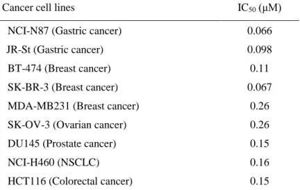
absence of significant ATP competitive binding (DiscoveRx, Fremont, CA, USA) against 400 kinases at 10 μM. Firstly, the sensitivity of various cancer cell lines to CH5164840 with in vitro cell growth assays was evaluated. The IC50 values of CH5164840 were found to be 66–260 nM in nine
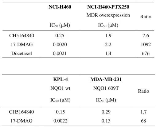
cancer cell lines (Table 1-1). CH5164840 also showed in vitro sensitivity against 17-DMAG-resistant cell lines, NCI-H460-PTX250, which overexpress MDR1 and MDA-MB-231 with NQO1 C609T mutation (Table 1-3). Secondly, CH5164840-induced client protein degradation in cancer cells was examined. CH5164840 was found to significantly reduce phosphorylation and protein levels of HER2, HER3, and EGFR, and inhibit downstream signals in NCI-N87 (HER2-overexpressing gastric cancer) and BT-474 (HER2-(HER2-overexpressing breast cancer) cells. Furthermore, activated caspase-3/7 levels (Figs. 1-2b, d) and cleaved poly (ADP-ribose) polymerase (Figs. 1-2a, c) increased following exposure to CH5164840, indicating that CH5164840 induces apoptosis. HSP70 induction was examined as it is a known response marker of HSP90 inhibition. The results confirmed that like 17-DMAG, HSP70 expression were induced by CH5164840 in vitro (Figs. 1-2a, c). In a time-course study, CH5164840 and 17-DMAG caused client protein degradation and HSP70 induction (Fig. 1-2e). These results indicate that CH5164840 selectively inhibited HSP90, induced key client protein degradation, and suppressed both the PI3K⁄AKT survival and RAF⁄MEK⁄ERK signaling pathways, leading to apoptosis induction and inhibition of tumor proliferation.
3- 2. CH5164840 exhibits tumor-selective HSP90 inhibition.
Based on the observation of tumor HSP90 in super-chaperone complexes with high ATPase activity and its high binding affinity to the HSP90 inhibitor
17-allylamino-17-demethoxy-11
geldanamycin (17-AAG) (7), the tumor selectivity of CH5164840 was assessed using the following procedure. Firstly, the anti-proliferative activity of CH5164840 in normal fibroblast (NHDF-Ad) cells was compared to that in NCI-N87 tumor cells, revealing that NHDF-Ad cell proliferation was moderately inhibited by CH5164840 treatment (IC50, 1.2 μM), and that the cells were less sensitive to
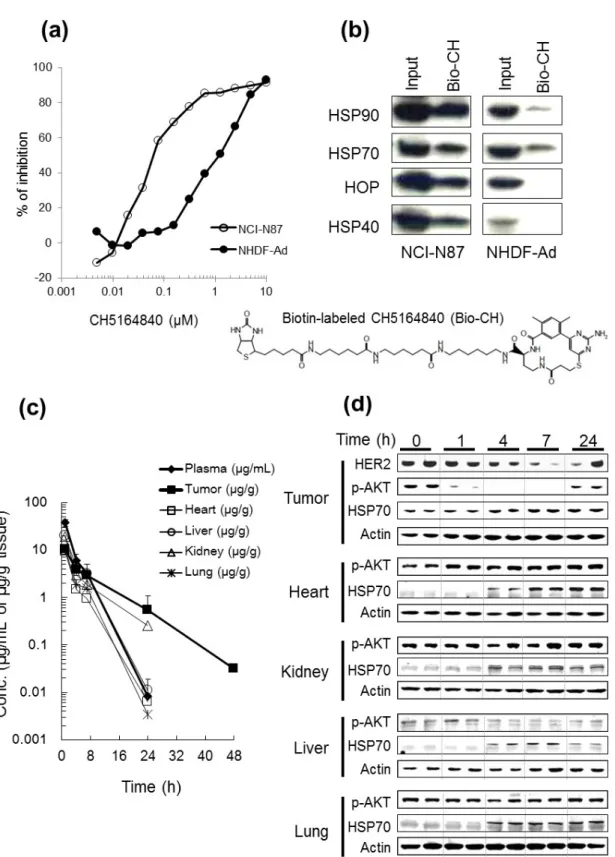
CH5164840 than NCI-N87 and other cancer cells (Table 1-1; Fig. 1-3a). Next, to examine whether CH5164840 binds selectively to tumor HSP90, pull-down assays were conducted with a biotin-labeled CH5164840 (Bio-CH) probe using cell extracts prepared from NCI-N87 or NHDF-Ad cell lines. As shown in Fig. 1-3b, Bio-CH preferentially bound to HSP90 when it formed a super-chaperone complex with the co-chaperones HSP70, HOP, and HSP40, in NCI-N87 cells, rather than to HSP90 in NHDF-Ad cells, which remained mainly free from these co-chaperones. These data indicate that CH5164840 selectively binds to tumor HSP90, which forms a super-chaperone complex. Further, tumor-selective distribution of CH5164840 after a single administration in mice, was explored. CH5164840 was only detected in tumor samples 48h after oral administration of one dose of 50 mg/kg CH5164840 in the NCI-N87 xenograft model. Greater CH5164840 retention was observed in tumor tissues (half-life = 6.3 h) compared to normal tissues and plasma (Fig. 1-3c). Finally, AKT phosphorylation level in tumor and normal tissue was measured to determine CH5164840 associated to tumor-selective HSP90 inhibition in vivo in NCI-N87 models. The results indicate that CH5164840 reduces AKT phosphorylation in tumor tissues, but not in normal tissue (Fig. 1-3d). Additionally, HSP70 induction in tumor and normal tissue was confirmed. Collectively, these results indicate that preferential binding to tumor HSP90 and increased retention in tumors lead to tumor-selective HSP90 inhibition by CH5164840.
3- 3. CH5164840 demonstrates potent antitumor efficacy in xenograft models.
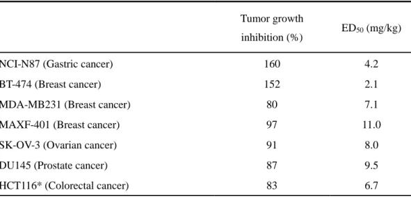
The in vivo antitumor efficacy of CH5164840 against human tumor xenograft models in mice, was investigated. Daily oral administration of CH5164840 resulted in significant dose-dependent antitumor efficacy in the NCI-N87 and BT-474 xenograft models, with a maximum TGI of 160% and 152%, respectively (Figs. 1-4a, b), without any significant loss of body weight. Additionally, HER2, HER3, EGFR, cyclinD1, and phospho-AKT protein levels significantly decreased in the NCI-N87 xenograft model, resulting in CH5164840 inhibition of downstream signals (Fig. 1-4c). Further, CH5164840 demonstrated significant antitumor efficacy against various xenograft models (Table 1-2). These results are consistent with those regarding the in vitro antitumor activity and tumor-selective HSP90 inhibition of CH5164840.3- 4. CH5164840 enhances the antitumor efficacy of HER2-targeted therapy with
trastuzumab or lapatinib.
12
of a combination of 25 mg/kg CH5164840 and 30 mg/kg trastuzumab to the trastuzumab-sensitive NCI-N87 and BT-474 xenograft models resulted in the enhancement of the antitumor efficacy of trastuzumab by CH5164840, increasing its TGI as a single agent from 56% to 167% in the NCI-N87 model (Fig. 1-5a), and from 136% to 240% in the BT-474 xenograft model (Fig. 1-5b). With regards to lapatinib, co-administration of 100 mg/kg lapatinib and 12.5 mg/kg CH5164840 to the NCI-N87 and BT-474 xenograft models resulted in significant enhancement of the antitumor efficacy of lapatinib by CH5164840 in both the NCI-N87 (TGI = 45% lapatinib only vs 155% in combination; Fig. 1-5c) and BT-474 xenograft (TGI = 88% lapatinib only vs 214% in combination; Fig. 1-5d) model. In all cases, the co-therapy-enhanced efficacy was statistically significant. These results suggest that CH5164840 potentiates the efficacy of HER2-targeted agents.
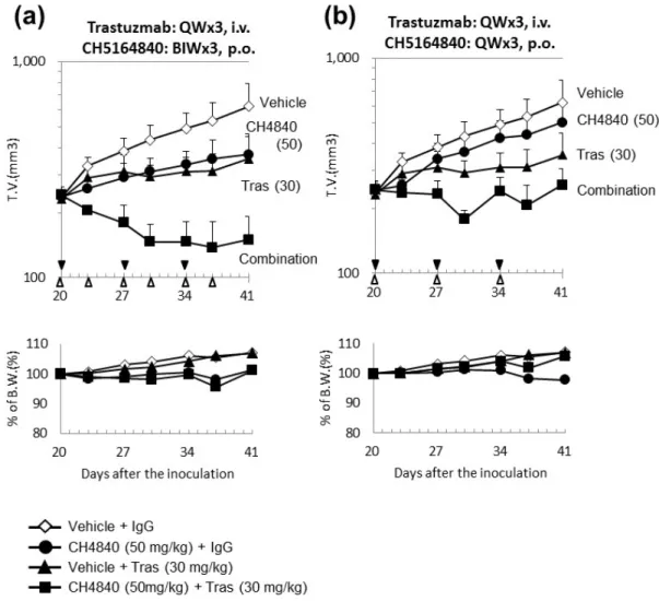
Furthermore, when the efficacy of intermittent dosing was examined in the form of once- or twice-weekly regimens of 50 mg⁄ kg CH5164840 in the NCI-N87 xenograft model, CH5164840 was found to enhance the antitumor efficacy of trastuzumab (TGI = 69% with trastuzumab only vs 97% or 123% with combined trastuzumab and once- or twice-weekly CH5164840 administration, respectively; Fig. 1-7).
3- 5. CH5164840 suppresses lapatinib treatment-induced HER3 signal activation in
vitro.
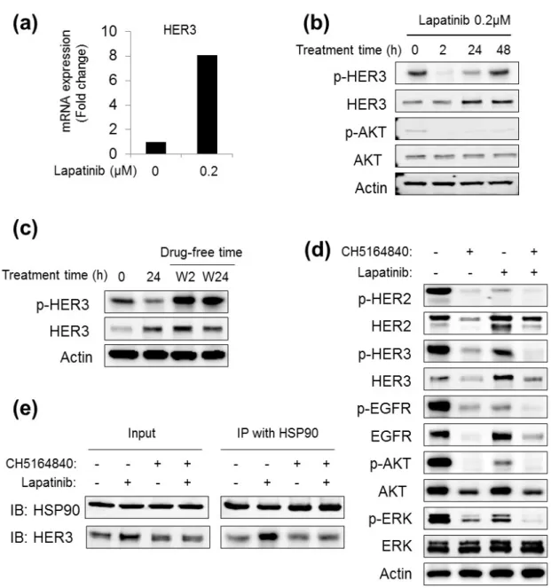
The mechanism underlying the synergistic efficacy of the combination of CH5164840 and lapatinib was examined, owing to elucidation of this synergism. Firstly, HER3 expression was examined after lapatinib treatment in NCI-N87 cells, as lapatinib treatment reportedly induces HER3 expression in SK-BR-3 cells (41). Quantitative RT-PCR revealed that lapatinib induced HER3 mRNA expression (Fig. 1-6a). HER3 protein levels consistently increased in a time-dependent manner (Fig. 1-6b). Although HER3 phosphorylation was transiently inhibited by lapatinib, it was not long lasting, despite continuous lapatinib treatment (Fig. 1-6b). Additionally, lapatinib discontinuation after 24 h of treatment, followed by lapatinib-free incubation, resulted in hyper-phosphorylation of HER3 (Fig. 1-6c). Next, to determine whether HSP90 inhibition induced the degradation of lapatinib-induced HER3 and other clients, the effect of CH5164840 was investigated in NCI-N87 cells. As shown in Fig. 1-6d, HER2 and EGFR were degraded, and their phosphorylation and downstream signals significantly decreased by CH5164840 and lapatinib co-therapy. Interestingly, lapatinib-induced HER3 returned to basal levels after the addition of CH5164840. Finally, HER3 was observed to be strongly co-precipitated with HSP90 in lapatinib-treated cells compared to that in non-treated control cells, and importantly, was returned to basal levels by the addition of CH5164840 (Fig. 1-6e).
4. Discussion
13
polymorphic metabolism by NQO1, and efflux by P-glycoprotein. They are therefore undesirable for therapeutic use and have not yet been approved for cancer therapy. To overcome these drawbacks, a novel HSP90 inhibitor CH5164840, with a unique chemical structure, was generated through virtual screening based on a 3-D structure. Its in vitro sensitivity against the 17-DMAG-resistant cell lines NCI-H460-PTX250, which overexpress MDR1 and MDA-MB-231, which have NQO1 C609T mutation, was confirmed. An attractive feature of HSP90 as an antitumor target is the possibility of tumor selectivity. As shown in Fig. 1-3b, a higher binding affinity of bio-CH to tumor HSP90 compared to normal HSP90, was observed. In this pull-down assay, the biotin label was positioned based on the co-crystal structure of HSP90α with CH5164840, to prevent influence on CH5164840– HSP90 binding. This selective binding of CH5164840 to HSP90 in tumor tissues and its long retention in tumor tissues in the mouse model explain why tumor tissue phosphorylated AKT levels, but not that of normal tissues, were observed to be lower in the pharmacodynamics study, indicating the tumor-selectivity of HSP90 inhibition by CH5164840.
Furthermore, CH5164840 enhanced the antitumor efficacy of the HER2-targeted therapies trastuzumab and lapatinib, in the NCIN87 and BT-474 tumor models. In combination with trastuzumab in the NCI-N87 xenograft model, antitumor efficacy was sustained over the follow-up period without any additional administration. Furthermore, in the form of once- or twice-weekly regimens of 50 mg⁄ kg CH5164840 in the NCI-N87 xenograft model, CH5164840 was found to enhance the antitumor efficacy of trastuzumab. In other words, variety schedule regimen of CH5164840 is possible in combination therapy. The efficacy of the combination of CH5164840 and trastuzumab observed in the present study is similar to the findings of a preclinical study (39) and a phase I study in which a combination of 17-AAG and trastuzumab was well tolerated and demonstrated clinical efficacy in HER2-overexpressing breast cancer patients whose tumors had progressed during trastuzumab treatment (40). Additionally, as shown in Figs. 1-5(c, d), when a reduced-dosage regimen was evaluated by combining 12.5 mg⁄ kg CH5164840 (one-quarter of the MTD dose for single-agent administration) with lapatinib, the reduced dosage of CH5164840 still enhanced the antitumor efficacy of lapatinib. These results suggest that CH5164840 administration at reduced frequencies and doses, which might aid the avoidance of possible side-effects, still provides a high level of antitumor efficacy in combination with HER2-targeted agents.
As shown in Fig. 1-6, the molecular mechanisms behind CH5164840-induced enhancement of the antitumor efficacy of agents was examined by elucidating HER3 induction by lapatinib in the NCI-N87 cells, as previously reported in SK-BR-3 cells (41). Lapatinib therapy cessation as found to lead to HER3 hyper-phosphorylation during a previous study regarding gefitinib-induced p-HER3 (42). These data suggest that lapatinib-induced HER3 confers resistance to lapatinib itself in NCI-N87 cells in a preclinical setting, and possibly in a clinical setting as well. Interestingly, this lapatinib- induced HER3 is strongly degraded by CH5184840, suggesting that lapatinib-induced HER3 is a preferred
14
client of HSP90, and is therefore sensitive to HSP90 inhibition. This hypothesis is strongly supported by the preferred binding of induced HER3 to HSP90 and its suppression by CH5164840. Overall, the results suggest that the HSP90 inhibitor CH5164840, could enhance the antitumor efficacy of lapatinib by effectively targeting lapatinib-induced HER3. This possibly unique feature of HSP90 inhibition is strongly supported by a recent report by Chandarlapaty et al. (43), who showed that an HSP90 inhibitor prevented the production of AKT inhibitor-induced receptor tyrosine kinases, resulting in a high efficacy. Conclusively, the characteristics of CH5164840, particularly its selective binding to tumor HSP90 and relatively long retention in tumor tissue, allow it to exert a potent antitumor activity, even when administered as a single agent. In this examination of the antitumor efficacy of CH5164840 both
in vitro and in vivo, the combination of CH5164840 with HER2-targeted agents, especially lapatinib,
was found to enhance the antitumor efficacy of the agent by degradation of lapatinib-induced HER3. These observations indicate that possibility of treating tumors whose growth and survival depend on HSP90 with a combination therapy of the HSP90 inhibitor CH5164840, and HER2-targeted therapies.
15
5. Tables
Table 1- 1. In vitro antitumor spectrum of CH5164840
Cancer cell lines IC50 (μM)
NCI-N87 (Gastric cancer) 0.066
JR-St (Gastric cancer) 0.098
BT-474 (Breast cancer) 0.11
SK-BR-3 (Breast cancer) 0.067
MDA-MB231 (Breast cancer) 0.26 SK-OV-3 (Ovarian cancer) 0.26 DU145 (Prostate cancer) 0.15
NCI-H460 (NSCLC) 0.16
16
Table 1- 2. Spectrum of the antitumor efficacy of CH5164840 in various xenograft
models
Tumor growth
inhibition (%) ED50 (mg/kg)
NCI-N87 (Gastric cancer) 160 4.2
BT-474 (Breast cancer) 152 2.1
MDA-MB231 (Breast cancer) 80 7.1
MAXF-401 (Breast cancer) 97 11.0
SK-OV-3 (Ovarian cancer) 91 8.0
DU145 (Prostate cancer) 87 9.5
HCT116* (Colorectal cancer) 83 6.7
17
Table 1- 3. In vitro antitumor activity of CH5164840 vs 17-DMAG
NCI-H460 NCI-H460-PTX250 MDR overexpression Ratio IC50 (μM) IC50 (μM) CH5164840 0.25 1.9 7.6 17-DMAG 0.0020 2.2 1092 Docetaxel 0.0021 1.4 676 KPL-4 MDA-MB-231 Ratio NQO1 wt NQO1 609T IC50 (μM) IC50 (μM) CH5164840 0.15 0.29 1.7 17-DMAG 0.0022 0.13 68
18
6. Figures
Fig. 1- 1
Chemical structure and binding affinity of CH5164840 to HSP90 protein.
(a) Chemical structure and binding affinity of CH5164840 to HSP90. (b) X-ray co-crystal structure of CH5164840 and HSP90α. (Modified finger. in ref. (33))20
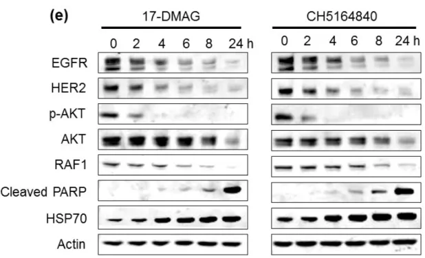
Fig. 1- 2. CH5164840 induces the degradation of multiple Hsp90 client proteins and
apoptosis.
CH5164840 induces the degradation of multiple heat shock protein 90 (HSP90) client proteins and caspase-3/7-dependent apoptosis in HER2-positive cells. (a) (c) NCI-N87 or BT-474 cells were treated with the indicated concentrations of CH5164840 for 24 h before lysing and analysis using Western blotting. (b) (d) NCI-N87 and BT-474 cells were treated with 0, 0.2, or 1 μM of CH5164840 for 48 h. Caspase-3/7 activity and cell viability were measured with the CaspaseGlo3/7 assay and CellTiter-Gro, respectively, using EnVision High Throughput Screening. Caspase activity was normalized by cell viability. (e) NCI-N87 cells were treated with the 0.5 μM of 17-DMAG and 2 μM of CH5164840 for the indicated times before lysing and analysis using Western blotting. p-, phospho; PARP, poly (ADP-ribose) polymerase.
21
Fig. 1- 3. CH5164840 displays higher affinity to HSP90 with multi-chaperone
complexes in tumor cells and tumor-selective retention of CH5164840 and
pharmacodynamics.
22
(a) Normal fibroblast (adult normal human dermal [NHDF-Ad]) and tumor (NCI-N87) cell lines were incubated with different concentrations of CH5164840 for 4 days. Then, Cell Counting Kit-8 solution was added, and after incubation for several more hours, absorbance at 450 nm was measured with a microplate reader. The vertical axis shows the percentage of inhibition relative to the DMSO control. (b) NHDF-Ad and NCI-N87 cell lysates were precipitated with biotin-labeled CH5164840 (Bio-CH) and analyzed using Western blotting with the indicated antibodies. (c) Mice with established NCI-N87 xenografts were orally administered a single dose of 50 mg/kg CH5164840 and killed at the indicated times. Plasma and sample tissues were removed and homogenized, and the CH5164840 concentration of plasma, heart, kidney, liver, lung, and tumor analyzed using liquid chromatography/tandem mass spectrometry (LC/MS/MS). Data are shown as mean ± SD (n = 3). (d) Lysates were prepared from the tumor and organs and analyzed using Western blotting.
23
Fig. 1- 4. CH5164840 efficacy against HER2-overexpressing NCI-N87 and BT-474
xenograft models.
Mice bearing HER2-overexpressing tumors were orally administered the indicated doses of CH5164840 daily for 11 consecutive days. Mean tumor volume is shown. (a) NCI-N87 and (b) BT-474. Data are shown as mean ± SD (n = 4-5). (c) Lysates were prepared from the tumor and analyzed using Western blotting. p-, phospho.
24
Fig. 1- 5. Combination therapy with CH5164840 enhanced antitumor efficacy
against HER2-overexpressing NCI-N87 and BT-474 xenograft models.
25
consecutive days (indicated by black arrows) and/or intraperitoneally injected with 30 mg/kg trastuzumab once weekly for 2 weeks (indicated by white arrows). Mean tumor volumes were measured for over 4 weeks from treatment commencement. (b) Mice bearing BT-474 tumors were orally administered 25 mg/kg CH5164840 daily for 11 consecutive days (indicated by black arrows) and/or intraperitoneally injected with 30 mg/kg trastuzumab once weekly for 2 weeks (indicated by white arrows). (c, d) Mice bearing the HER2-overexpressing tumors NCI-N87 (c) or BT-474 (d), were orally administered 12.5 mg/kg CH5164840 daily for 11 consecutive days (indicated by black arrows) and/or orally administered 100 mg/kg lapatinib (indicated by white arrows). Data are shown as mean ± SD (n = 5).
26
Fig. 1- 6. CH5164840 suppressed lapatinib treatment-induced HER3 signal
activation in vitro.
(a) The relative expression of HER3 mRNA in NCI-N87 cells treated with 0.2 μM lapatinib was evaluated via real-time RT-PCR analysis of total RNA with an HER3-specific primer and normalized to the housekeeping gene (GAPDH). Relative expression is displayed as the fold change relative to the untreated control of the value for DMSO control lysate. (b) NCI-N87 cells were treated with 0.2 μM lapatinib for 2, 24, or 48 h. The cells were then lysed and analyzed via Western blotting with the indicated antibodies. (c) NCI-N87 cells were treated with 0.2 μM lapatinib for 24 h, washed with PBS, and then incubated with fresh medium for 2 or 24 h. The cells were then lysed and analyzed via Western blotting with the indicated antibodies. (d) NCI-N87 cells were treated with 1 μM CH5164840
27
and/or 0.2 μM lapatinib for 48 h, lysed, and analyzed via Western blotting with the indicated antibodies. (e) NCI-N87 cells were treated with 1 μM CH5164840 and/or 0.2 μM lapatinib for 24 h. The cells were then lysed, subjected to immunoprecipitation with the HSP90 antibody, and analyzed using Western blotting with the indicated antibodies. IP, immunoprecipitation.
28
Fig. 1- 7. Study of the CH5164840 administration schedule during combination
therapy with trastuzumab against HER2-overexpressing NCI-N87 xenograft models.
Mice bearing NCI-N87 tumors were orally administered (a) 50 mg/kg CH5164840 (CH4840) twice weekly (BIW) (indicated by white arrows) or (b) 50 mg/kg CH5164840 once weekly (QW) (indicated by white arrows), and intraperitoneally injected with 30 mg/kg trastuzumab (Tras) once weekly for 3 weeks (indicated by black arrows). Mean tumor volumes were measured for over 3 weeks from treatment commencement. Data are shown as mean ± SD (n = 4).
29
Chapter 2
Enhanced antitumor activity of erlotinib in combination with the HSP90 inhibitor
CH5164840, against non-small-cell lung cancer
1. Introduction
Epidermal growth factor receptor (EGFR) plays a key role in non-small-cell lung cancer (NSCLC) development and progression. Two tyrosine kinase inhibitors (TKI) against EGFR – erlotinib and gefitinib – are currently available for NSCLC therapy. Although targeted inhibition of EGFR has shown promising initial clinical efficacy in the treatment of patients with NSCLC, which have exon 19 (DE 746-A750) and 21 (L858R)-activating EGFR mutations (44-46), primary and acquired resistance to these drugs in some patients is an obstacle to treatment efficacy (47). The major mechanisms of resistance to TKI are EGFR mutations (48), MET amplification (49), hepatocyte growth factor (HGF) activation (50), phosphatase and tensin homolog (PTEN) loss (51) and other genomic alterations with wild-type EGFR, including KRAS mutations (48, 52) and ALK-fusion proteins (53). Additionally, adenocarcinoma with sensitive EGFR mutations can transform into small cell lung cancer (SCLC) in the process of acquiring resistance to EGFR-TKIs (54). A secondary mutation in the EGFR gene (T790M) is the major cause of resistance to gefitinib and erlotinib (55, 56), and is found in 50% of clinically resistant patients (57). Due to the potential for resistance to TKI in EGFR-addicted tumors, identification of an effective treatment using rationalized combinations of agents is particularly promising.
EGFR is one of the most potent oncogenic client proteins of HSP90. Furthermore, mutated EGFR seems to be more sensitive than wild-type EGFR to degradation from HSP90 inhibition (23, 24). These unique features are expected to overcome the problem of resistance to TKI. STAT3 is also an HSP90 client protein (58) and an important signaling mediator for EGFR. It is activated in approximately 50% of NSCLC primary tumors and lung cancer cell lines (59-61), and this pathway is upregulated in NSCLC cells harboring EGFR mutations (62). Janus kinase (JAK), upstream of STAT3, is also an HSP90 client, and the JAK-STAT pathway is involved in immune function, cell growth, differentiation, hematopoiesis, and tumor growth.
Recently, a novel potent HSP90 inhibitor: CH5164840, was identified. The present study elucidates the in vitro and in vivo properties of CH5164840 alone and in combination with erlotinib, including its antitumor activity in EGFR overexpressing and erlotinib-resistant NSCLC xenograft models.
30
2. Materials and Methods
2- 1. Compounds
CH5164840 was synthesized by Chugai Pharmaceutical Co., Ltd (Kanagawa, Japan) (33). Erlotinib was obtained from Chugai Pharmaceutical ⁄ F. Hoffmann-La Roche (Basel, Switzerland).
2- 2. Cell lines
The human NSCLC cell lines HCC827, NCIH292, NCI-H1781, A549, NCI-H1650, NCI-H1975 and NCI-H441, were obtained from the American Type Culture Collection (Manassas, VA, USA). The human NSCLC cell line EBC-1 was obtained from RIKEN Cell Bank (Ibaraki, Japan). The human NSCLC cell line PC-9 was obtained from Immuno-Biological Laboratories (Fujioka, Japan). All cell lines were maintained according to the supplier’s instructions.
2- 3. Cell culture
For proliferation assays, tumor cells were seeded into microtiter plates containing compounds and incubated at 37°C in 5% CO2. After incubation for 4 days, a Cell Counting Kit-8 solution
(Dojindo Laboratories, Kumamoto, Japan) was added and absorbance measured at 450 nm with a Microplate- Reader iMark (Bio-Rad Laboratories, Hercules, CA, USA). Anti-proliferative activity was calculated using the formula: (1−T/C) × 100 (%), where T represents the absorbance of drug-treated cells and C represents that of undrug-treated control cells at 450 nm. The IC50 values were
calculated using Microsoft Excel 2007 (Redmond, WA, USA). Caspase-3⁄7 activity and cell viability were measured with the Caspase-Glo 3⁄7 Assay Kit (Promega, Madison, WI, USA) and CellTiter-Glo Luminescent Cell Viability Assay (Promega), respectively, using EnVision High Throughput Screening (PerkinElmer, Waltham, MA, USA).
2- 4. Western blotting
Western blotting was performed as described in chapter 1. Primary antibodies were used for pY1068-EGFR, EGFR (L858R), pS473-AKT, AKT, pT202/Y204-ERK, ERK, pY705-STAT3, STAT3, JAK1, JAK2 and tyrosine kinase 2 (TYK2) (Cell Signaling, Beverly, MA, USA), EGFR, HER2, MET, RAF1, actin, GAPDH and Histone H1 (Santa Cruz Biotechnology, Santa Cruz, CA, USA) and HSP70 and HSP90 (Stressgen, Victoria, Canada). Signals were developed using ECL Plus (GE Healthcare, Buckinghamshire, UK), followed by LAS- 4000 (Fujifilm, Tokyo, Japan) or the Odyssey Infrared Imaging System (LI-COR Biosciences, Lincoln, NE, USA).
2- 5. RNA interference
Cells were seeded onto a 96-well plate and transfected with Human STAT3, JAK1, JAK2 and TYK2 ON-TARGETplus SMARTpool ORF and ON-TARGETplus Non-targeting Pool (Thermo
31
Scientific, Dharmacon, Waltham, MA, USA) using Lipofectamine RNAiMAX (Invitrogen, Carlsbad, CA, USA), and treated with 0.2 μM erlotinib. At 96 h after siRNA transfection, cell viability was determined using the CellTiter-Glo luminescent cell viability assay (Promega).
2- 6. Xenograft models and efficacy studies
All animal studies were approved by the Chugai Institutional Animal Care and Use Committee. Cancer cells (0.5–1.0 x 10⁷) were implanted subcutaneously into the right flank of athymic nude
(BALB ⁄ c nu ⁄ nu) mice (Charles River Laboratories, Kanagawa, Japan). Tumor volume (TV) was calculated using the formula TV = ab2 ⁄ 2, where a and b represent tumor length and width, respectively. Once the tumors reached a volume of approximately 200–300 mm3, THE animals were
randomized into each group (n = 4–5) and treatment was initiated. CH5164840 was dissolved in a vehicle of 10% DMSO⁄10% Cremophor EL⁄ 0.02 N HCl in water. CH5164840 and erlotinib were orally administered once daily for 11 days. Tumor growth inhibition (TGI) was calculated using the formula TGI = (1−[Tt−T0]/[Ct−C0]) × 100 (%), where T (TV of treated group; T0 on day 0 or Tt on
day t) and C (TV of control group; C0 on day 0 or Ct on day t) represent mean tumor volume. The
maximum tolerated dose was defined as the dose that resulted in neither lethality nor more than 20% bodyweight loss. For pharmacodynamic studies, blood was collected 4 h after the last administration of CH5164840 and PBMC were prepared with M-SMF (JIMRO, Gunma, Japan), lysed, and analyzed using Western blotting.
2- 7. Statistical analysis
For in vivo combination studies, SAS preclinical package v5 software (SAS Institute, Cary, NC, USA) was used for statistical analyses (Tukey’s test). For in vitro experiments, statistical values were defined using the Student’s t-test.
3. Results
3- 1. Novel HSP90 inhibitor CH5164840, has potent growth inhibitory activity
against NSCLC in vitro and antitumor activity in vivo
Recently, through virtual screening and structure-based drug design, CH5164840 was identified as an HSP90 inhibitor with a novel chemical structure (32, 33). It was demonstrated to show antitumor-activity against HER2 positive-tumors in mono- and combination-therapies with HER2-targeted agents (63). In this study, the potency of CH5164840 against NSCLC was examined. In an initial evaluation, the in vitro cell growth inhibition of CH5164840 or erlotinib was examined to determine the sensitivity of NSCLC cell lines with various genotypes, including PC-9 and HCC827 (EGFR ΔE746-A750), NCI-H292 (wild-type EGFR overexpression), NCI-H1975 (EGFR T790M and L858R mutant), NCI-H1650 (EGFR ΔE746-A750, PTEN null), NCI-H1781 (HER2
32
G776insV_G/C mutant), and A549 (KRAS mutant). The IC50 values of CH5164840 were found to
be 140–550 nM in seven NSCLC cell lines, regardless of genetic mutations, although sensitivities to erlotinib were more variable (IC50 values, 4.7–13000 nM; Table 2-1). Next, the degradation of
HSP90 client proteins in NSCLC cell lines by CH5164840 was examined. CH5164840 was found to significantly reduce EGFR, HER2, and MET protein levels, and suppress downstream AKT and ERK signaling (Fig. 2-1). The induction of HSP70, a marker of HSP90 inhibition, was also confirmed. To evaluate the in vivo antitumor activity of CH5164840, several xenograft studies were conducted using NSCLC cell lines in nude BALB/c mice. Daily oral administration of CH5164840 resulted in a significant dose-dependent antitumor activity in the NCI-H1650 (EGFR mutant, PTEN null) xenograft models, with a maximum TGI of 131% (Fig. 2-2a, Table 2-2). Additionally, we confirmed the dose-dependent induction of HSP70 in murine peripheral blood mononuclear cells (PBMC), which is a marker of HSP90 inhibition (Fig. 2-2b). Like NCI-H1650 antitumor activity, CH5164840 showed substantial antitumor activity in NCI-H292 (wild-type EGFR overexpression), NCI-H1975 (EGFR mutant) and NCI-H441 (wild-type EGFR, MET overexpression) xenograft models (Table 2-1). In all in vivo studies, the doses of CH5164840 tested were well tolerated, with no gross toxicity observed in the treated animals. Collectively, these data show that CH5164840 inhibited HSP90, resulting in the inhibition of in vitro cell growth and in vivo antitumor activity against multiple NSCLC cell lines.
3- 2. CH5164840 enhances the antitumor activity of erlotinib in a NCI-H292
wild-type EGFR overexpression, erlotinib-halfway sensitive model
Due to the moderate activity of erlotinib against NCI-H292 NSCLC cells, the effect of CH5164840 on erlotinib activity against NCI-H292 cells, which overexpress wild-type EGFR, was investigated. To confirm the cellular effects of this combination treatment in vitro, cell growth inhibition and caspase-3/7 activity were evaluated. As shown in Fig. 2-3a, CH5164840 enhanced the cell growth inhibitory activity of erlotinib. Further, when NCI-H292 cells were treated with a combination of erlotinib and CH5164840, CH5164840 significantly enhanced the caspase-3/7 activity of erlotinib (Fig. 2-3b). To examine the combined antitumor activity of CH5164840 and erlotinib in vivo, 25 mg/kg of erlotinib and/or 12.5 mg/kg of CH5164840 was administered to the NCI-H292 xenograft models. CH5164840 synergistically enhanced the antitumor activity of erlotinib (Fig. 2-3c). Erlotinib and CH5164840 co-therapy produced a statistically significant effect on TGI, compared with monotherapy with either, causing tumor regression. These data support the effectiveness of a combination strategy using erlotinib and CH5164840 against EGFR-overexpressing NSCLC. Next, the molecular mechanisms of the synergistic effect of erlotinib and CH5164840 co-therapy in NCI-H292 cells were examined. Firstly, the effect of these agents on cell signaling was analyzed via Western blotting. Erlotinib and CH5164840 co-therapy reduced EGFR and phospho-EGFR protein levels and
33
suppressed downstream AKT and ERK signaling (Fig. 2-4a). Additionally, erlotinib resulted in a dose-dependent increase in STAT3 phosphorylation compared with DMSO control (Fig. 2-4b), which was mainly present in the nucleus (Fig. 2-4c). Interestingly, co-treatment with CH5164840 and erlotinib reduced the phosphorylation level of STAT3 (Fig. 2-4c).
JAK1reportedly phosphorylates STAT3 in NSCLC cells (64). It was confirmed that siRNA knockdown of JAK1, but not its family protein JAK2 or TYK2, inhibited erlotinib-induced STAT3 phosphorylation in NCI-H292 cells (Fig. 2-4d). Additionally, HSP90 inhibition by CH5164840 induced degradation of the JAK1 protein (Fig. 2-4a). Further, to explore the effect of JAK1 and STAT3 on cell growth, their knockdown by siRNA was examined. Inhibition of STAT3 expression by siRNA specifically enhanced cell growth inhibition by erlotinib (Fig. 2-4e), producing statistically significant results. Combined knockdown of erlotinib and JAK1 led to more effective cell growth inhibition compared to STAT3 (Fig. 2-4e). Collectively, these findings suggest that in addition to AKT and ERK signaling, suppression of the JAK1-STAT3 signaling pathway by CH5164840 enhanced the antitumor effects of erlotinib in NSCLC.
3- 3. CH5164840 enhances the antitumor activity of erlotinib in an erlotinib-resistant
NCI-H1975 model
The effect of erlotinib and CH5164840 co-treatment were further explored using NCI-H1975 NSCLC cells. NCI-H1975 cells harbor the EGFR T790M gatekeeper mutation, which modulates the accessibility of the kinase ATP-binding pocket, a secondary EGFR mutation that is a major cause of resistance to erlotinib. To investigate the combined effect of erlotinib and CH5164840, the anti-proliferative effect of the agents on NCI-H1975 cells were examined. CH5164840 enhanced the cell growth-inhibitory and caspase-3/7 activities of erlotinib (Figs. 2-5a, b). The in vivo antitumor activity of this combination therapy against human tumor xenograft models in mice was then investigated. When a combination of 25 mg/kg erlotinib and 25 mg/kg CH5164840 was orally administered to a NCI-H1975 xenograft model, tumor growth was significantly inhibited, despite erlotinib treatment alone having no effect (Fig. 2-5c). The enhanced antitumor activity with combination therapy was statistically significant compared with monotherapy with either, and no gross toxicity was observed in any of the treated animals. Additionally, combination therapy resulted in reduced protein levels of mutated EGFR, JAK1, phospho-STAT3, phospho-AKT, and phospho-ERK (Fig. 2-5d). These results suggest that CH5164840 potentiates the efficacy of erlotinib in an erlotinib-resistant model harboring the EGFR T790M gatekeeper mutation.
4. Discussion
EGFR is an attractive anticancer therapy target because it plays an important role in lung carcinogenesis. Moreover, its expression is correlated with poor prognosis (65) and EGFR-TKI,
34
erlotinib, and gefitinib, have shown a clinical response in NSCLC with activating EGFR mutations. However, the development of primary and acquired resistances to these drugs over several years has become a major clinical problem. Recently, many resistant mechanisms of EGFR-TKI have been investigated (66), including EGFR mutations T790M (48, 67), MET amplification (49), hepatocyte growth factor (HGF) activation (50), PTEN loss (51, 68) and AXL activation (69). To overcome TKI resistance, corresponding strategies for each TKI-resistant mechanism are needed. The combination of a TKI and an HSP90 inhibitor is considered particularly promising because the inhibition of HSP90 induces degradation of molecules involved in TKI resistance, including mutant EGFR, MET, and EML4-ALK. HSP90 inhibitors have shown efficacy against established EGFR-TKI–resistant cells, EGFR T790M (70), and HGF overexpression (71). In fact, the HSP90 inhibitor IP-504, has shown clinical activity in patients with ALK rearrangement in a phase 1 ⁄ 2 study (72). CH5164840, an HSP90 inhibitor with a novel chemical structure, can be taken orally and exhibits highly potent antitumor efficacy (63). In the present study, it inhibited cell growth in NSCLC cell lines with different oncogenic drivers, induced degradation of representative HSP90 client proteins: EGFR, HER2, MET and RAF1, and inhibited the phosphorylation of downstream signaling proteins AKT and ERK. Treatment with CH5164840 also induced HSP70 expression. Generally, studies on HSP90 inhibitors focus on the degradation of client proteins or induction of HSP70 as a biomarker for the HSP90 inhibitor. However, induction of HSP70 by HSP90 inhibition has been shown to reduce the antitumor effect of HSP90 inhibitors, and knockdown of HSP70 by siRNA increased 17-AAG-induced apoptosis (73). Therefore, abrogating HSP70 induction approaches are raised to increasing the sensitivity of HSP90 inhibitors (74).
Furthermore, CH5164840 monotherapy showed antitumor activity against NSCLC xenograft models; CH5164840 showed antitumor activity against a NCI-H441 (wild-type EGFR and MET overexpression) model. Therefore, it is considered useful for patients who exhibit MET-associated TKI resistance. In fact, as shown in Fig. 2-6, this was confirmed in NCI-H441 and EBC-1 (MET amplification) cells, in which erlotinib did not show cell growth inhibition, which CH5164840 enhanced the cell growth inhibitory activity of erlotinib with IC50 values of erlotinib in the
individual cell lines of >20 μM (EBC-1) and 10.0 μM (NCI-H441) in vitro. Furthermore, combination treatment resulted in degradation of phosphorylated and total EGFR or MET in these cells. Collectively, these results show that the HSP90 inhibitor is a potential therapeutic option in combination with erlotinib against TKI-resistant NSCLC with aberrant c-MET. Similarly, it may also be effective in patients with PTEN loss, which is one of the mechanisms of TKI resistance, because of the antitumor activity of CH5164840 in the NCI-H1650 (EGFR DE746-A750, PTEN null) model. These results show that the novel HSP90 inhibitor CH5164840, has potent antitumor activity in vitro and in vivo against clinically relevant NSCLC, including erlotinib-resistant models. The association between EGFR protein expression levels and clinical response to EGFR-TKI has
35
been variable (75, 76), and, in fact, erlotinib elicits only a moderate response against wild-type EGFR-overexpressing NCI-H292 cells. In the present study, treatment of NCI-H292 cells with a combination of EGFR-TKI erlotinib and the HSP90 inhibitor CH5164840, was shown to lead to synergistic cell growth inhibition and apoptosis induction. These in vitro results were consistent with the regression of antitumor activity by combination therapy in a NCI-H292 xenograft model. Further, the molecular mechanisms by which CH5164840 enhanced the efficacy of erlotinib was examined. Combination therapy with erlotinib and CH5164840 reduced EGFR and phospho-EGFR levels, as well as that of downstream signaling proteins AKT and ERK. Interestingly, it was noticed that erlotinib treatment increased phospho-STAT3 in NCI-H292 cells, an effect that was strongly suppressed by CH5164840. The sensitivity of erlotinib-induced phospho-STAT3 is sensitive to HSP90 inhibition is most likely because STAT3 is a client protein of HSP90 (58). Therefore, in NCI-H292 cells, the efficacy of erlotinib might be increased through blocking STAT3 signaling by HSP90 inhibition. STAT3 can be activated through multiple pathways, including EGFR, the IL-6 ⁄ gp130 receptor family, platelet-derived growth factor receptor (PDGFR), Src kinase, and JAK. JAK1 reportedly causes STAT3 activation in NSCLC cells (64), which was confirmed in the present study, together with the fact that both basal- and erlotinib-induced phospho-STAT3 were inhibited by JAK1 knockdown with siRNA, but not JAK2 and TYK2 knockdown (Fig. 2-4d). Although a combination of erlotinib treatment and JAK1 knockdown is more effective for cell growth inhibition than that of erlotinib treatment and STAT3 knockdown, the former does not completely inhibit cell growth. Therefore, additional blocking of the AKT and ERK pathways by an HSP90 inhibitor might be an important therapeutic approach in NCI-H292 NSCLC cells. However, the mechanism by which phospho-STAT3 is increased by erlotinib-induced EGFR inhibition remains unknown. Further investigation is needed to clarify the upstream pathways of EGFR TKI treatment-induced STAT3 activation. Acquired resistance to TKI, usually owed to the development of a second point mutation (T790M) in EGFR, is also a serious clinical problem. Consistent with other HSP90 inhibitors (77, 78), CH5164840 monotherapy shows antitumor activity against NCI-H1975 NSCLC cells harboring EGFR L858R and T790M mutations. Additionally, erlotinib and CH5164840 co-therapy showed significant synergism, despite the lack of an antitumor activity by erlotinib monotherapy in this model. Further, combination therapy simultaneously induced degradation of mutant EGFR, phospho-EGFR, and downstream AKT, ERK, and STAT3 signaling. As such, combination therapy with TKI and an HSP90 inhibitor might be useful in preventing TKI resistance development, as most TKIs are at potential risk of encountering TKI resistance and addicted-oncogenic proteins are degraded by HSP90 inhibition (55). In conclusion, these data highlight the benefits of using an HSP90 inhibitor in combination with EGFR-targeted therapies for the treatment of EGFR-addicted NSCLC, and supports the clinical development of this co-therapy.
36
5. Tables
Table 2- 1. Anti-proliferative activity of CH5164840 in non-small-cell lung cancer
cell lines
Cell lines EGFR status CH5164840
IC50(μM) Erlotinib IC50(μM) PC-9 E746_A750del 0.16 0.0048 HCC827 E746_A750del 0.14 0.0047 NCI-H292 WT overexpression 0.49 0.16 NCI-H1781 WT 0.55 1.7 A549 WT 0.19 3.4 NCI-H1650 E746_A750del 0.16 4.1 NCI-H1975 L858R & T790M 0.30 13
Table 2- 2. Antitumor efficacy of CH5164840 in tumor xenograft models at MTD
Models Tumor types Status Client
MTD (mg/kg)
TGI (%)
NCI-H292 Mucoepidermoid EGFR
overexpression EGFR 25 92
NCI-H441 Papillary adeno MET overexpression MET 50 115
NCI-H1975 Adenocarcinoma EGFR mutant EGFR
L858R & T790M 50 112
NCI-H1650 Adenocarcinoma EGFR mutant EGFR
E746-A750del 50 131
EGFR, epidermal growth factor receptor; MET, met proto-oncogene; MTD, maximum tolerated dose; TGI, tumor growth inhibition. CH5164840 was orally administered once daily for 11 days.
37
6. Figures
Fig. 2- 1. CH5164840 induces degradation of multiple HSP90 client proteins in
non-small-cell lung cancer (NSCLC) cell lines.
NSCLC cell lines were treated with 0, 0.04, 0.2, 1, or 5 μM CH5164840 for 24 h, lysed, and analyzed using Western blotting with indicated antibodies.
38
Fig. 2- 2. Antitumor activity of CH5164840 in a NCI-H1650 erlotinib-resistant
xenograft model and the pharmacodynamic response.
(a) Mice bearing NCI-H1650 tumors were orally administered the indicated doses of CH5164840 daily for 11 consecutive days. Mean tumor volume is shown. Data are shown as mean ± SD (n = 4-5). (b) Four hours after the final administration in (a), murine PBMC were isolated from blood, lysed, and analyzed using Western blotting.
39
Fig. 2- 3. Antitumor activity of CH5164840 in combination with erlotinib on
NCI-H292 EGFR-overexpressing NSCLC in vitro and in vivo.
(a, b) NCI-H292 cells were treated with 0.2 μM erlotinib and/or 0.5 μM CH5164840 for 48 h (for caspase-3/7 activity and cell viability) and 96 h (for cell viability). Caspase-3/7 activity and cell viability were measured with the Caspase-Glo 3/7 assay and CellTiter-Glo, respectively, using EnVision High Throughput Screening. Caspase-3/7 activity was normalized to cell viability. (c) Mice bearing NCI-H292 tumors were orally administered 12.5 mg/kg CH5164840 daily for 11 consecutive days and/or 25 mg/kg erlotinib. Data are shown as mean ± SD (n = 5). Tukey's test: ***P < 0.001.
40
Fig. 2- 4. CH5164840 treatment suppressed erlotinib-induced STAT3 signaling in
NCI-H292 cells.
(a) NCI-H292 cells were treated with 0.2 μM erlotinib and/or 0.5 μM CH5164840 for 48 h, lysed, and analyzed using Western blotting. (b) NCI-H292 cells were treated with the indicated concentration of erlotinib for 24 h and their STAT3 and phospho-STAT3 protein content analyzed. (c) NCI-H292 cells were treated with 0.2 μM erlotinib and/or 0.5 μM CH5164840 for 48 h, and their cytosol and nuclear fractions analyzed using Western blotting. Histone H1 indicates loading control for nuclear fraction. (d) NCI-H292 cells were transfected with 10 nM STAT3, JAK1, JAK2, TYK2, or control siRNA. At 24 h post-transfection, cells were treated with 0.2 μM erlotinib, cultured for 24 h, lysed, and analyzed using Western blotting. (e) NCI-H292 cells were transfected with 10 nM STAT3 siRNA, JAK1 siRNA, or control siRNA, and treated with DMSO or 0.2 μM erlotinib. At 96 h post-transfection, cell viability was determined using CellTiter-Glo. Data are shown as mean ± SD (n = 3). Student's t-test: *P < 0.05; **P <0.01; ***P < 0.001. c-PARP, cleaved poly (ADP-ribose) polymerase; p, phospho; −, DMSO; +, compound.
41
Fig. 2- 5. Antitumor activity of CH5164840 in combination with erlotinib in a
NCI-H1975 erlotinib-resistant model.
(a, b) NCI-H1975 cells were treated with 1 μM erlotinib and/or 1 μM CH5164840 for 48 h (for caspase-3/7 activity and cell viability) and 96 h (for cell viability). Caspase-3/7 activity and cell viability were measured with the Caspase-Glo 3/7 assay and CellTiter-Glo, respectively, using EnVision High Throughput Screening. Caspase activity was normalized to cell viability. (c) Mice bearing NCI-H1975 tumors were orally administered 25 mg/kg of CH5164840 and/or 25 mg/kg of erlotinib daily for 11 days. (d) Four hours after the final administration in (c), tumors were resected, lysed, and analyzed using Western blotting. p, phospho. Data are shown as mean ± SD (n = 4-5) Tukey's test: ***P < 0.001.
42
Fig. 2- 6. Antitumor activity of CH5164840 in combination with erlotinib on NSCLC
with aberrant c-MET in vitro.
(a) EBC-1 cells were treated with 1 μM erlotinib and/or 0.2 μM. CH5164840 and NCI-H441 cells were treated with 1 μM erlotinib and/or 0.5 μM CH5164840 for 96 h. Cell viability was measured with the CellTiter-Glo, using EnVision High Throughput Screening. Data are shown as mean ± SD (n = 3) (b) EBC-1 cells were treated with 1 μM erlotinib and/or 0.2 μM CH5164840, and NCI-H441 cells were treated with 1 μM erlotinib and/or 0.5 μM CH5164840 for 48 h, lysed, and analyzed using Western blotting.