Posted at the Institutional Resources for Unique Collection and Academic Archives at Tokyo Dental College, Available from http://ir.tdc.ac.jp/
Author(s)
Alternative
Tsuji, M; Ueda, T; Sawaki, K; Kawaguchi, M;
Sakurai, K
Journal
Gerodontology, 33(4): 539-544
URL
http://hdl.handle.net/10130/4988
Right
Biocompatibility of a titanium dioxide coating method for denture base acrylic resin
Masashi Tsuji*, Takayuki Ueda*, Kohei Sawaki♯
, Mitsuru Kawaguchi‡ and Kaoru Sakurai*
*Department of Removable Prosthodontics & Gerodontology, Tokyo Dental College, Tokyo, Japan ♯
Laboratory of Pharmacotherapy, Yokohama College of Pharmacy, Yokohama, Japan
‡Department of Pharmacology, Tokyo Dental College, Tokyo, Japan
Running title: Biological evaluation of TiO2-coated acrylic resin
Article category: Original research
Correspondence: Dr. T. Ueda,
Department of Removable Prosthodontics & Gerodontology, Tokyo Dental College,
2-9-18 Misaki-cho, Chiyo da-ku, Tokyo 101-0061, Japan
Email: [email protected]
Abstract
Objectives: Ease-of-denture-cleaning is of paramount importance in geriatric patients and those with
limited dexterity. We have previously investigated methods of coating dentures with titanium dioxide
(TiO2) and reported the effects (self-cleaning and antibacterial) of such treatments in in vitro studies.
This study was to verify the biocompatibility of a TiO2-coated acrylic resin produced by the new coating
method with spray-coating technique.
Methods: Specimens were prepared from denture base acrylic resin and polished up to grit #1000.
The TiO2 coating agent was sprayed onto the specimens using an air-brush gun. Specimens were then
divided into ‘PMMA’, ‘primer-coated PMMA’ and ‘TiO2-coated PMMA’ groups to be evaluated for
biological safety using a hamster oral mucosa irritation test, a guinea pig skin sensitization test and a
rabbit intracutaneous test. The biological reaction was scored.
Results: Reaction scores were considerably less than 1.0, the acceptable limit set by the ISO, in all
three tests. Indeed, in most samples there was no deleterious effect at all.
Conclusion: These results tested on animals demonstrate that denture base resin coated with TiO2 by
this method does not cause irritation or sensitization of the oral mucosa, skin or intracutaneous tissue,
Introduction
Polymethyl methacrylate (PMMA) resin has long been used as a denture base material1,
because of its hardness and rigidity under masticatory pressure, ease of handling, good esthetics and
low price. However, despite its obvious suitability as a denture base material2,3, PMMA is also
susceptible to deterioration4, surface roughness following fatigue, microbial adherence5,6,7 and
colonization by bacteria due to water absorption1.
Microbe adhesion to the denture surface befouls the oral cavity and can cause systemic infections
(e.g. aspiration pneumonitis)8,9. Geriatric patients and those with limited dexterity (e.g. maniphalanx
dysfunction or rheumatoid arthritis) find oral hygiene (OH) troublesome and require assistance from
family or nursing staff10. However, although various mechanical, ultrasonic and chemical cleaning
methods have been proven efficacious11,12, these helpers often lack the OH knowledge to apply them.
Hygienic denture materials would be invaluable in improving OH in denture wearers. We have
previously reported on the coating method of titanium dioxide (TiO2) application, which has
photocatalytic effect (i.e., oxidation decomposition and super-hydrophilicity), and the viability of making
dentures that can be cleaned simply by rinsing in water13,14,15. TiO
2 can be incorporated into the resin11,16,17 to achieve the photocatalytic effect but requires ~5wt% TiO
2, which can weaken the material and cause internal decomposition through its photocatalytic effect. A preferable option is to use TiO2
coating method, which does not alter the resin itself. Coating can be by brushing, dipping or spraying,
technique using an air-brush gun, which produces a thin coating13 and reportedly changes the surface
characteristics of the acrylic resin, making it less likely to accumulate bolus residue and subsequently
easier to clean13. This TiO
2 coating of acrylic resin also inhibits the adhesion of microbes14, and is sufficiently durable to withstand normal oral function15. Unlike the mixing method, the spray-coating
technique is not material-dependent and can be applied to all denture components, including the base,
artificial teeth and major and minor connectors. Finally, because the coat is concentrated at the surface,
where it is most useful, the photocatalytic effect can be achieved with much lower amounts of TiO2.
The TiO2 coating material contains TiO2 and organic material (primer) 15. This organic material,
and the solvent in which it is solubilized, are the most likely candidates for inducing an inflammatory
response in tissues. Additionally, it has been reported that TiO2 nanoparticles may develop a
cytotoxicity 19. The application protocol minimizes the risk of deleterious effects from these components,
but although the material has been validated in vitro, the biological safety of the TiO2 coat remains to be
verified. The purpose of this study was to investigate the biocompatibility of this TiO2 spray-coating
Materials and methods
Plate specimens
PMMA specimens were prepared from wet-heat-curing acrylic resin (Acron, GC Corporation,
Tokyo, Japan). The volume ratio of powder : liquid was 1 : 0.43. The resulting mixture was packed into
a plaster mold and placed in 60 °C water for 60 min (for primary polymerization) and then boiling water
for 60 min (for secondary polymerization). Specimens were polished up to the equivalent of grit #1000
by wet abrasive paper.
TiO2 coating
Specimens were pre-treated with a primer agent (Paltitan PTI5603S; Nihon Parkerizing,
Kanagawa, Japan), whose main component is acryloxypropyltrimethoxysilane in ethanol, sprayed for 2
s from an air-brush gun (Super Airbrush Advance; WAVE, Tokyo, Japan) then dried for 10 min at 70 °C
in air. TiO2 coating agent (Paltitan PTI5603S, Nihon Parkerizing, Kanagawa, Japan), containing 2.0%
anatase-type TiO2 in water and ethanol, was then sprayed onto the substrate for 2 s and dried in an
oven for 10 min at 70 °C.
Animals
All animals used in these experiments were obtained from Japan SLC, Inc. (Hamamatsu, Japan),
cycle (lights on: 6:00 am – 6:00 pm) and maintained on commercial laboratory chow and water for at
least one week before being used. Animals were treated according to the Guidelines for the Treatment
of Experimental Animals approved by the Tokyo Dental College (No.242607, No.232606, No.242606).
Oral mucosa irritation test
The oral mucosa irritation test20, 21 was performed using 5-week-old male Syrian hamsters.
Animals were divided into 4 groups as follows: ‘positive control’, ‘PMMA’, ‘primer-coated PMMA’ and
‘TiO2-coated PMMA’. Cheek pouches were everted and rinsed with physiological saline before suturing
a specimen (a 5 mm diameter 0.8 mm height disc or cotton wool with liquid of resin as positive control) into the right-hand cheek pouch with non-resorbable nylon thread. The left cheek pouch was
sutured without a specimen insertion as negative control. After 24 hours, animals were euthanized with
an excess of pentobarbital and the cheek pouches were excised, fixed with formalin and embedded in
paraffin. Serial sections, stained with hematoxylin and eosin, were examined for microscopic evidence
of epithelial and connective tissular reaction. Every challenge site was scored (0–4 points) for erythema
by 2 judges independently. The judges were well trained and discuss each other about criteria of
scoring the before the test. If the scores from two judges were not same, the worse score was used to
make the conservative decision.
Sensitization test
divided into 5 groups as follows: ‘negative control’, ‘positive control’, ‘PMMA’, ‘primer-coated PMMA’
and ‘TiO2-coated PMMA’. Hair was clipped from the back region and 0.1 ml of emulsified Freund's
complete adjuvant was applied intradermally at the four corners of a 2×4 cm area before a criss-cross
lattice of abrasions was made at the injection site. Specimens (as for the mucosal irritation test, above)
or 0.1 ml of 0.5% 2,4-dinitrochlorobenzene (as positive control) in acetone were applied to the abraded
area of skin for 24 hours. Abrasion and patch application was repeated on each of the following two
days. Sodium lauryl sulphate (10%) was applied to the injection site after six days, and again on the
seventh day in combination with specimen or 0.2 ml 0.5% 2,4-dinitrochlorobenzene. This was then left
for 48 hours. Negative control animals were similarly treated, but without the test materials. At fourteen
days after the last induction, all animals were again challenged with the specimen or 0.01 ml of 0.1%
2,4-dinitrochlorobenzene in acetone together. Every challenge site was scored (0–4 points) for
erythema and swelling respectively at 24 hours and 48 hours after application by 2 judges
independently.
Intracutaneous test
The intracutaneous test20, 24 was performed using male rabbits, weighing 2.5–2.8 kg, divided into
4 groups as follows: ‘positive control’, ‘PMMA’, ‘primer-coated PMMA’ and ‘TiO2-coated PMMA’.
Extraction liquid from each group of specimens was acquired by soaking a disc of each material (18
mm diameter, 1.0 mm height) in polar solvent (physiological saline) or non-polar solvent (sesame oil)
oil. Back fur was closely clipped, allowing sufficient space either side of the spine for injection of 0.2 ml
of test liquid (injected intracutaneously at five sites on one side) and solvent only (injected at five sites
on the contralateral side as negative control). Each injection site was evaluated for signs of skin
reaction immediately and at 24, 48 and 72 hours after injection by 2 judges independently, with reaction
scored on a scale of 0–4 points for erythema and swelling respectively. A net score was obtained by
subtracting the negative control score from the test score. A score of 1.0 or less was deemed to be
Results
Oral mucosa irritation test
Table 1 shows the results of the oral mucosa irritation test. The reaction was scored with regard
to focused on erythema (no erythema: 0point, less than exposure area: 1point, all exposure area: 2
points, beyond exposure area: 3 points and eschar formation: 4 points). Marked erythema was found in
all animals in the positive control group, with a mean score of 2. In contrast, no erythema was apparent
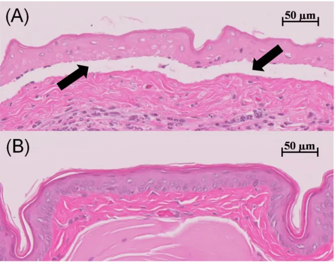
in the other groups (mean score: 0). An example of histological evaluation of hamster cheek pouch
mucosa exposed to liquid of resin as positive control (A) and TiO2-coated PMMA (B) are shown in
Figure 1. Abnormal finding on the (B) was not observed. However, the coagulation necrosis both
epithelium and connective tissue layers, and pressure bulla were observed on the (A).
Sensitization test
Table 2 shows skin sensitization rates. In the negative control group, the score in each animal
was zero. The reaction was scored with regard to focused on erythema (no erythema: 0point, less than
exposure area: 1point, all exposure area: 2 points, beyond exposure area: 3 points and eschar
formation: 4 points) and swelling (no swelling: 0point, barely tactile thickness: 1 point, less than 1 mm
thickness: 2 points, approximately 1mm thickness: 3 points and more than 1 mm thickness: 4 points). In
the positive control (2,4-dinitrochlorobenzene), marked erythema and swelling were apparent in all
Overall, there was no evidence of significant skin reaction in the ‘PMMA’, ‘primer-coated PMMA’ or
‘TiO2-coated PMMA’ groups (mean scores: 0.2, 0 and 0.2, respectively), although minor erythema (1
point) was observed in one animal in each of the ‘PMMA’ and ‘TiO2-coated PMMA’ groups at 24 hours
after application. However, this had disappeared at 48 hours.
Intracutaneous test
Table 3 shows the intracutaneous reactivity data. The reaction was scored with regard to
focused on erythema (no erythema: 0point, less than exposure area: 1point, all exposure area: 2 points,
beyond exposure area: 3 points and eschar formation: 4 points) and swelling (no swelling: 0point,
barely tactile thickness: 1 point, less than 1 mm thickness: 2 points, approximately 1mm thickness: 3
points and more than 1 mm thickness: 4 points). The each net score was obtained by subtracting the
mean negative score from the mean extract score. Marked erythema and swelling were found in all
positive control animals (kappa-carrageenan in saline or liquid of resin in oil; net scores: 5.93 and 5.18,
respectively). No significant reaction was observed in any of the test groups (net scores: <<1.0, the
Discussion
Despite evidence of functional efficacy, the biocompatibility of this coating material remained
unknown. The denture base resin contacts not only the wearer’s oral mucosa, but also his or her skin
and that of the dentist and dental technician1 so a guarantee of high safety had to be established prior
to clinical use. Many studies have evaluated the safety of denture base resins25,26,27, mostly by
evaluating in vitro cellular reactions to the resin monomer. However, reactions at the cellular level are
not representative of the systemic biological inflammatory reaction, so we evaluated our TiO2-coated
denture base material in three whole-animal models to assess its biocompatibility.
The safety evaluation was planned in accordance with ISO10993-10, which requires an “oral
mucosa irritation test”, a “sensitization test”, and an “intracutaneous test”20. The oral mucosa irritation
test determines the reaction against the material in direct contact with the oral mucosa21,22, while the
sensitization test evaluates irritation reaction and sensitization effects in contact with skin23. However,
these tests do not take account the risk of elution of unpolymerized organic components and residual
organic solvent into the saliva, hence the inclusion of the intracutaneous test to assess component
penetration into tissues.
There are differences among the surface texture and character of PMMA, primer-coated PMMA
and TiO2-coated PMMA. We have previously reported that the surface roughness (Rmax) of the polished
water contact angle of the resin plate was dropped from 70° to 10° after TiO2 coating, i.e. the wettability
of the surface was increased13. These differences might have any effect on biological reaction.
Therefore, in the oral mucosa irritation test, the specimens should not over-expand the cheek pouches,
so were kept relatively small (5 mm diameter, 1 mm thickness). Also, to eliminate irritation related to
friction between the specimen and oral mucosa, the test period was kept relatively short (24 hours),
allowing chemical-induced reaction to be studied selectively.
In the sensitization test, guinea pigs were tested using Freund’s Complete Adjuvant (to amplify
antibody production) and focal stimulation with sodium lauryl sulfate to induce sensitization23. With a
mean score of 0.2 (“negligible (0–0.4)”, according to the ISO skin irritation index), these low-grade
reactions do not alter the conclusion that the PMMA, primer-coated PMMA and TiO2-coated PMMA do
not cause significant skin irritation.
The intracutaneous test employed liquids infused with any leached unpolymerized monomer from
the specimens24, and tested this on rabbit back skin, which is known to be highly sensitive. Extraction of
monomer from the specimens was performed at 37 °C for 72 hours, more severe than the clinical
situation. In the oral cavity, the solvent is saliva, which is polarized due to its electrolyte content.
Moreover, lipophilic components elute into organic salivary elements (e.g., proteins and amino acids),
hence our study considered both polar and non-polar solvents. To eliminate the irritation of the
intracutaneous injection and by the solvent itself, the negative control received an injection of solvent
the ‘non-irritant’ requirements of the ISO test (mean net score: <1.0). The results indicate that neither
the primer nor the TiO2 components of the coating material (residual reactive component and organic
solvent) produce any reaction above that caused by PMMA itself, which is recognized as a highly
biocompatible denture base material. This suggests that the TiO2-coated PMMA material should also
be highly biocompatible.
This TiO2 coating agent contains nano size TiO2 particles (5-10 nm in diameter). If the nano
particles were released from the coating, it might cause cytotoxicity19. However, TiO
2 particles were bound to each other and to primer agent chemically. The most likely deleterious components in this
coating method were the organic material in the primer agent and the organic solvent required to adapt
the TiO2 particles for spray application. Acryloxypropyltrimethoxysilane, which is main component in the
primer agent, binds TiO2 on acrylic resin, but has irritant property. However, the specimens were
warm-dried for 10 min after both the primer application and the TiO2 coating, and the absence of
obvious reaction in our three safety tests suggests that this protocol leaves negligible amounts of
residual reactive components. Indeed, the coating can be as thin as 2 μm, so most organic solvent will
be volatilized during the drying stage, minimizing the amount of potentially injurious solvent in the
coating layer.
Conclusion
irritation to the oral mucosa, nor does it cause skin sensitization. Any elution of components from the
coating has no deleterious effects for tissues.
Disclosures
Conflicts of interest: No conflicts of interest declared.
References
1 Pfeiffer P, Rosenbauer EU.
Residual methyl methacrylate monomer, water sorption, and water solubility of hypoallergenic denture
base materials.
J Prosthet Dent. 2004; 92: 72–78.
2 Cimpan MR, Cressey LI, Skaug N, Halstensen A, Lie SA, Gjertsen BT, Matre R.
Patterns of cell death induced by eluates from denture base acrylic resins in U-937 human
monoblastoid cells.
Eur J Oral Sci. 2000; 108: 59–69.
3 Silva Cde S, Machado AL, Chaves Cde A, Pavarina AC, Vergani CE. Effect of thermal cycling on denture base and autopolymerizing reline resins.
J Appl Oral Sci. 2013; 21: 219–224.
4 Sato M, Ohshima T, Maeda N, Ohkubo C.
Inhibitory effect of coated mannan against the adhesion of Candida biofilms to denture base resin.
Dent Mater J. 2013; 32: 355–360.
5 Marra J, Paleari AG, Rodriguez LS, Leite AR, Pero AC, Compagnoni MA.
Effect of an acrylic resin combined with an antimicrobial polymer on biofilm formation.
J Appl Oral Sci. 2012; 20: 643–648.
6 Nam KY, Lee CH, Lee CJ.
Antifungal and physical characteristics of modified denture base acrylic incorporated with silver
Gerodontology. 2012; 29: e413–419.
7 Sahin C, Ergin A, Ayyildiz S, Cosgun E, Uzun G.
Effect of biofilm formation, and biocorrosion on denture base fractures.
J Adv Prosthodont. 2013; 5: 140–146.
8 Sumi Y, Kagami H, Ohtsuka Y, Kakinoki Y, Haruguchi Y, Miyamoto H.
High correlation between the bacterial species in denture plaque and pharyngeal microflora.
Gerodontology. 2003; 20: 84–87.
9 Ryu M, Ueda T, Saito T, Yasui M, Ishihara K, Sakurai K.
Oral environmental factors affecting number of microbes in saliva of complete denture wearers.
J Oral Rehabil. 2010; 37: 194–201.
10 De Visschere LM, Grooten L, Theuniers G, Vanobbergen JN.
Oral hygiene of elderly people in long-term care institutions--a cross-sectional study.
Gerodontology. 2006; 23: 195–204.
11 Sawada T, Sawada T, Kumasaka T, Hamada N, Shibata T, Nonami T, Kimoto K. Self-cleaning effects of acrylic resin containing fluoridated apatite-coated titanium dioxide.
Gerodontology. Published online atApril 15 2013. doi: 10.1111/ger.12052.
12 Cruz PC, Andrade IM, Peracini A, Souza-Gugelmin MC, Silva-Lovato CH, de Souza RF, Paranhos Hde F.
The effectiveness of chemical denture cleansers and ultrasonic device in biofilm removal from
J Appl Oral Sci. 2011; 19: 668–673.
13 Kado D, Sakurai K, Sugiyama T, Ueda T.
Evaluation of cleanability of titanium dioxide (TiO2)-coated acrylic resindenture base.
Prosthodont Res Pract. 2005; 4: 69–76.
14 Arai T, Ueda T, Sugiyama T, Sakurai K.
Inhibiting microbial adhesion to denture base acrylic resin by titanium dioxide coating.
J Oral Rehabil. 2009; 36: 902–908.
15 Amano D, Ueda T, Sugiyama T, Takemoto S, Oda Y, Sakurai K.
Improved brushing durability of titanium dioxide coating on polymethylmethacrylate substrate by prior
treatment with acryloxypropyl trimethoxysilane-based agent for denture application.
Dent Mater J. 2010; 29: 97–103.
16 Shibata T, Hamada N, Kimoto K, Sawada T, Sawada T, Kumada H, Uemoto T, Toyoda M. Antifungal effect of acrylic resin containing apatite-coated TiO2 photocatalyst.
Dent Mater J. 2007; 26: 437–444.
17 Cheng Y, Sakai T, Moroi R, Nakagawa M, Sakai H, Ogata T, Terada Y. Self-cleaning ability of a photocatalyst-containing denture base material.
Dent Mater J. 2008; 27: 179–186.
18 Eri Makihara, Shinichi Masumi, Masahiro Arita, Toshihiro Takeuchi, Toshihiro Kawano, Shuichiro Howashi, Yukio Tsubaki
J Kyushu Dent Soc. 2007; 61: 82–86, in Japanese.
19 Sayes CM, Wahi R, Kurian PA, Liu Y, West JL, Ausman KD, Warheit DB, Colvin VL.
Correlating nanoscale titania structure with toxicity: a cytotoxicity and inflammatory response study with
human dermal fibroblasts and human lung epithelial cells.
Toxicol Sci. 2006; 92: 174-185.
20 International Organization for Standardization. ISO10993-10:2010(E) Biological evaluation of medical devices. Part 10 Tests for irritation and skin sensitization. Switzerland, International
Organization for Standardization, 2010, 7–26, 33–36.
21 Emoto M, Tomita K, Kanemaru N, Tokuda M, Torii M.
Development of surface coating material for discolored tooth equipped with bleaching effect.
Dent Mater J. 2012; 31: 797–805.
22 Zheng J, Su Q, Wang C, Cheng G, Zhu R, Shi J, Yao K.
Synthesis and biological evaluation of PMMA/MMT nanocomposite as denture base material.
J Mater Sci Mater Med. 2011; 22: 1063–1071.
23 Yanagi M, Hoya M, Mori M, Katsumura Y.
Modified short-term guinea pig sensitization tests for detecting contact allergens as an alternative to the
conventional test.
Contact Dermatitis. 2001; 44: 140–145.
24 Geetha CS, Sabareeswaran A, Mohanan PV.
Toxicol Mech Methods. 2012; 22: 144-150.
25 Acosta-Torres LS, Mendieta I, Nuñez-Anita RE, Cajero-Juárez M, Castaño VM. Cytocompatible antifungal acrylic resin containing silver nanoparticles for dentures.
Int J Nanomedicine. 2012; 7: 4777–4786.
26 Jorge JH, Giampaolo ET, Vergani CE, Machado AL, Pavarina AC, Carlos IZ.
Effect of post-polymerization heat treatments on the cytotoxicity of two denture base acrylic resins.
J Appl Oral Sci. 2006; 14: 203–207.
27 Ebrahimi Saravi M, Vojdani M, Bahrani F.
Evaluation of cellular toxicity of three denture base acrylic resins.
Table 1. Irritation scores in the oral mucosa of hamsters responding to the polymethyl methacrylate (PMMA) resin, the primer-coated PMMA resin and the titanium dioxide (TiO2)
coated PMMA resin.
Group
Mean (SD) of irritation score
With specimen
Without specimen
Positive control
a2 (0)
0 (0)
Polymethyl methacrylate (PMMA) resin
0 (0)
0 (0)
Primer-coated PMMA resin
0 (0)
0 (0)
TiO2-coated PMMA resin
0 (0)
0 (0)
Reaction of hamster oral mucosa to materials was measured by mucosal irritation test.
Table 2. Sensitization scores on the back skin of guinea pig responding to the polymethyl methacrylate (PMMA) resin, the primer-coated PMMA resin and the titanium dioxide (TiO2)
coated PMMA resin.
Group
Mean (SD) of sensitization score
24 hours
48 hours
Negative control
a0 (0)
0 (0)
Positive control
b5.8 (0.84)
6.2 (0.45)
Polymethyl methacrylate (PMMA) resin
0.2 (0.45)
0 (0)
Primer-coated PMMA resin
0 (0)
0 (0)
TiO2-coated PMMA resin
0.2 (0.45)
0 (0)
Reaction of guinea pig skin to materials was measured by skin sensitization test.
a Without specimen b 2,4-dinitrochlorobenzene
Table 3. Irritation scores in the back skin of rabbits responding to components eluted from the polymethyl methacrylate (PMMA) resin, the primer-coated PMMA resin and the titanium dioxide (TiO2) coated PMMA resin.
Group
Solvent
Mean (SD) of irritation score
Net of irritation score
With extract
Without extract
Positive control
ab
Saline
5.93 (1.01)
0 (0)
5.93
Oil
5.18 (1.05)
0 (0)
5.18
Polymethyl methacrylate (PMMA) resin
Saline
0.04 (0.08)
0 (0)
0.04
Oil
0.07 (0.12)
0.07 (0.12)
0
Primer-coated PMMA resin
Saline
0.11 (0.14)
0 (0)
0.11
Oil
0.04 (0.08)
0.04 (0.08)
0
TiO2-coated PMMA resin
Saline
0.04 (0.08)
0 (0)
0.04
Oil
0.33 (0.42)
0.11 (0.10)
0.22
Reaction of rabbit skin to components eluted from materials was measured by intracutaneous test.
a Kappa-carrageenan b Liquid of resin
Fig. 1. An example of histological evaluation of hamster cheek pouch mucosa exposed to liquid of resin as positive control (A) and TiO2-coated PMMA (B) was exhibited. Tissue exposed to material specimens for 24 hours were fixed with formalin, embedded in paraffin, sectioned, and stained with
hematoxylin and eosin. Cell poor fibrous connective tissue was covered by stratified squamous
epithelium with hyperkeratosis. Abnormal finding was not observed TiO2-coated PMMA case. The coagulation necrosis both epithelium and connective tissue layers, and pressure bulla were observed