要旨:〔目的〕IGRA の検査法の一つである T-SPOT®.TB が,結核菌培養陽性である活動性結核の診 断に有用であることの報告はあるが,T-SPOT®.TB 陽性症例を T-SPOT®.TB のスポット数の違いで比 較検討することが,結核の日常診療に有効であるかを検討した。〔対象と方法〕2013 年 4 月から 2015 年 7 月までに当院呼吸器内科にて,肺結核が否定できない症例で T-SPOT®.TB を施行した中で T-SPOT®.TB 陽性であった 92 例を対象とし,T-SPOT®.TB 陽性群をスポット数により T-SPOT®.TB 強 陽性群 35 例と弱陽性群 57 例に分類した。これらの症例の臨床的な相違を retrospective に検討した。 スポット数での比較を明瞭化するために,T-SPOT®.TB のパネル A(ESAT-6)もしくはパネル B (CFP-10)の値が 50 以上のものを T-SPOT®.TB 強陽性とし,50 未満で 8 以上のものを T-SPOT®.TB 弱 陽性と今回の報告では記載した。スポット数を 50 で分けた理由としては,スポット数 50 以上は T-SPOT®.TB が陽性であることが明らかであり,それ以上を測定していないため,50 以上と 50 未満 とを区別した。〔結果〕T-SPOT®.TB 強陽性群 35 例のうち結核菌培養陽性は 10 例,T-SPOT®.TB 弱陽 性群 57 例のうち結核菌培養陽性は 7 例であり,T-SPOT®.TB 強陽性群は T-SPOT®.TB 弱陽性群と比 較して,結核菌培養陽性であった症例が有意に多かった(P<0.05)。〔結論〕T-SPOT®.TB のスポッ ト数は活動性結核の補助的診断の一つとして有用な検査であると考えられる。 キーワーズ:結核,診断,インターフェロンγ遊離試験,T-SPOT®.TB 東邦大学医療センター大橋病院呼吸器内科 連絡先 : 小高倫生,東邦大学医療センター大橋病院呼吸器内科, 〒 153 _ 8515 東京都目黒区大橋 2 _ 17 _ 6 (E-mail: norio.kodaka@med.toho-u.ac.jp) (Received 1 Mar. 2017/Accepted 25 Jul. 2017)
活動性結核の診断における T-SPOT
®
.TB の
有用性の検討
小高 倫生 岸本久美子 押尾 剛志 松瀬 厚人
緒 言 わが国の結核罹患率は欧米諸国と比較すると高く,い まだ中蔓延国であるとされている1)。そのため活動性結核 や潜在性結核感染症(latent tuberculosis infection : LTBI) の早期発見,治療が重要視されている。免疫学的診断法 であるインターフェロン γ遊離試験(interferon-γγ release assays : IGRA)は,LTBI を含む結核診断や接触者健診等 では欠くことのできない検査法の一つとなっている。 一般的に結核診断は,結核患者との接触のほか,他疾 患での診療中や健康診断,また持続する気道症状や体重 減少などの自覚症状を認め,胸部 X 線が施行され結核が 疑われることが多い。一般的な結核診断手順としては, 結核が疑われた場合,確定診断に必要な菌を証明するた めに 3 日連続で喀痰検査を行う。喀痰が出ない場合など は,胃液検査や気管支内視鏡検査を施行し,できるだけ 菌を証明するための努力をする。喀痰の塗抹検査や培養 検査,また核酸増幅法検査が施行され結核診断となるが, どうしても菌を同定できない場合があるため,細菌検査 と同時に血液検査で IGRA が行われることが多い。その 場合,画像所見や IGRA をもとに臨床的に結核と診断し て治療を施行することがある。 現在,本邦で施行されている IGRA は,ESAT-6 およ び CFP-10 に加え TB7.7 が結核菌特異抗原として用いら れるクォンティンフェロン®TB ゴールド(QFT®-3G)と, ESAT-6 および CFP-10 が結核菌特異抗原として用いられ る T- スポット®.TB(T-SPOT®.TB)があり2) ∼ 4),LTBI の診 断や接触者健診,活動性結核等での有用性が報告されて いるが,われわれは T-SPOT®.TB のスポット数に着目し, 活動性結核に対する診断の有用性を検討した。Table 1 Baseline characteristics of T-SPOT®.TB positive patients between April 2013 and July 2015 at our hospital (n=92)
Age (years) Male/Female (n)
Previous history of tuberculosis (yes/no) Calcifi ed mediastinal lymph nodes (yes/no) Pleural thickening (yes/no)
MGIT culture-confi rmed tuberculosis (positive/negative) Non-tuberculous mycobacteriosis (n) 73.9±17.9† 60/32 23/69 24/68 52/40 17/75 12 M.avium 5 M.intracellulare 2 M.kansasii 1 M.gordonae 1 M. abscessus 2 unknown 1
†Data are presentd as means±SD (standard deviation).
MGIT: mycobacterial growth indicator tube
③結核菌培養陽性群 17 例のうち T-SPOT®.TB 強陽性群 10 例と,T-SPOT®.TB 弱陽性群 7 例 に分類し,それぞれ対象患者の年齢,性別,細菌学的所 見,TP 値,Alb 値,画像所見(縦隔リンパ節石灰化,胸 膜肥厚)に関して比較検討した。 T-SPOT®.TB のパネル A(ESAT-6)もしくはパネル B (CFP-10)の値が 50 以上のものを T-SPOT®.TB 強陽性と し,50 未満で 8 以上のものを T-SPOT®.TB 弱陽性と今回 の報告では記載した。スポット数を 50 で分けた理由と しては,スポット数 50 以上は T-SPOT®.TB が陽性である ことが明らかであり,それ以上を測定していないため, 50 以上と 50 未満とを区別した。 ④また①に関して,胸部 CT 所見(胸水貯留,スリガラ ス影,空洞影,結節腫瘤影,粒状影,浸潤影)を,患 者情報を blind にして呼吸器内科専門医 2 名以上によ り両群間で比較した。 〔統計〕 統計は IBM SPSS statistics(version 22)を用いた。 2 群 の比較では単変量解析 Mann-Whitneyの U 検定,Fisher’s exact test を用い P 値を求めた。いずれの解析も P<0.05 を有意とした。 結 果 T-SPOT®.TB 陽性 92 例で培養陽性であったのは 17 例 であり,培養陽性であった 17 例はすべて TB-PCR 陽性で あった。 〔T-SPOT®.TB 陽性症例の患者背景〕 T-SPOT®.TB が陽性であった対象症例 92 例の患者背景 を Table 1 に示した。T-SPOT®.TB 陽性 92 例中,結核菌培 養陽性は 17 例(M. avium の重複感染 1 例を含む),結核 菌培養陰性は 75 例(非結核性抗酸菌症 12 例含む)であ った。 対象と方法 〔対象患者〕 2013 年 4 月から 2015 年 7 月までの期間に東邦大学医 療センター大橋病院呼吸器内科で結核(肺結核,結核性 胸膜炎)が否定できないすべての症例で,T-SPOT®.TB を施行した症例 539 例のうち,T-SPOT®.TB 陽性 92 例を 対象とした。 結核が否定できない症例とは,検診や当院での胸部 X 線写真や胸部 CT 画像検査にて結核を疑わせる所見があ るもの,結核患者との接触歴があるもの,原因不明の持 続する咳嗽や血痰また 怠感などの自覚症状があり呼吸 器内科医が結核を否定できない症例とした。 結核が否定できない場合,複数回の喀痰検査とともに T-SPOT®.TB を施行した。喀痰検査ができない場合は, 胃液検査,気管支内視鏡検査また胸水検査を施行した。 抗酸菌培養検査は当院臨床検査部で施行し,T-SPOT®. TBおよび抗酸菌培養陽性の場合の TB-PCR は㈱ LSI メ ディエンス中央総合ラボラトリーに即日検査委託した。 T-SPOT®.TB の判定基準は ESAT-6 および CFP-10 の双 方,あるいはいずれか一方が 8 スポット以上の場合を陽 性とし,ESAT-6 および CFP-10 の双方が 4 スポット以下 の場合を陰性とした。また ESAT-6 および CFP-10 の双 方の最大値が 5 ∼ 7 の場合を判定保留とし,陰性コント ロールのスポット数が 10 を超える場合および陽性コン トロールのスポット数が 20 未満となる場合は判定不可 とした。 本研究は,東邦大学医療センター大橋病院倫理委員会 の承認のもとに行った(東邦大学倫理委員会 承認番号: H16052)。 〔臨床像の検討〕 対象患者の年齢,性別,T-SPOT®.TB のスポット数,細 菌 学 的 所 見,TP(Total Protein) 値,Alb(Albumin) 値, 画像所見(縦隔リンパ節石灰化,胸膜肥厚,胸水貯留, スリガラス影,空洞影,結節腫瘤影,粒状影,浸潤影) をカルテから retrospective に検討した。画像所見は,結 核が否定できず T-SPOT®.TB を含めた採血検査を施行さ れた同時期の検査を参照した。結核菌の同定は塗抹およ び培養検査で施行され,検体は喀痰,気管支鏡検体,胸 水を用いた。結核菌培養検査は液体培地である MGIT® が使用された。 また T-SPOT®.TB 陽性症例 92 例を, ①結核菌培養陽性群 17 例と結核菌培養陰性群 75 例 ② T-SPOT®.TB のパネル A(ESAT-6)もしくはパネル B (CFP-10)の値が 50 以上(以下 T-SPOT®.TB 強陽性群) 35 例 と 50 未 満 8 以 上( 以 下 T-SPOT®.TB 弱 陽 性 群 ) 57 例
Table 2 Comparison between MGIT culture-confi rmed tuberculosis positive and negative patients between April 2013 and July 2015 at our hospital (n=92)
MGIT culture-confi rmed tuberculosis positive (n=17)
MGIT culture-confi rmed
tuberculosis negative (n=75) P-value Male/ Female, n (%)
Age (years)
Previous history of tuberculosis, n (%) Calcifi ed mediastinal lymph nodes, n (%) Pleural thickening, n (%)
TP (g/dl) Alb (g/dl)
Spot counts of T-SPOT
7/10 (41.2%/58.8%) 64.3±26.3† 3 (17.6%) 6 (35.3%) 11 (64.7%) 7.41±0.98† 3.47±1.01† 41.4±12.9† 53/22 (70.7%/29.3%) 75.3±14.9† 20 (26.7%) 18 (24.0%) 41 (54.7%) 7.27±0.76† 3.58±0.73† 29.8±16.8† *P<0.05α P=0.22β P=0.43α P=0.33α P=0.58α P=0.31β P=0.98β *P<0.05β †Data are presented as mean±SD (standard deviation).
P-value :αFisher’s exact test, βMann-Whitney U test, *P<0.05
TP : total protein, Alb : albumin
Fig. 1 Comparison of the spot counts of T-SPOT®.TB between MGIT culture-confi rmed tuberculosis positive and negative. Results were compared between MGIT culture-confi rmed tuberculosis positive (n=17) and negative (n=75) patients between April 2013 and July 2015 at our hospital. P-value: Mann-Whitney U test, *P<0.05
50 40 30 20 10 0 (spot count) Culture negative Culture positive *P<0.05 17 9
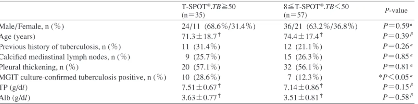
Table 3 Comparison based on the spot counts of T-SPOT®.TB patients between April 2013 and July 2015 at our hospital (n=92)
T-SPOT®.TB≧50 (n=35) 8≦T-SPOT®.TB<50 (n=57) P-value Male/Female, n (%) Age (years)
Previous history of tuberculosis, n (%) Calcifi ed mediastinal lymph nodes, n (%) Pleural thickening, n (%)
MGIT culture-confi rmed tuberculosis positive, n (%) TP (g/dl) Alb (g/dl) 24/11 (68.6%/31.4%) 71.3±18.7† 11 (31.4%) 9 (25.7%) 20 (57.1%) 10 (28.6%) 7.51±0.67† 3.63±0.77† 36/21 (63.2%/36.8%) 74.4±17.4† 12 (21.1%) 15 (26.3%) 32 (56.1%) 7 (12.3%) 7.14±0.86† 3.51±0.81† P=0.59α P=0.39β P=0.26α P=0.85α P=0.81α *P<0.05α P=0.15β P=0.58β †Data are presented as mean±SD (standard deviation).
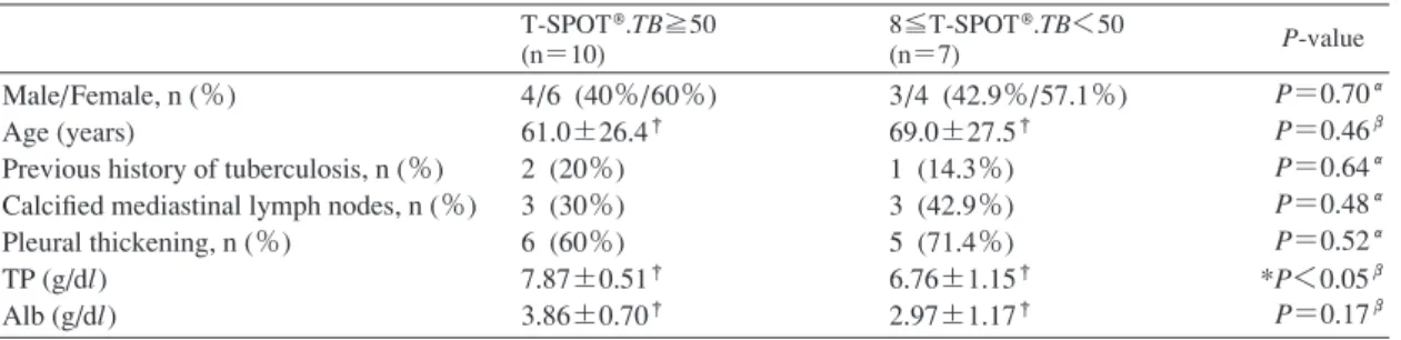
P-value :αFisher’s exact test, βMann-Whitney U test, *P<0.05 結核菌培養陰性であった 75 例中,結核菌培養陰性で あるが臨床的に結核の活動性があると診断し治療を行っ た症例は 3 例であった。その 3 例の詳細は,画像上左肺 尖部に tree in bud を示したもの,胸膜炎+縦隔気腫を認 めたもの,右下肺に結節影を認めたもの,であった。そ れぞれの T-SPOT®.TB は,パネル A スポット数 19,19, 50 以 上,パ ネ ル B ス ポ ッ ト 数 3 ,50 以 上,50 以 上 で, T-SPOT®.TB 弱陽性または強陽性例であった。この 3 例 は治療後に,陰影の改善を認めた。 ( 1 )T-SPOT®.TB 陽性 92 例における結核菌培養陽性群 17 例と結核菌培養陰性群 75 例の比較 結核菌培養陰性群は結核菌培養陽性群と比較し,男性 が有意に多かった。T-SPOT®.TB のスポット数は結核菌 培養陽性群で有意に多かった(T-SPOT®.TB のスポット 数が 50 以上のものは 50 として評価した)。胸膜肥厚やリ ンパ節石灰化,血清 TP,Alb 値は 2 群間で有意差は認め られなかった(Table 2)(Fig. 1)。 ( 2 )T-SPOT®.TB 陽性 92 例における T-SPOT®.TB 強陽 性群 35 例と T-SPOT®.TB 弱陽性群 57 例の比較 T-SPOT®.TB 強陽性群は,弱陽性群と比較すると,結 核菌培養陽性である症例が有意に多かった(P<0.05)。 胸膜肥厚やリンパ節石灰化,血清 TP,Alb 値は 2 群間で 有意差は認められなかった(Table 3)。 ( 3 )結核菌培養陽性 17 例中における T-SPOT®.TB 強 陽性群 10 例と T-SPOT®.TB 弱陽性群 7 例の比較 結核菌培養陽性群 17 例を T-SPOT®.TB 強陽性群と
T-Table 4 Comparison based on the spot counts of T-SPOT®.TB in MGIT culture-confi rmed tuberculosis positive patients between April 2013 and July 2015 at our hospital (n=17)
T-SPOT®.TB≧50 (n=10) 8≦T-SPOT®.TB<50 (n=7) P-value Male/Female, n (%) Age (years)
Previous history of tuberculosis, n (%) Calcifi ed mediastinal lymph nodes, n (%) Pleural thickening, n (%) TP (g/dl) Alb (g/dl) 4/6 (40%/60%) 61.0±26.4† 2 (20%) 3 (30%) 6 (60%) 7.87±0.51† 3.86±0.70† 3/4 (42.9%/57.1%) 69.0±27.5† 1 (14.3%) 3 (42.9%) 5 (71.4%) 6.76±1.15† 2.97±1.17† P=0.70α P=0.46β P=0.64α P=0.48α P=0.52α *P<0.05β P=0.17β †Data are presented as mean±SD (standard deviation).
P-value :αFisher’s exact test, βMann-Whitney U test, *P<0.05
Fig. 2 Comparison of CT fi ndings between MGIT culture-confi rmed tuberculosis positive and negative. Chest CT fi ndings of T-SPOT®.TB positive patients (n=92) were retrospectively analyzed. Results were compared between MGIT culture-confi rmed tuberculosis positive (n=17) and negative (n=75) patients between April 2013 and July 2015 at our hospital.
MGIT culture-confirmed tuberculosis negative MGIT culture-confirmed tuberculosis positive
Infiltration shadow Small nodular shadow Nodular shadow
Cavity Ground-glass opacity Pleural effusion
P-value: Fisher’s exact test *P<0.05 0 5 10 15 20 25 30 35 40 45 50% P=0.44 P=0.54 P=0.58 * * * SPOT®.TB 弱陽性群で比較検討したところ,T-SPOT®.TB 弱陽性群は T-SPOT®.TB 強陽性群と比較して有意に TP 値が低値であった(P<0.05)(Table 4)。 ( 4 )T-SPOT®.TB 陽性 92 例における結核菌培養陽性群 17 例と結核菌培養陰性群 75 例の画像所見の比較 T-SPOT®.TB 陽性症例の結核菌培養陽性群と結核菌培 養陰性群で CT 画像所見をそれぞれ比較したところ,結 核菌培養陽性群 17 例は,結核菌培養陰性群 75 例と比較 して,空洞影(23.5% vs 6.7%,P<0.05),粒状影(47.1% vs 9.3%,P<0.05),浸潤影(47.1% vs 16.0%,P<0.05)が それぞれ有意に高い傾向を認めた(Fig. 2)。 考 察 結核の診断は,喀痰などの臨床検体や組織などから結 核菌を証明することが基本であるが,実際の結核症例で は結核菌の検出が困難な例や長期間を要する症例もとき に経験する。ツベルクリン反応のほか,IGRA が開発さ れ,活動性結核の診断だけでなく,LTBI や接触者健診に も使用されている。現在わが国では IGRA として QFT® -TB と T-SPOT®.TB が行われている。T-SPOT®.TB は LTBI に対する陽性率において,QFT®-3G と比較すると劣って いるという報告もあるが5),活動性結核診断の有用性の 報告においては両者に差異はほとんどないとされてい る4) 6) ∼ 8)。実際の診療において,T-SPOT®.TB はスポット 数で表現されるが,T-SPOT®.TB のスポット数が明らか に多い時にはどれくらいの頻度で結核であるのか,少な い時にはどれくらいの頻度で結核なのかを考えることが ある。 今回の検討では,T-SPOT®.TB のスポット数で差異を 比較するためにスポット数を 50 で分け,強陽性群,弱陽 性群と定義したが,一般的にそのような定義はない。ま たスポット数を 50 で分けた理由としては,スポット数 50 以上は T-SPOT®.TB が陽性であることが明らかであ り,それ以上を測定していないため,50 以上と 50 未満 とを区別した。 今回の検討で結核菌培養陽性の症例では T-SPOT®.TB のスポット数が有意に多く,また T-SPOT®.TB 強陽性群 35 例は弱陽性群 57 例と比較して有意に結核菌培養陽性 が 多 く(T-SPOT®.TB 強 陽 性 28.6% vs T-SPOT®.TB 弱 陽 性 12.3%),活動性結核の補助的診断として有用である 可能性が示唆された。しかしあくまでも今回の報告では 活動性結核=結核菌培養陽性とした場合の統計結果であ
り,現状では結核菌培養陰性でもまた T-SPOT®.TB 陰性 でも,少なからず活動性結核が一定数いることを理解し ておく必要がある。今回の検討でも , 結核菌培養陰性で あるが T-SPOT®.TB 陽性のため,臨床的に活動性結核と 診断し,治療を施行した症例を 3 例認め,そのうち 2 例 の T-SPOT®.TB は強陽性であった。また T-SPOT®.TB 陰 性で結核菌培養陽性であった症例に結核性胸膜炎を 1 例 認め,その 1 例の T-SPOT®.TB スポット数はパネル A: 3 ,パネル B: 3 であった。 性差に関しては,T-SPOT®.TB 陽性で結核菌培養陰性 例が男性に有意に多く認められたが,結核罹患率は高齢 になってくると男性が多いため1),相対的に男性が潜在 性の結核既感染者が多いことが疑われた。 結核菌培養陽性の症例のうち T-SPOT®.TB 弱陽性群は T-SPOT®.TB 強陽性群と比較して有意に TP 値が低値で あった。この結果は,T-SPOT®.TB は高齢者などの低栄 養患者では結核菌培養陽性であったとしても T-SPOT®. TB陰性となってしまうことが多いとされる既存の報 告9) 10)と矛盾しない。今回の検討では,結核菌培養陽性 の症例のうち T-SPOT®.TB 弱陽性群は T-SPOT®.TB 強陽 性群と比較し平均年齢が高い傾向はあったが,TP 値以 外の有意差は認めなかったため,T-SPOT®.TB 弱陽性群 で TP 値が低値であった原因は同定できなかった。免疫 低下の要因に関しては,HIV,糖尿病,腎不全,ステロ イドや免疫抑制剤投与している症例が,結核菌培養陽性 の中で非常に少数であり,評価が困難であった。 画像所見では,T-SPOT®.TB 陽性の検討で,結核菌培 養陽性群であった症例は,結核菌培養陰性群と比較して 粒状影,空洞影,浸潤影を示し多様な画像を呈すること が多かった。多様な画像を呈したことは,尾形ら11)の報 告と比較しても矛盾しない。本研究で結核菌培養陽性群 において浸潤影が多かった原因としては,活動性結核で 一般に認められる tree in bud sign 様の陰影を粒状影+浸 潤影と判断した症例があったことがあげられる。tree in bud sign 様の陰影のように結節腫瘤影など,単一で表現 できない陰影が多いことが,多様な画像所見を呈するこ とが多かった理由の一つと考えられた。 T-SPOT®.TB は結核菌陰性の活動性結核の補助診断と して使われることがあり,早期発見にもつながる。その 際には画像検査が重要であることは周知の事実である が,T-SPOT®.TB のスポット数と組み合わせることで, さらに活動性結核の補助診断に有効であると期待しうる。 以上のことから T-SPOT®.TB 強陽性であり,画像所見 で空洞影や粒状影,浸潤影を認める場合は活動性結核を 疑うが,低栄養を呈する症例では9) 10) 12),今までの報告 と同様に T-SPOT®.TB 弱陽性や陰性になることを留意す る必要があると思われた。 結 論 結核菌培養陽性結核の症例は T-SPOT®.TB のスポット 数 が 有 意 に 多 く,T-SPOT®.TB 強 陽 性 群 は T-SPOT®.TB 弱陽性群と比較して培養陽性であった症例が有意に多か った。 T-SPOT®.TB のスポット数が 50 以上である陽性であ り,画像所見で空洞影や粒状影,浸潤影を認める場合は 活動性結核を疑う必要がある。T-SPOT®.TB は陽性/陰 性だけでなく,スポット数も併せて評価し,かつ胸部 CT などの画像診断結果と組み合わせることで活動性結 核の有用な補助的診断であると考えられた。 本論文の要旨は第 91 回日本結核病学会総会で発表し た。
著者の COI(confl icts of interest)開示:本論文発表内 容に関して特になし。 文 献 1 ) 結核予防会:「結核の統計 2015」. 結核予防会, 東京, 2015. 2 ) 日本結核病学会予防委員会:インターフェロンγ遊離 試験使用指針. 結核. 2014 ; 89 : 717 725.
3 ) Danielsen AV, Fløe A, Lillebaek T, et al.: An interferon-gamma release assay test performs well in routine screening for tuberculosis. Dan Med J. 2014 ; 61 : A4856.
4 ) Sester M, Sotgiu G, Lange C, et al.: Interferon-γγ release assays for the diagnosis of active tuberculosis, a systematic review and meta-analysis. Eur Respir J. 2011 ; 37 : 100 111. 5 ) 山田全啓,
村井孝行:結核集団感染事例におけるQFT-3G検査とT-SPOT検査の比較検討. 結核. 2016 ; 91 : 531 536.
6 ) Diel R, Loddenkemper R, Nienhaus A: Evidence-based comparison of commercial interferon-γγ release assays for detecting active TB. Chest. 2010 ; 137 : 952 968.
7 ) Fan L, Chen Z, Hao XH, et al.: Interferon-gamma release assays for the diagnosis of extrapulmonary tuberculosis: a systematic review and meta-analysis. FEMS Immunol Med Microbiol. 2012 ; 65 : 456 466.
8 ) Azghay M, Bouchaud O, Mechaï F, et al.: Utility of QuantiFERON-TB Gold In-Tube assay in adult, pulmonary and extrapulmonary, active tuberculosis diagnosis. Int J Infect Dis. 2016 ; 44 : 25 30.
9 ) Pan L, Jia H, Liu F, et al.: Risk factors for false-negative T-SPOT.TB assay results in patients with pulmonary and extra-pulmonary TB. J Infect. 2015 ; 70 : 367 380. 10) Han YM, Kim HS, Kim CH, et al.: Analysis of patients with
positive acid-fast bacilli culture and negative T-SPOT.TB results. Korean J Lab Med. 2010 ; 30 : 414 419.
11) 尾形英雄:肺結核の CT 画像と病理所見. 結核. 2009 ; 84 : 559 568.
12) Iwagaitsu S, Naniwa T, Maeda S, et al.: A comparative analysis of two interferon-γγ releasing assays to detect past
tuberculosis infections in Japanese rheumatoid arthritis patients. Mod Rheumatol. 2016 ; 26 : 690 695.
Abstract [Objectives] We investigated the usefulness of T-SPOT®.TB, an interferon-gamma release assay (IGRA), for diagnosis of active tuberculosis (TB) disease. We espe-cially focused on the spot counts of T-SPOT®.TB.
[Methods] The present study included 92 patients who were suspected as pulmonary TB and showed positive results for T-SPOT®.TB between April 2013 and July 2015 at our department. These 92 patients were further divided into T-SPOT®.TB strong positive group (n=35) and T-SPOT®.TB weak positive group (n=57) based on the spot counts of T-SPOT®.TB. Clinical parameters were retrospectively analyzed.
[Results] Mycobacterium tuberculosis were determined by mycobacterial growth indicator tube (MGIT) culture in respiratory samples of 10 of 35 T-SPOT®.TB strong positive group and 7 of 57 T-SPOT®.TB weak positive group patients, indicating active TB was found signifi cantly higher in the T-SPOT®.TB strong positive group (n=35) compared to the T-SPOT®.TB weak positive group (n=57) (P<0.05). In all
T-SPOT®.TB positive patients, chest CT fi ndings including cavity/small nodular/infi ltration were found signifi cantly higher in those with positive results for MGIT culture-confi rmed tuberculosis than those with negative results for MGIT culture-confi rmed tuberculosis (P<0.05).
[Conclusion] The present study indicates that the spot counts of T-SPOT®.TB is useful of supportive diagnostic methods for active TB disease.
Key words: Tuberculosis, Diagnosis, Interferon-gamma re-lease assays, T-SPOT®.TB
Division of Respiratory Medicine, Department of Internal Medicine, Toho University Ohashi Medical Center
Correspondence to: Norio Kodaka, Division of Respiratory Medicine, Department of Internal Medicine, Toho University Ohashi Medical Center, 2_17_6, Ohashi, Meguro-ku, Tokyo 153_8515 Japan. (E-mail: norio.kodaka@med.toho-u.ac.jp) −−−−−−−−Original Article−−−−−−−−