はじめに
脳アミロイドアンギオパチー(cerebral amyloid angiopathy; CAA)は,脳卒中,アルツハイマー病(Alzheimer disease; AD)
という老年発症の神経疾患に関連する1).高齢化社会で増加
が予想される疾患であるが,これらの CAA の臨床表現型の予 防に関しては,今のところ脳出血予防としての降圧療法しか
ない2).CAA のマーカーとして,MRI 上の脳小血管病所見,す
なわち “脳表限局性微小出血(cerebral microbleeds; CMBs)” 3)4), “脳表ヘモジデリン沈着(cortical Superficial Siderosis; cSS)” 5)6), “半卵円中心周辺の(MRI 上確認可能な)多発拡大血管周囲腔 (enlarged perivascular spaces; EPVS)” 7)8)が重要視されている.
今回我々は,これら全ての MRI 所見が CAA 関連出血以前に 得られていた AD 合併例を経験したため報告する. 症 例 患者:85 歳女性 主訴:頭痛,嘔吐 既往歴:以前より物忘れがあり,当院入院の 1 ヶ月前に近 医の物忘れ外来を受診した.エピソード記憶障害,遂行機能 障害,視空間失認をみとめ,Mini Mental State Examination は
15点であった.頭部 MRI で海馬の軽度萎縮をみとめ,AD と 診断された.その他に高血圧症(未加療),脂質異常症(加療 中),逆流性食道炎があり,内服薬としてガランタミン,ロス バスタチン,ラベプラゾールが処方されていた.抗血栓薬の 内服はなかった. 家族歴:脳卒中,認知症の家族歴はなし. 嗜好歴:飲酒歴,喫煙歴はなし. 現病歴:2015 年 1 月某日,頭痛,嘔気,嘔吐が出現した (第 1 病日).一旦は軽快したものの,翌日後に増悪し,当院 へ救急搬送された(第 2 病日). 入院時現症:身長 151.0 cm,体重 49.0 kg,体温 35.5°C,血 圧 187/59 mmHg,脈拍 76 回 / 分・整であった.一般身体所見 では異常所見はなかった.意識レベルは呼びかけによる開眼 をみとめ,Japan Coma Scale 10,Glasgow Coma Scale E2 V4 M6 であった.他の神経学的所見として,左半側空間無視,病態 失認,左上下肢不全麻痺をみとめ,National Institute of Health Stroke Scale(NIHSS)は 17 であった.家族から認知症に関す る病歴を聴取し評価したInformant Questionnaire on Cognitive Decline in the Elderly(IQCODE)9)のスコアは 107 であった(最 高 130.健常人推定平均は 87.6 ± 13.3; AD 推定平均は 121.4 ± 10.1)9). 検査所見:血算に異常はなかった.凝固系検査ではプロト ロンビン時間,活性化部分トロンビン時間は正常で,D-Dimer 1.95 μg/ml(施設基準値 0.00~1.00 μg/ml)は軽度上昇してい た.血液生化学では LDL コレステロール 140 mg/dl(0~ 119 mg/dl)と高値をみとめたが,他の脂質系,空腹時血糖,ヘ モグロビン A1C は正常であった.尿検査で,蛋白尿(221 mg/ 日), 微量アルブミン尿(30 mg/ 日)をみとめた.12 誘導心電図は
症例報告
発症前に多彩な脳アミロイドアンギオパチー関連 MRI 所見が
描出されていたアルツハイマー病合併脳葉型出血の 1 例
野中 俊宏
1)藥師寺祐介
1)*
井手 俊宏
1)伊藤 寛
2)河本 和裕
1)原 英夫
1) 要旨: 症例は未加療の高血圧があった 85 歳女性である.脳葉型出血を発症し,救命目的で開頭血腫除去術が施 行された.血腫の病理標本で脳アミロイドアンギオパチーと診断された.1 ヶ月前に近医でアルツハイマー病と診 断された際の脳 MRI では,脳アミロイドアンギオパチーに関連する所見,すなわち“脳表現局型微小脳出血”,“脳 表ヘモジデリン沈着”,“半卵円中心周囲の重度拡大血管周囲腔”が見られていた.認知症診療においてアルツハイ マー病患者の心血管リスク管理を考慮する上で,これらの所見を包括的に捉えることが有用かもしれない. (臨床神経 2016;56:338-343)Key words: 脳アミロイドアンギオパチー,MRI,微小脳出血,脳表ヘモジデリン沈着,拡大血管周囲腔
*Corresponding author: 佐賀大学医学部内科学講座神経内科〔〒 849-8501 佐賀市鍋島 5 丁目 1-1〕
1)佐賀大学医学部内科学講座神経内科
2)佐賀大学医学部内科学講座脳神経外科
(Received December 25, 2015; Accepted February 28, 2016; Published online in J-STAGE on April 28, 2016) doi: 10.5692/clinicalneurol.cn-000860
12 mm)をみとめた. 画像所見:頭部 CT にて,右側頭葉から頭頂葉にかけて周 囲に浮腫を伴う皮質下出血をみとめた(Fig. 1).ABC/2 法10) による CT 上の推定血腫量は 63 ml で,右シルビウス裂から 右大脳半球脳溝にクモ膜下出血を伴い,血腫による圧迫で 左方への正中偏位をみとめていた.頭部 CT アンギオグラ フィーでは出血源となる異常所見はみとめなかった. 入院後経過:入院同日(第 2 病日)に家族同意の下,救命 目的としての右前頭側頭開頭血腫除去術を施行した.以後, では,susceptibility-weighted imaging(SWI)上で両側後頭- 頭頂葉,小脳半球に複数の CMBs と,出血巣の対側大脳半球 に cSS をみとめた(Fig. 2A, B).T2強調画像上の EPVS は, 基底核領域(左)に 10 個程度,半卵円中心周辺のものは 20 本以上と推測できたがアーチファクトで詳細な判断はできな かった(Fig. 2C, D).
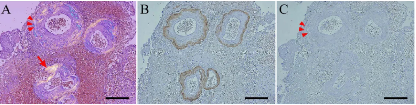
術中に採取された血腫病理標本の Congo-red 染色では細動 脈の血管壁が赤色に染色され,CAA に特徴的とされる血管壁 の二重化(double barrel vessel)をみとめた(Fig. 3A).偏光
Fig. 1 Brain CT (obtained on admission).
Brain CT demonstrates the hematoma (estimated volume 63 ml) in the right temporo-parietal lobe with subarachnoid hemorrhage, edema and midline shift (A, axial view; B, coronal view). A right parasagittal extra-axial calcified lesion is considered as a meningioma (A: asterisk).
Fig. 2 Brain MRI (obtained 15 days after the cerebral hemorrhage development).
MRI is performed with a 3.0-T scanner. Susceptibility-weighted imaging (A, B: TR, 27 ms; TE, 20 ms; FA; 15°; Slice thickness, 3 mm) shows two strictly lobar CMBs in left occipital lobe (A: arrow heads) and focal cortical superficial siderosis in the left central sulcus (B, arrow). T2-weighted imaging (C, D: TR, 4,500 ms; TE, 89 ms; FA; 180°; Slice thickness, 6 mm) shows dot-like hyperintensities (about 10 or more
lesions) characteristic of enlarged perivascular spaces in the left basal ganglia (C) and linear hyperintensities (about 20 or more) typical of enlarged perivascular spaces in the left centrum semiovale. However, those enlarged perivascular spaces are not clearly seen because of the patientʼs motion artifact.
顕微鏡下では同部位に,淡黄色の偏光を呈した(Fig. 3A).脳 アミロイド β(Aβ)の免疫染色では,Aβ40 は強陽性で,Aβ42 はわずかに染色されるのみであった(Fig. 3B, C).α 平滑筋と 汎 Aβ の蛍光二重染色では菲薄化した平滑筋と外膜へのアミ ロイド沈着をみとめた(Fig. 4).追加検査で行ったアポリポ プロテインのフェノタイプは ε3/ε3 であった.Enzyme-Linked ImmunoSorbent Assay法による髄液中のAβ40濃度は1,631 pg/ml (参考値 4,003±1,185 pg/ml 11)),Aβ42 濃度は 88.1 pg/ml(参考 値 838±253 pg/ml 11))と各々低下し,それらの比率 Aβ40/Aβ42 は 18.51(参考値 4.91±1.13 11))と高値を示した.これらの所 見は髄液中 Aβ40,42(特に後者)の低下を示唆するものであっ た.前医での AD 診断時(脳出血発症 1 か月前)の頭部 MRI を取り寄せたところ,T2*強調 MRI 上,後頭葉に複数の CMBs をみとめ,また左中心溝(今回の脳出血と対側)に cSS をみ とめていた(Fig. 5A, B).T2強調 MRI では基底核領域に 12 個(左側),半卵円中心領域に 44 個(右側)の EPVS をみと め,後者優位であった(Fig. 5C, D).
以上の経過,所見より,AD を合併した CAA 関連脳出血が 示唆された.AD に関しては,前医の診断に加え当院入院後 に得られた IQCODE スコアも考慮した上で National Institute on Aging-Alzheimerʼs Association(NIA/AA)の診断基準上の Probable AD dementia with evidence of the AD pathophysiological process12),CAAに関しては,Boston criteriaに基づいてProvable CAA with supporting pathologyと診断した13).入院中再出血や
CMBsの増悪所見もなかった.第 36 病日の転院時には,左上 下肢不全麻痺が残存し,NIHSS は 4 であった. 考 察 CAAは遺伝性と孤発性に分けられる.臨床的に遭遇する頻 度の高い孤発性 CAA は加齢とともに増加し,60 歳以上の 10~ 50%にみられ,90 歳以上では 74%に達する14)15).Aβ 関連疾 患の代表である AD の 80~90%には CAA がみられ14)15),認 知症診療では CAA の潜在に留意する必要がある.一方で CAA は脳血管障害の原因の一つであり,脳表型出血を呈し16),70 歳以上の年間脳出血発症例の約 1/3 が CAA による17).本症例 は 85 歳と高齢の AD 患者であり,脳表型脳出血をきたしてい ること,病理・生化学的な裏付けもあることから CAA 関連脳 出血の臨床的確実例である. 本例の特徴は,発症前 MRI で近年 CAA のバイオマーカー としての認識されてきた脳小血管病所見,すなわち “脳表限 局性 CMBs”,“cSS”,“半卵円中心周辺の多発 EPVS” を全て有 していた点にある.これらを有した CAA は,いわば典型例と いうべき症例である.しかし CAA 関連脳小血管病全体を包括 して論じた症例報告は少なく,神経内科医師を含めた臨床医 全般に広く認知されているとは言えない.本症例は,認知症 診療で撮像される MRI に脳出血のリスク管理に影響しうる Fig. 3 Pathological findings (1).
Serial sections (A, Congo red; B, Aβ40 immune stain; C, Aβ42 immune stain) of the evacuated hematoma show amyloid depositions with bright yellow color birefringence in polarized light microscopy (A: arrow indicates representative lesion) affecting the whole circumference of thickened small vessels and double barrel vessel (A: arrowheads), definitely positive amyloid-β40 (B), and weak positive amyloid-β42 (C). Scale bars represent 100 μm.
Fig. 4 Pathological findings (2).
Confocal microscopy of double-immunolabeled vessel (green, smooth muscle actin; red, pan-amyloid) shows focal disappearance of smooth muscle cells at the site of amyloid burden (B). Scale bar represents 20 μm.
CAAに関連した所見が存在することを示唆している. 本症例では SWI 上で両側後頭-頭頂葉,小脳半球に多発 CMBsをみとめていた.CMBs 近傍の細動脈の病理は,高血 圧性変化と CAA に大別され18),前者は主に脳深部・テント 下に,後者は脳表限局性に見られる3)19)20).MRI 上の脳表限 局性 CMBs と CAA の関連は,各々がアポリポプロテイン ε4 と関連することにより支持されている3.本症例では同フェノ タイプは ε3/ε3 であったが,以下に述べる Boston criteria に則 り CAA 関連の CMBs であることが示唆される.
CAAの臨床診断を目的に開発された Boston criteria は,脳 表限局性 CMBs(小脳の CMBs の有無は問わない)と脳表型出
血を主要な評価項目としている13).本症例は,臨床データお
よび病理組織(血腫吸引標本)から probable CAA with supporting
pathologyと診断した.日常臨床では病理所見が得られないこ
との方が多く,そのような場合に MRI を含めた画像所見で probable CAAや possible CAA の診断を行うことができるよう になっている.
近年,CAA の新たな画像マーカーとして cSS が重要視され
てきている5)6).CAA 診断において,cSS を診断項目に追加し た Modified Boston Criteria は,原版と比較して特異度に差は 無いが,感度では原版の 89.5%から 94.7%に上がる5).また 欧州の多施設縦断的研究では,cSS は CAA 関連脳出血の独立 したリスクであることが示され,そのハザード比は 2.53 で あった21).本症例でも発症 1 ヶ月前に撮像された MRI におい て対側大脳半球に cSS をみとめていた. Virchow-Robin腔として知られる EPVS の観察も有用で,基 底核領域や半卵円中心周辺で評価し,数の多い側を判定に用 いる7)8).半卵円中心周辺で 21 個以上見られる場合は病理学
的にCAAである可能性が高い22).Boston criteriaに同領域EPVS 重症度を組み合わせると,CAA の診断率が増すことも報告さ れている22). 脳卒中患者における CAA 関連脳出血予防については,高血 圧性脳出血同様に,降圧療法が有効であることが示されてい る2).本症例では未加療の高血圧があり,尿蛋白,心筋肥大 などの高血圧性臓器障害があったが,介入はされていなかっ た.認知症外来は MRI 上の「脳表限局性 CMBs」,「cSS」,「半 Fig. 5 Brain MRI obtained one month before the onset of symptomatic intracerebral hemorrhage at a regional memory clinic. MRI is performed with a 1.5-T scanner. T2*-weighted imaging (TR, 600 ms; TE, 25 ms; FA, 20°; Slice thickness, 6 mm) shows
multiple strictly lobar CMBs in occipital lobes (A: arrow heads) and focal cortical superficial siderosis in the left central sulcus (B, arrow). T2-weighted imaging (TR, 4,000 ms; TE, 106 ms; FA, 180°; Slice thickness, 6 mm) shows dot-like hyperintensities
(12 lesions) characteristic of enlarged perivascular spaces in the left basal ganglia, as well as dot-like (arrow) and linear (arrowhead) hyperintensities (44 lesions) typical of enlarged perivascular spaces in the centrum semiovale.
卵円中心周辺 EPVS」の検出を契機に脳出血未発症 CAA 症例 を見出す良い機会と思われる23).認知症外来ではこれらの CAA関連 MRI 所見を包括的に捉え,もし所見が存在した場 合には脳出血未発症例においても積極的な血圧管理を考慮す べきかもしれない. 本報告の要旨は,第 210 回日本神経学会九州地方会で発表し,会長 推薦演題に選ばれた. ※本論文に関連し,開示すべき COI 状態にある企業,組織,団体 はいずれも有りません. 文 献
1) Vinters HV. Cerebral amyloid angiopathy. A critical review. Stroke 1987;18:311-324.
2) Arima H, Tzourio C, Anderson C, et al. Effects of perindopril-based lowering of blood pressure on intracerebral hemorrhage related to amyloid angiopathy: the PROGRESS trial. Stroke 2010;41:394-396.
3) Vernooij MW, van der Lugt A, Ikram MA, et al. Prevalence and risk factors of cerebral microbleeds: the Rotterdam Scan Study. Neurology 2008;70:1208-1214.
4) 藥師寺祐介,原 英夫.脳出血治療の最新治験 微小脳出 血:その意義と管理.臨床神経 2012;52;1106-1109.
5) Linn J, Halpin A, Demaerel P, et al. Prevalence of superficial siderosis in patients with cerebral amyloid angiopathy. Neurology 2010;74:1346-1350.
6) Charidimou A, Jager RH, Fox Z, et al. Prevalence and mechanisms of cortical superficial siderosis in cerebral amyloid angiopathy. Neurology 2013;81:626-632.
7) Charidimou A, Meegahage R, Fox Z, et al. Enlarged perivascular spaces as a marker of underlying arteriopathy in intracerebral haemorrhage: a multicentre MRI cohort study. J Neurol Neurosurg Psychiatry 2013;84:624-629.
8) Yakushiji Y, Charidimou A, Hara M, et al. Topography and associations of perivascular spaces in healthy adults: The Kashima Scan Study. Neurology 2014;83:2116-2123.
9) Jorm AF, Jacomb PA. The Informant Questionnaire on Cognitive Decline in the Elderly (IQCODE): socio-demographic correlates, reliability, validity and some norms. Psychol Med 1989;19:1015-1022.
10) Kothari RU, Brott T, Broderick JP, et al. The ABCs of measuring intracerebral hemorrhage volumes. Stroke 1996;27:1304-1305.
11) Verbeek MM, Kremer BP, Rikkert MO, Van Domburg PH, Skehan ME, Greenberg SM. Cerebrospinal fluid amyloid beta(40) is decreased in cerebral amyloid angiopathy. Ann Neurol 2009;66:245-249.
12) McKhann GM, Knopman DS, Chertkow H, et al. The diagnosis of dementia due to Alzheimer’s disease: recommendations from the National Institute on Aging-Alzheimer’s Association workgroups on diagnostic guidelines for Alzheimer’s disease. Alzheimers Dement 2011;7:263-269.
13) Knudsen KA, Rosand J, Karluk D, et al. Clinical diagnosis of cerebral amyloid angiopathy: validation of the Boston criteria. Neurology 2001;56:537-539.
14) Yamada M. Cerebral amyloid angiopathy: an overview. Neuropathology 2000;20:8-22.
15) Zhang-Nunes SX, Maat-Schieman ML, van Duinen SG, et al. The cerebral beta-amyloid angiopathies: hereditary and sporadic. Brain Pathol 2006;16:30-39.
16) Hirohata M, Yoshita M, Ishida C, et al. Clinical features of non-hypertensive lobar intracerebral hemorrhage related to cerebral amyloid angiopathy. Eur J Neurol 2010;17:823-829. 17) Schutz H, Bodeker RH, Damian M, et al. Age-related spontaneous
intracerebral hematoma in a German community. Stroke 1990; 21:1412-1418.
18) Schrag M, McAuley G, Pomakian J, et al. Correlation of hypointensities in susceptibility-weighted images to tissue histology in dementia patients with cerebral amyloid angiopathy: a postmortem MRI study. Acta Neuropathol 2010;119:291-302. 19) Yakushiji Y, Noguchi T, Hara M, et al. Distributional impact of
brain microbleeds on global cognitive function in adults without neurological disorder. Stroke 2012;43:1800-1805.
20) Yakushiji Y, Yokota C, Yamada N, et al. Clinical characteristics by topographical distribution of brain microbleeds, with a particular emphasis on diffuse microbleeds. J Stroke Cerebrovasc Dis 2011; 20:214-221.
21) Charidimou A, Peeters AP, Jager R, et al. Cortical superficial siderosis and intracerebral hemorrhage risk in cerebral amyloid angiopathy. Neurology 2013;81:1666-1673.
22) Charidimou A, Jaunmuktane Z, Baron JC, et al. White matter perivascular spaces: An MRI marker in pathology-proven cerebral amyloid angiopathy? Neurology 2014;82:57-62. 23) Charidimou A, Ni J, Martinez-Ramirez S, et al. Cortical
superficial siderosis in memory clinic patients: further evidence for underlying cerebral amyloid angiopathy. Cerebrovasc Dis 2016;41:156-162.
Pre-critical MRI findings of an Alzheimer’s disease patient
with pathologically proven cerebral amyloid angiopathy related lobar hemorrhage
Toshihiro Nonaka, M.D.
1), Yusuke Yakushiji, M.D., Ph.D.
1), Toshihiro Ide, M.D.
1),
Hiroshi Ito, M.D.
2), Kazuhiro Kawamoto, M.Eng.
1)and Hideo Hara, M.D., Ph.D.
1)1)Division of Neurology, Department of Internal Medicine, Saga University Faculty of Medicine 2)Department of Neurosurgery, Saga University Faculty of Medicine