Acta med. Nagasaki. 21 : 38-44
Radioimmunoassay of Plasma 17α‑Hydroxyprogesterone
Tadayuki ISHIMARU, Atsumi MORI, Yasuaki KASE
and Seiran MIURA*
Department of Obstetrics and Gynecology, Nagasaki University School Of Medicine
Nagasaki, Japan
Received for publication, November 18, 1976
A radioimmunoassay for the measurement of 17α‑hydroxyprogesterone (17α‑OHP) in plasma was investigated utilizing antiserum produced by the introduction of 17α‑hydro‑
xyproges‑terone‑3‑oxim BSA. Accuracy was such that within‑assay variance was 16.2%
and between‑assay variance 18.3%.
The 17α‑OHP mean plasma levels were 930±201 pg/ml(n=8)for normal adult males and 402±186 pg/ml(n=11)in the follicular phase and 1,190±662 pg/ml(n=12)in the luteal phase of females with a normal menstrual cycle.
The 17α‑OHP levels in some tissues and tissue fluid also were measured, being 18.06±12.91 ng/ml(n=6) in the normal trophoblastic tissue, 1.7±0.5 ng/ml(n=17) in the trophoblastic tissue of a hydatidiform mole and 855.5±507.4 ng/ml(n=5)in lutein cyst fluid.
INTRODNCTION
We investigated a radioimmunoassay for the measurement of 17a-hydroxyprogesterone (17a-OHP) in plasma utilizing antiserum resulting from the introduction of 17a-hydroxy- progesterone. Levels of 17a-OHP in some tissues and plasma also were measured. The results are reported herein.
MATERIALS AND METHODS
1) Specificity of the Antiserum
The specificity of the antiserum used was tested by cross reaction studies with various
* 石 丸 忠 之 ,森 淳 躬 加 瀬 泰 昭,三 浦 清 轡
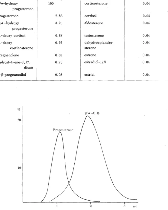
Table 1. Cross reactions of various steroids with antiserum Produced by 17a-OHP-BSA
steroid compounds % cross reaction steroid compounds % cross reaction
17a-hydroxy 100 corticosterone 0.04
progesterone
progesterone 7.85 cortisol 0.04
20a -hydroxy 3.23 aldosterone 0.04
progesterone
11-deoxy cortisol 0.88 testosterone 0.04
11-deoxy 0.66 dehydroepiandro- 0.04
corticosterone sterone
pregnenolone 0.52 estrone 0.04
androst-4-ene-3,17, 0.25 estradiol-17(3 0.04
dione
5-P-pregnanediol 0'.08 estriol 0.04
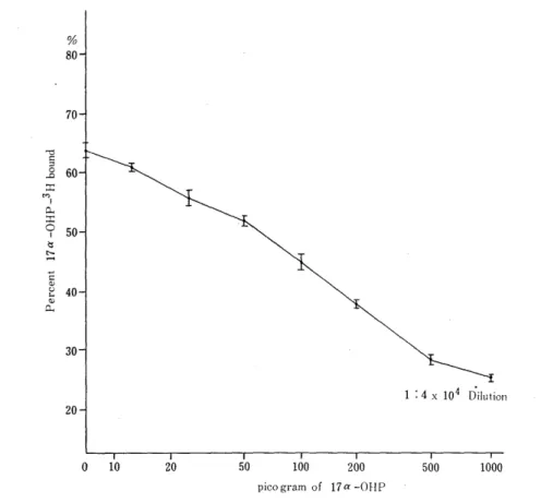
Fig. 1 Chromatographic pattern of progesterone
and 17a-OHP in Sephadex LH-20 microcolumn
steroids (Table 1). The cross reaction of 17a-OHP was taken as 100%. Cross reaction of progrsterone and 20a-OHP-progesterone was 7.85% and 3.23%, respectively, but that of all other steroids in the test was less than 1 % .
2) Plasma Extraction, Separation and Purification
1 x 10³ dpm volume of 3H-17a-OHP was added to plasma to produce an internal standard for recovery estimations. Extraction of 17a-OHP was carried out with 4ml of ether, following which the etheral extract was transferred to other tubes and evaporated to dryness at 37-40°C. The dried residue was dissolved in a benzenemethanol (95 : 5 solvent) and then chromatographic separation of 17a-OHP was accomplished by Sephadex LH-20 microcolumn chromatography (Fig. 1).
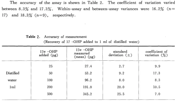
3) Standard Curve
Titration of the antiserum revealed that a dilution of 1 : 4 x 10⁴ was most suitable for assay. Namely, when the antiserum was diluted 40,000 times, the calibration curve became an almost straight line in the range of 0-1,000 pg of 17a-OHP (Fig. 2). In this range, the 17a-OHP percent bound to antiserum displayed a standard deviation of ±0. 1-
±0.7 and a coefficient of variation of 0.2-3.7%.
Fig 2 Standard curve with antiserum produced by 17a-OHP-BSA and diluted 1/4X 104
4) Measurement Procedure
The measurement procedure is as shown in Figure 3. The concentration of 17a-OHP in plasma was calculated by the following formula :
E x (M -m) x 100 x 1 where
R Re, Plasma sample (ml)
E=affluent from column chromatography
R=volume of effluent used in RIA
M=17a-OHP value estimated by standard curve
m=value of water sample for testing blank of system Re% -recovery percentage
Serum 0.01^-1.0ml
added 1 x lO3dpm 3H-17a-OHP
(for recovery)
Extraction
4 ml Ethyl Ether
twice
Dry
37-V40°C
under N2 gas spray Sephadex LH-20 column chromatography
2/3 1/3
Assay Recovery
counted for 3H 3H-17a-OHP 1 x 10⁴ dpm added
dried by N2 gas spray at 45°C
Incubation with diluted antiserum
(0.3ml antiserum 1/4 x 104 dilution) (room temp. 30 min.)
Separation of "free" and "bound" forms
0.3ml of dextran-coated charcoal added in ice bath.
Incubated 5^-10 min.
2500-3000 rpm. centrifuged for 10 min.
Supernatant 0.3 ml
3H counted counted
Calculation of concentration
from calibration curve.
Fig. 3 Measurement procedure
5) Accuracy and Precision
The accuracy of the assay is shown in Table 2. The coefficient of variation varied between 8.3% and 17.3%. Within-assay and between-assay variances were 16.2% (n=
17) and 18.3% (n=9), respectively.
Table 2. Accuracy of measurement
(Recovery of 17 -OHP added to 1 ml of distilled water)
17a -OHP 17a -OHP standard coefficient of
added (pg) (mean) easue(pg) deviation (±) variation (%)
25 27.4 2.7 9.9
Distilled 50 53.2 9.2 17.3
water 100 96.2 8.0 8.3
lml 200 191.0 20.0 10.5
500 345.2 25.5 7.0
RESULTS
The plasma 17a-OHP levels determined by the assay were 930±201 pg/ml (n=8) in normal adult males, and 402±186 pg/ml (n=11) in the follicular phase and 1,190±662
pg/ml (n=12) in the luteal phase of females with a normal menstrual cycle.
Plasma levels of LH, estradiol, progesterone and 17a-OHP during the menstrual cycle of the same female were examined, the results of this being shown in Fig. 4. In a normal pregnancy, the plasma 17a-OHP level was observed to be 2.03±0.65 ng/ml (n
=5) at the 7th to 17th weeks.
We next examined tissue levels of 17a-OHP. Normal trophoblastic tissue and the
tissue of a hydatidiform mole were homogenized and the levels of 17a-OHP in the super-
natants determined. The levels were 18.06±12.91 ng/ml (n=6) and 1.71±0.5 ng/ml,
respectively. Lutein cyst fluid obtained during surgery involving a destructive mole and
chorioepithelioma also was examined and the 17a-OHP level was found to be 855.5 ±507.4
ng/ml (n = 5).
Fig. 4 The plasma levels of LH, 17a-OHP, estradiol and progesterone simul- taneously measured in one person during normal menstrual cycle.