Japanese Joumal of Tropical Medicine and Hygiene
第9巻 第1号 昭和56年6月15日
内 容 原 著
高知県における大榎殖門条虫症の第4例………熊沢 秀雄,鈴木 了司,、大倉 俊彦 1−7 免疫抑制剤およぴD班usion Chamber移植法によるラットの日本産カンテツに対
する防御免疫効果の検討(英文)………一・……・・…………・……・…一「…一…・橋口 義久 9−22 高温環境下における連続7日間の運動が高温に対する生理的反応および耐熱性にお
よぼす影響(英文)………一一…・一・………・………一一…・・………一・堀 清記 23−29 狂犬病ウイルス免疫マウス脾臓細胞によるウイルス螢光フォーカス形成抑制(英文)
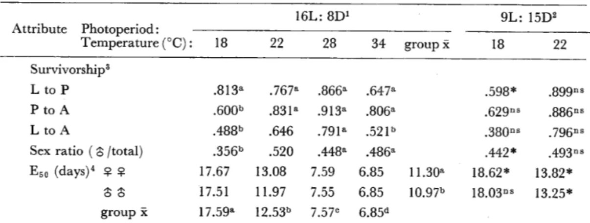
一牧野 芳大,三舟求真人 31−36 コガタアカイエカの幼虫の発育と成虫の属性に及ぽす温度と日長の影響
・・Niaz,Sh.and Reisen,W.K.37−47 会 報
昭和55年度第2回幹事会記録一・…………・…・・…………一…・……一…………一・………… 49 昭和55年度評議員会記録・・……一……一・………・・…・………・・…・………・50 第22回会務総会記録………一…一・・ 51−53 会則…………・…一・・………一…・…・…………一・………・一・・54−57 投稿規定…・……・………・………… 58−59
会員名簿………一…… 61−8g
日熱医会誌
Japan.J.T.M.H.
日 本熱帯医学会
lications which belong to Hidco Kumazawa,Ryoji Suzuki,Toshihiko Okura,,and Yoshihisa Hashiguchi published in voL9,No.1respectively. Please correct the
mistakes fbllowing the errata.
編集事務の誤りで9巻1号掲載の著者に御迷惑をおかけしました。別表正誤表に従って御
訂正下さい。
1. 高知県における大複殖門条虫症の第4例 熊沢秀雄,鈴木了司,大倉俊彦(和文論文)
訂 正 箇 所 1頁 左段本文下から 3行目
〃 上から 11行目 右段本文上から 1行目 〃 〃 3行目 〃 下から 10行目 2頁 左段本文下から 9行目 〃 〃 7行目 〃 〃 5行目 3頁 右段本文下から 11行目 〃 〃 1行目 4頁 右段本文上から 3行日 5頁 左段本文下から 4行目 右段〃上から 1行目
誤
特続 1571 判断
同月29日午前7時頃一 6彩 縦走節 横断図 腔 1981
D.9鰯n伽 海産類 下痢腹鳴 2例
正
文 献
1)
5)
9)
12)
14)
16)
目目目目目目行行行行行行22﹃⊥2﹃⊥− (Cestoda;Pserdophyllidea)
0.加1伽80がθ7σ召 大複殖門条虫成熟虫寄生例 k892
Z)ゆhツJJo60∫h7」 ¢
Additional II cases
持続 1971 診断
同月29日午前3時頃から複鳴持 続し,午前7時頃に
0彪
縦走筋,横走筋髄層の 横断面 膣 1891
五).970n4ゴs
海産魚 下痢,腹鳴 2列
(Cestoda;Pseudophyllidea)
0.わα」α8%0ρ∫87σ召
大複殖門条虫未成熟虫寄生例 1892
1)ゴ1)h夕〃060渉h短%吻 Additional ll cases
rats, using immunosuppressants or diffusion chamber techniques
YOSHIHISA HASHIGUCHI
p p p p p p p p p p
lO ll 12 14 14 15 16 16 17 19 21 22
ll
ll
ll
ll
ll
ll
Seventh line from the bottom
Fifth line ll
Twentieth line from the Top Eighth line ll
Figure 1
Column i.n the Table 3 Fifteenth line from the Top Column in the Table 5 Figure 2
Fourth line from the bottom REFERENCE 25)
l !> : J ) ) I ft
// // 1l F̲..
// T ) 8 ff
// // 6 ff // // 5 tEl
// 4fT
ll// 2 T
llerror
batach 50 minutes C‑1 X 100 blbumin, C: betaglobulin, without chambers
copared No. serum sampler
B : alphaglobulin theat
Fasciola hepatic Fasciola Repatica
Ouchterlong : l i
Dexamethazdne
j 1 e;(Z)
Ouchterlong r‑globulin
correct
batch 50 minutes.
C‑IjC X 100 albumin,
C : beta‑globulin,
without chamber compared No. serum samples
B: alpha‑globulin that
Fasciola hepatica Fasciola hepatica
Ouchterlony
Dexamethasone 1 & o Ouchterlony
r‑globulin
高知県における大複殖門条虫症の第4例
熊沢秀雄1,鈴木了司1,大倉俊彦2 和和56年2月9日 受付
緒 口
大複殖門条虫 Z)ψあgoηψor郷 g順n漉5
(BZαηchαr4,1894)mhe,1899の人体寄生例は静 岡,烏取,長崎をはじめとする19府県から,すで に85例が知られている。しかしこの中には断片的 な記録しか残っていないものも多く,感染経路を 含む疫学上の諸問題や,この条虫をめぐる分類学 上の問題は未解決のままであり,個々の症例を精 確に記録してゆく必要性は今も変わらない。
筆者らはすでに高知県から,本条虫の未熟虫体 寄生の2例を報告したが,本条虫の感染源が小型 の海産魚と推定されること(Kamoθ≠磁,1571)
などから,高知県における本条虫症例は少なくな いことが予想された(熊沢ら,1981)。最近,ひ とりの患者が前後少なくとも3回に亘って成熟片 節を自然排出した1症例を得たので,ここに報告
する。
症 例
○岡O。45歳。男。高知県安芸市赤野在住。農 業のかたわらで養鰻および沿岸漁業に従事。
既往歴,家族歴には特記すべきことはない。
現病歴。生来格別病気をしたことはなかったが,
飲酒後下痢をするのが常であった。1980年1月26 日から連夜飲酒,同月27日,下痢便とともに虫体
(目測で約1m)を自然排出したが捨てたという。
この頃から腹鳴が特続するようになり,時折腹痛 もあったが放置。同月29日早朝,虫体(標本番号 1)を下痢便とともに排出し,虫体を携えて受診
した。虫体から大複殖門条虫症と判断し,駆虫を 勧めたが患者多忙のためその後来院しなかった。
同年2月下旬より飲酒の機会多く,同月29日午前 7時頃に軟便とともに虫体を排出(標本番号2)。
さらに同年3月18日午前8時30分頃,下痢便とと もに標本番号3a,3b,3cの虫体断片をこの順序 で排出した。但し標本3bは患者が持参しなかっ たので測定することはできなかったが,患者によ れぱ約20cmの長さであったという。いずれの場 合も虫体排出後,腹鳴は治まったが,軟便傾向は 持続した。駆虫を勧めたがその後患者からの連絡 がなく現在に至っている。
理学的所見。特記すべきことはない。
尿検査所見。比重1.015。pH6.0。蛋白,糖,ケ トン体,潜血反応,ピリミジン,いずれも(一)。
ウロビリノーゲン,正常。
糞便検査は行なわなかった。
血液学的検査。赤血球数452×104/cmm.Hb
15.Og/d1.血色素指数1.04。白血球数9,100/cmm
(好中球,1核3%,2核16勉,3核21%,他20%,
リンパ球25%,単球7%,好酸球8勉,好塩基球
6勉)。
血液化学検査。LDH210単位。GOT7単位。
GPT8単位。A1−P6.6単位。CHE O.65∠。ZTT 70単位。黄疸指数7単位。総蛋白量6.69/d1。
患者の嗜好。自宅の前の海で捕れたアジ,サバ の刺身やイワシの酢物を常食し,好物のカッオを 除き市場の海産魚介類は食べない。川魚は食べな い。豚肉は好食するが牛肉は特に好まない。養鰻 のためイトミミズを誤まって口にすることがあっ
た。
1高知医科大学寄生虫学教室 南国市岡豊町小蓮 2 高知学園短期大学 高知市旭天神町
虫体観察所見
回収した虫体は次の4断片で,これらはすべて,
クロロホルム加冷水道水で弛緩させて計測したの ち,5%ホルマリンで固定した。
標本番号1長さ494cm,最大幅19.5mm(前
端から300cm)。
標本番号2長さ352cm.最大幅20.5mm(前
端から280cm)。
標本番号3a長さ310cm.最大幅21.5mm
(前端から260cm)。
標本番号3c長さ111cm.最大幅13.Omm
(後端)。
このうち3aと3cは連続して排出されたもの で,3cの後端の幅は3aの前端の幅(16.Omm)
よりやや小さい。いずれの断片も2列の生殖器列 を有するほか,2,3a,3cの断片では1〜3個の過 剰生殖器を含む片節(Figure1)が見られる。い ずれの断片にも頭節はない。どの断片でも片節は 縦径よりも横径が大で,ストロビラの縁はやや鋸 歯状を呈し,体表面には,生殖器と神経幹にそれ ぞれ対応する部位にやや深い縦溝があるほか,両 生殖器間に5〜7条,生殖器と神経幹の間に片側 2〜4条,神経幹より外側に片側5〜7条の極めて 弱い縦溝が認められる。両神経幹の間の縦溝は背 腹で必ずしも一致せず,縦溝の数は腹側がやや多
いo
Table1は虫体断片1の最大幅付近の切片標本 についての計測値である。片節の幅径は縦径の 18.6倍,生殖口間の距離は片節幅の25鰯となる。
虫体の断面は外側から角皮,角皮下細胞,皮質層,
縦走節,髄層の各層から成る(Figure2)。
1対の神経幹と2本以上の排泄管が髄層内にあ り,前者の横断図は背腹にやや扁平な楕円形であ る。貯精嚢は陰茎嚢の背後方に,これと鋭角をな して接続する。輸精管と腔は共に生殖賓に開口し,
後者は生殖口として片節縦径の前端から約1/3の 部位に開口する。子宮口はそれより約200μ後方 にある(Figure3)。
精巣は陰茎嚢の周辺を除く髄層内のやや背側寄
Table1 Measurements(μ)of the DψJo−
80吻o驚specimen*。
segment
length 955
width 17800
thickness 600
distance between
two genital organs 4500 genital organ and nerve trunk 2240 9・o・and excretory canal l390 thickness of
tegument
tCgUment Cell layCr cortical parenchyma longitudinal muscle tranSVerSe mUSCle
medullary parenchyma nerve trunk,diameter
dorsoventral horizontal cirrussac
length width
wall thickness Seminal VeSiCle
length width
wall thickness testis,diameter
dorsoventral horizonta1
士95 士271
±35
±95
±141 士190
1.9± 0.6 44.3ニヒ 7.0 89.2二L 20.5 74.7ニヒ 7.3 35.0士 8.4 108.〇ニヒ 21.8
69.4土 6.5 86.4土 9.1
286.1土 240.4土 6.5±
117.3士 87.3土 19.2土
18.1 6.9 3.4
18﹃︶
589
45.5士 7.5 62.1ニヒ 10.7
*expressed as mean±standard deviation.
りに一層に配列し,横断切片では背腹に扁平な楕 円形で,両陰茎嚢の間に28〜31個,陰茎嚢より外 側に片側6〜31個が数えられる。精巣の配列は片 節間で中断することなく連続している。
卵黄腺は角皮下細胞近くの皮質層内にあり,圧 平標本では径32〜81μ(平均52μ)の不規則な 円形をなし,その分布は陰茎嚢や子宮と重なるこ とはなく,また陰茎嚢の前や子宮の後で左右が連 続することはない。他の部分では卵黄腺は均一に 分布する。子宮ループは片側で2〜5本が認めら
撚 難
灘灘 難一灘
鰻 轍尋翼 ■
,鑓、 騒…
麟
Figure1 Figure2 Figure3
1、・欝纏
譲
Apartofthestrobi拾窺o.2ne轟ritsmaximumwidthshowingfburrowsofgenital
organs. Stained w三th alum carm圭ne、 ×23・
Tran呂verse scction of the strobila n甑L Stained wi皇h Mayer五ematoxylin鋤d eosin、 ×67.
Sagittal section of the strobi1包uo.L Stained with M&yer hematoxylin and eosin.
x67.
Abb童eviations c,cortic拭1parenchyma; co,cirrus openingl csラcir罫u8言acl ga,genital aもrium; 9P,琶eniね1pore; 1m,10ngitudinal muscle3 m,medullary parenchyma; sv,餐eminal ves量cle茎 tうte藝ument l tc,tegument cell layer} tmコ transversemuscle;ts}testまsl uo,uterineopenin導l vt,vitellarial vo,vagまnal opening,
れる。子宮内には大きさ63.4〜77.9×3L2〜46、8μ
(平均71,7×36、8μ)の虫卵がある。
考 察
大複殖門条虫の人体症例は197i年までに55例が 知られていた(Kamo就認.,1971)。その後の症例 をT&ble2に,主として発表順序に従って要約し た。本論文の症例を日本における第85例として扱 う。このほか静岡より1例が近く報告される筈で
(影井,私信),これを加えると全87症例となる。
高知からは197工年以前には岩田・安岡(1970)の 1例があるのみで,本論文の症例は高知の第4例
となる(高知の第2,3例はTable2)。
本症例でみられた下痢,腹鳴,腹痛の症状は,
従来もしぱしば報告されている (Kamo撹磁,
1971)が,これらの症状と飲酒との関連は,今回 の症例の場合明らかでない。今回の症例でも,本 条虫の寄生が貧血や炎症その他の症状をひきおこ すという証拠はない。
大複殖門条虫はヒゲクジラ類寄蛋のD,b認昭ル 砂孟ぴα8(Lδ無berg,1981)と形態的に区別できな いといわれる(Rausch,19641岩田,ig67)、鰭脚 類やラッ瓢寄生のDj8俄ψ≠εr欝(v、Siebold,
i848)が人体寄生する可能性もある(Rausch,
1964)。今回の標本は虫体が大きいこと,卵黄腺 の分布が子宮ループと重ならず,片節の前縁や後 縁で連続しないこと,卵黄腺が大きいことなどの 点でn観rヒ膨εr螂よりはD.ゐα伽塑ρ魏α8に似
る。人体寄生のものを後者と同一視することには 未だ疑問があるので,今回の患者から得たものは 大複礁門条虫D遭灘ηぬ5と同定した。
条虫の成長は速く,たとえば広節裂頭条虫は犬 体内で毎日,前日の約1.4倍の長さになり,人体 内での絶対成長速度は犬でのそれの3倍を越える といわれる(Wardle and Green,1941)。このこ とから,大きな虫体断片が相次いで排出されたと しても,それらはすべて同一個体由来かもしれな い。特に虫体断片2,3a,3cは過剰生殖器の出現 の様子が類似しているのでその可能性は十分に存 する。一方,患者の嗜好から言って,複数寄生の
可能性も全く否定はできない。
感染源については従来の各症例報告と同様に明 らかではないが,本報告の患者が新鮮な海産類を 常食していたという事実は,過去の大複殖門条虫 症例が西南日本の海に面した府県に集中して分布 していることと対応している(Figure4)。感染 源がカッオかアジなら初夏,サバかイワシなら秋 から冬に感染する機会が多いものと思われる。感 染から虫体排出までの期間は,広節裂頭条虫では Table2 Cases of1)ψ10goηψ01謝infヒction,recognized after197L
n。. re琵rcnce patient
locality age sex
ユ
1ength
(cm)
width
(mm) scolex
678901234567890123456789012346555566666666667777777777888888 733323666666666664111111115009
111 11111111111 11111111111灘鮮㎞藷鞭㎞羅灘㎞灘灘解 SNNNSESTTTTTTSTTSESSSSSSSSNKKO 23081188P658906302P2160211501263434463 343243557 54454534775 mf
mm mm mm?
mm mm mm mm
m?f
mm mm mm
mff
m
15
04880020864630276072005665054
05653317351117332619151911987?314411 31 1 34122 14351 12 2 500 05 5ワヘ6翫289753410831851758802777557? 111 1111 12 1 21 21 12十
十
十十
十
d
〃
6 15
5
1分
1
494
1
3
2 1 4
4
41
1
1レ
2 1 20 1
Figure4 Distribution by prefbcture incidence of diplogonoporiasis.
約80日という事例があり(吉田ら,1979),卵検 出までの期間は条虫類一般にもっと短かい(An・
dersen,1978).Kamo8君α乙(1971)は大複殖門条 虫の症例数のピークが南から北に次第に遅くなる
ことと関連して,魚の消費量が秋から冬に最大と なることに注目している。今後も,感染源を明ら かにするために,個々の症例をさらに検討する努 力が必要であろう。
要 約
1. 高知県安芸市在住の45歳男子より1980年1 月29日,2月29日,3月18日の3回に亘り自然排
出された4虫体断片を大複殖門条虫と同定した。
2.下痢腹鳴,腹痛のほかは,特別な症状はな かった。
3.虫体断片は長さがそれぞれ494cm,352cm,
310cm,111cm,最大幅が19.5mm,20.5mm,21.5
mm,13.Ommで,いずれも2例に配列した完成 した生殖器を有し,後3者の断片では1〜3個の 過剰生殖器を備えた片節があった。どの断片にも 頭節はなかった。
4.本症例は高知県における第4例目である。
謝 辞
本研究の逐行に当り終始ご協力いただいた高知 医科大学寄生虫学教室,橋口義久博士と吾妻健博 士,症例数にっきご教唆いただいた国立予防衛生 研究所,影井昇博士,種々ご助言いただいた高知 医科大学第二内科学教室,宇賀茂敏博士に深謝す
る。
本論文の要旨は第36回日本寄生虫学会西日本支 部大会において発表した。
文 献
1) Andersen,K.(1978):The development of the tapeworm Dψゐ郵060≠hr伽御1α如勉(L1756)
(Cestoda;Pserdophyllidea)in its definitive hosts,with special references to the growth pat−
terns of1》 4εn4ブ露づo% 2(Nitzsch,1824) and Z).4髭アr8辮% z(Creplin,1827)ン Parasitology,77,
U1一↓20
2)Hatsushika,R,Yamane,呈,Maejima,J.and Kanno,S.(1974):The tenth case of human infection with D弼ogonψo欄g厩π廊,Yonago Acta Med.,18,78−83
3) Hatsushika,R.,Yamane,Y.and Yoshida,R.(1977):A case of human infection with D弼ogoηqρo朋grα顧5(Cestoda)in Ehime Prefecture,Kawasaki Med.J.,3,231−236
4) 平井和光,酒井雅博,阿波井五郎(1976):大複殖門条虫について,愛媛県における大複殖門条 虫寄生の第4例,農村医学,25,599−603
5) 岩田正俊(1967):大複殖門条虫1)ψZogoηqρorμ59耀n4ま3(Blanchard,1894)LUhe,1899は,鯨 条虫D.わαZαηθρρ∫87αθLdnnberg,1892とすべきである,動物分類誌,3,20−24
6) 岩田正俊,本田龍三,鈴木 卓(1972):複殖門条虫の人体寄生1例,寄生虫誌,21(2,補),40 7) 岩田正俊,安岡五良(1970):複殖門条虫の高知県における人体寄生1例,寄生虫誌,19,417−418 8) Kamo,H.,Hatsushika,R.and Yamane,Y.(1971):Diplogonoporiasis and diplogonadic cestodes in Japan,Yonago Acta Med.,15,234−246
9) 木船悌二,辻 浩一(珍80): 大分県から見出された大複殖門条虫成熟虫寄生例,日本寄生虫学 会西・南支部合同大会講演要旨,p.19
10) 熊沢秀雄,鈴木了司,近藤慶二,塩見文俊,田口博國,紙谷晋吾,井上文之(1981): 高知県に おける大複殖門条虫症2例,寄生虫誌,30(印刷中)
11) 望月 久(1976):静岡県における大複殖門条虫について,静岡衛研報告,19,41−44
12) Rausch,R、L.(1964):Studies on the helminth fauna of Alaska XII.Observations on cestodes of the genus Z)ψZogoηoρorμ3L6nnberg,k892(Diphyllobothriidae), Canad.J,ZooL,42,1049−
1069
13) 坂口祐二,野島尚武,提 恒雄,中島昌夫,中島正敏(1974):大複殖門条虫の人体寄生例追加,
熱帯医学,16,27−33
14)Wardle,R.A.and Green,N.K(1941):The rate of growth of the tapeworm Dゆhメ励o孟hr魏
」 z伽規(L.), Canad.J.Res.,D,19,245−251
15) 山本真志,今岡 誠,松本慶蔵,中島康雄(1976):小腸透視にて確認した大複殖門条虫の人体 寄生例,熱帯医学,18,179−182
16)Yamane,Y.,Maejima,J.,Yazaki,S.and Fukumoto,S,(1977):Additional ll cases of diplo−
gonoporiasis in Sanin districts,Yonago Acta Med.,21,19−25
17) 吉田幸雄,松尾喜久男,猪飼 剛,山田 稔,竹内 滋,荻野賢二(1979):京都における広節 裂頭条虫症の研究,寄生虫誌,28(2,補),59
THE FOURTH CASE OF DIPLOGONQPORIASIS
IN KOCHI PREFECTURE
HIDEo KuMAzAwA1,NoRIJI SuzuKIl AND TosHIHIKo OKuRA2
Received fbr publication9February l981
A total offbur fragmented strobilae,evacuated spontaneously on January29,Febru−
ary29and March l8in1980,from a45year−old man living in KQchi Pre飴cture,werc
identified as Diplogonoporus grandis (Blanchard, 1894) Luhe, 1899. They were from 1 1 1 to 494 cm in length, and from 13.0 to 2 1 .5 mm in the maximum width, all with two rows of complete genital organs, and three containing proglottides exhibiting supernumerary genital organs. The scolex was absent. The patient suffered from diarrhea, abdominal pain and borborygmus. Fresh raw fish was taken frequently by the patient. This is the record of the fourth case from Kochi Prefecture, Japan.
1 Department of Parasitology, Kochi Medical School, Nankoku, Kochi 78 1‑5 1 , Japan Gakuen College, Asahi‑tenjin‑cho; Kochi 780, Japan.
2 Kochi
EXAMINATIONS ON THE PROTECTIVE IMMUNITY TO THE COMMON LIVER FLUKE, FASCIOLA IN RATS,
USING IMMUNOSUPPRESSANTS OR DIFFUSION CHAMBER TECHNIQUES
YOSHIHISA HASHIGUCHI
Received for publication 16 February 1981
Abstract: The current study was designed to obtain an information on the immune factors which could be underlying the induction of protective immunity to the genus Fasciola in rats, using immunosuppressive drugs and diffusion chamber technique. The rats became resistant to challenge with the Japanese species of Fasciola following an oral infection with metacercariae, indicating a significant degree of reduced worm burdens.
This protective effect in the immunized rats, however, was abrogated by the administration of immunosuppressants, dexamethasone and prednisolone. In rats treated with these drugs, no antigen‑antibody band in immunodiffusion plate was recognized throughout the infection, while many bands were observed in sera from the rats without treatment with the drugs. From the results obtained, the antigen‑antibody bands in diffusion plate would be a good indicator to estimate the degree of protective immunity to the genus Fasciola in host animals. By intraperitoneally implanting the immature worms in diffusion chamber, the rats were able to induce a significantly high degree of protective immunity when compared to the natural‑immunity control. However, the degree of protective effect was significantly high in rats which received free worm (without chamber) implan‑
tation. In the experimental rats, each fraction of their serum proteins was also examined by performing cellulose acetate electrophoresis. More detailed examination is required to investigate the immune factors in the protective immunity to the fluke in rats, using immunosuppressive drugs and/or diffusion chamber techniques in terms of host‑parasite relationship.
INTRODUCTION
The Japanese common liver fluke, Fasciola sp. indicates a similarity to F. hepatica and F. gigantica in the migration and development in the final hosts (Ono and Isoda, 1952; Mimura, 1961), though its taxonomical status has not yet been established (Watanabe, 1965). So far as is known, Iittle immunological study on the hosts infected with the Japanese Fasciola species has been carried out in terms of the pro‑
tective immunity to the fluke. The protective immunity of rats against F. hepatica.
on the other hand, has been widely recognized (Hayes et al., 1972 ; Goose and MacGregor, 1973 ; Armour and Dargie, 1974; Rajasekariah and Howell, 1977, 1978).
This immunity is induced not only by the oral infection of the normal or irradiated metacercariae but also by the implantation of the different ages of the liver fluke in
)epartment of Parasitolo Y, Kochi Medi̲cal School, NankQku City 781‑51, Kochi, Japan.
rats ,(Thorpe and Broome, 1962; Eriksen and Flagstad, 1974; Anderson et al., 1975;
Rajasekariah and Howell, 1978). Moreover, the resistance ofrats to reinfection with F. h̲ppatica is also stimulated by the transfer to naive animals with the serum or lymphoid cells from infected ones (Corba et al., 1971 ; Armour and Dargie, 1974;
Hayes et al., 1974a, b; Dargie et al., 1974; Howell et al., 1977; Rajasekariah and Howell, 1979). Thus, so far, the previous works have provided much important information on the irnmunological factors involved. The factors, however, should still be clarified to have a better understanding of the mechanisms which could be underlying the induction of protective immunity to Fasciola in host animals.
In the immunizing infection with the subcutaneous implantation of imlli'ature worms, 4 week‑old, Rajasekariah and Howell (1978) have observed a significant degree of resistance to F. h""patica in rats. According to Lang et al. (1972), moreover, the immunization with different ages (8 and 16 day‑old) of worms produced a sig‑
niflcant reduction in challenge worm burdens when compa,red to natural immunity (challenge) controls in mice. By subcutaneously implanting adult worms, on the other hand, Eriksen and Flagstad ( 1974) and Anderson et al. (1975) also showed some degree of protective immunity to challenge infection in rats, although Rajasekariah and Howell (1978) could not recognize any protection following subcutaneous implantation with the adults. In this immunity, but, the duration of liver migration by young worms may be of greater importance in stimulating the protective im‑
munity , than is worm age (Lang, 1974). From these facts, anyway, there may be several factors to induce protective immunity in relation to the age or liver migration of immunizing worms in the host.
In the series of experiments reported hitherto, the observation seems to indicate a complexity of the protective immune factors involved. The present study, there‑
fore, attempts to stimulate the hosts (rats) by introducing living parasite contained in a millipore diffusion chamber. In this method, a variety of soluble antigens wou]d become effective, but the inability of the liver fluke to infect the host would reduce or eliminate all of the pathology usually associated with the infection, without liver and/or bile duct migration of the worms. In addition, the current work is designed to determine the effects ofimmunosuppressants on the induction ofprotective immunity to the Japanese common liver fluke in rats.
MATERIALS AND METHODS
Animals. Male, around 250 g Wistar rats were used throughout the experiments.
They were fed a commercially prepared diet and received water ad libitum.
Parasites. A11 the Japanese species of the genus Fasciola metacercariae were from the same batach. They were col]ected from experimentally infected Austro‑
peplea ollula (Lymnea ollula) and administered per os to rats in normal saline, using an injection syringe with a slender vinyl tube.
Immunosuppressive drugs. Dexamethasone (Merck Decadrone) and prednisolone (Merck Codelcortone) were simultaneously administered into alternate thigh muscles of rats beginning one day before immunizing inoculation of the metacercariae, and continued every five days until necropsy. The dosage levels of these drugs are
1 mg/rat in Decadrone and 5 mg/rat in Codelcortone.
Preparation and implantation qf deffusion chambers into rats. Diffusion chambers (Millipore Filter Corp., Bedford, Massachusetts) were made of diffusion chamber ring (Plexiglass U‑100, diameter: 10mm, thickness : 2 mm) and millipore filter (pore size: 14 p). They wdre closed with millipore glue after introducing the flukes in sterile saline; the immature, 25 day‑old, worms isolated by peptic digestion from stock‑infected rats, were introduced aseptically into the chambers. Rats that IA ere to receive diffusion chambers A'ere anesthetized with ethyl ether during operation.
The abdominal area of the rats was washed with 700/0 ethyl alcohol and a small incision was made either to the right or left of center. A single chamber including two worms was then placed intraperitoneally into each rat with sterile forceps. The incision was closed with a sterile surgical suture and then the area was washed again with 700/0 ethyl alcohol including iodine tincture. It was observed that 100 , of the rat. s survived in these operations.
Experimental design. The experimental design consisted of three series. In the Ist series of the experiment, the degree of protective immunity to the Japanese species of Fasciola in rats was ascertained by the immunizing infection with the metacercariae per os. In the 2nd, an attempt to induce the protective immunity was made by intraperitoneally implanting the immature worms contained in diffusion chambers, and then the recipients were challenged with the metacercariae. In the 3rd, it was decided t,o assess the effects of immunosuppressants on the induction of protective immunity to the liver fluke in the animals infected experimentally.
Student's "t" test was used to compare worm burdens between groups. Probability values larger than 0.05 were not considered signiflcant. The standard deviation of the mean was calculated for mean worm burdens.
Autopsy. All rats were killed 2 weeks after challenge infection with the meta‑
cercariae, unless otherwise mentioned. In eac'h examination the peritoneal cavity was opened, and the surface of the liver and other visceral organs was inspected for haemorrhages. The peritoneal surface was flushed with normal saline and the washings were examined for free worms under a dissecting microscope. A11 of the internal organs were removed separately, then washed with normal saline severa]
times to recover free worms. The liver and bile ducts were examined for migrating immature worms and/or mature ones. The liver was minced with scissors in petri dishes. In order to obtain the penetrating worms, the minced liver in saline was incubated at 37 C for 2 hrs; the incubation revealed that the liver was fre̲.e from any worms.
Serum examination. Blood samples were collected weekly intervals by cutting the tail part of rats. All sera were analysed with a cellulose acetate electrophoresis cell (Model SE‑2, Toyo Kagakl4 Sangyo) supplying constant current. Serum samples were placed on a cellulose acetate membrane (Separax: Joko Sangyo Co. Ltd.), and then subjected to 0.8 mA per I cm membrane for 50 minutes The dried strips of cellulose acetate membranes were scanned in a Densitordl DMU‑2 (Toyo Kagaku Sangyo), and the relative percentages ofalbumin, alpha‑, beta‑ and gamma‑globulins were determined.
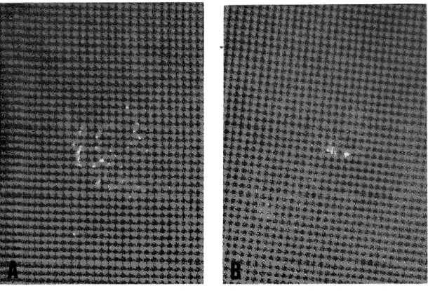
Double immunodlffusion. Ouchterlony plates were prepared by flooding 5 by
1 1 cm slides with 9.5 ml of 0.90/0 agar. The central well was filled with the Fasciola antigen and the other wells with serum samples of rats from each experirnent. The position of precipitin bands was recorded by drawings and photograph.s.
Worm antigen. Adult worms of the common liver fluke, recovered from the bile ducts ofstock‑infected rats and stored at ‑20 C, were added to 10 volumes (WjV) of phosphate buffer, pH 7.4. The worms were homogenized fbr 5 minutes at 4 C and centrifuged for 15 minutes at 3,000 rpm. The supernatant was stored at 20 C as the antigen for the agar double diffusion technique.
RESULTS
Worm recovery from immunized and non‑immunized rats 2 weeks ajler challenge. At 2 weeks after challenge all experimental rats had worms from immunizing infection in their bile ducts. In this series of the experiment, the immunized rats received challenge infection with the metacercariae 6, 7, 8, 9, I O and 1 1 weeks after immunizing (Ist) infection. As recognized in Table 1, the experimental rats harbored fewer challenge worms than the natural‑immunity control, apart from the rats received challenge 1 1 weeks after immunization. The challenge (natural‑immunity) controls harbored 3.6 worms in mean number, whereas the animals challenged during 6 to 9 weeks after immunizing infection harbored only 0.7 to I .5 worms. The percentage immunity to the Japanese species of Fasciola in rats was calculated by the following formula, C ‑ I x I OO, where C indicates a mean worm recovery of challenge control and I shows a mean recovery of worms from challenge inf'ection in immunized rats . The challenge infection from 6 to 9 weeks after immunization, revealed a higher percenta e immunity ranging from 58.3 to 80.6 than the natural‑immunity control.
These differences m worm bu den are highly srgmficant (Student's test, P < '' ,, 0.001), but the challenge at 10 weeks later had trended to indicate a gradual decrease in the degree of protective immunity to the liver fluke in rats. Even in the rats which received both immunizing and challenging infections, there was no difficulty in the
Table I Summary of the percentage immunity to the Japanese Fasciola in the rats im‑
munized with 4 metacercariae each per os before challenge infection.
Times (wks) of No. worms recovered from challenge ercentage No. rats No. Iarvae
challenge after used challenged rmmunity*
immunization ( o//o )
Total Mean s.d.6 8 9 lO 11
5 5 4 3 3 3
25 25 20 15 15 15
4 4 6 2 8 13
0.8 0.40
0.8 0.75
l.5 l. 12 O. 7 0.47
2.7 0.47
4・3 I .25
77.8 77.8 58.3 80.6 25.0 0.0
Challenge control 5 25 18 3.6 0.49
* Percentage immunity was calculated by the formula, C‑1/C x 100 (Miller and Smithers,
l 980)
differentiation of the worms between the two infections, because of autopsy at the early phase (2 weeks) of challenge infection. Thus, the results mentioned above have demonstrated a significant degree of worm reduction from challenge, by the immuni‑
zation with the Japanese Fasciola metac rcariae per os.
Immunization by intraperitoneally implanting worms with and z )ithout deffusion chambers.
The induction of protective immunity which was observed by the immunizing infection with the metacercariae per os, was examined using millipore diffusion chambers in relation to the liver migration and/or the ages of worms. The rat immunized by implanting worms was challenged 3 weeks after immunization, and autopsied 4 weeks after challenge. In the free worm implantation (without chambers), the differentiation of worms between the immunizing and challenging infections was performed according to the size of the worms recovered. In Table 2, the protective immunity to the Japanese species of Fasciola in rats is shown by performing im‑
plantation of the worms with or without chambers. In the experimental rats, the immunized groups indicated a significant degree of protective immunity by the immunizing infection (implantation) of the immature (25 day‑old) worms in both with and without chambers. The percentage immunity obtained by the before mentioned formula, was 50.00/0 in the immunization with chambers and 88.20/0 in that without chambers, respectively. In the worm burden, again, the immunized rats harbored only I .7 and 0.4 worms in the corresponding immunizations, whereas the rats in sham operation and challenge control showed the worm recovery of 4.2 and 3.4, respectively. The differences found in worm burdens between the im‑
munized, and the non‑immunized (sham) and challenge control are highly significant (P<0.001), but there is no significant difference between sham operation and chal‑
lenge control. With implantation of the worms contained in diffusion chambers, the current study demonstrated a significant degree of protective immunity to the Japanese Fasciola species when compared to the natural‑immunity control. The degree, however, was significantly higher (P<0.001) in the immunization with free worm implantation than that with enchambered worm ; the implanted worms could
Table 2 Results of the induction of protective immunity by intraperitoneally implanting the immature worms, 25 day‑old, with and without diffusion chambers in rats.
Group
Times (wks) of challenge
aft.
implantation
Times (wks) of autopsy
aft.
challenge No.
rats used
No. Iarvae challenged
No. worms from challenge Total Mean s.d.
Per‑
centage immunity
(o/o)
Immunized * With chamber Without chamber
(free worms) Non‑immunized Sham operation
(chamber only)
3 3
3
4 4
4
7 5
5
70 50
50
12 2
21
1 .7 0.45 0.4 0.49
4.2 0.75
50.0 88.2
0.0
Challenge control 4 7 70 24 3.4 0.49
* Viable 3 worms each were implanted intraperitoneally in rats.
survive 3 to 4 weeks or more in the diffusion chambers placed intraperitoneally in
rats.
The results of serological analysis on serum samples from the rats that received worm implantation with and without chambers are shown in Figure I , using cellulose acetate electrophoresis. In the case, the percentage value of each fraction in serum proteins was also determined in the samples from rats received sham operation, taking the blood 2 weeks after operation. The values obtained were 53.5 3.450/0 in blbumin, 13.9 1.900/0 in alpha‑globulin, 6.8 0.250/0 in beta‑globulin and 19.5 0.650/0 in gamma‑globulin, respectively, and the average of albumin per globulin (A/G) ratio was I .2 0.15. In comparison with the percentage values in sham operation, the values in both groups showed a remarkably higher or lower value in each fraction, demonstrating a great fluctuation. The weekly changes in the values, however, were similar in the serum samples from the two experimental groups. It was noted that the percentage value of albumin indicated an increase
(%) o 40 20 o 20 o 20
E
, ,,,,, ,b,,, D
c
S t
e
e
,
+
B o
60
40 't...,b ,,
lllt ""I"̲"̲'̲1't"̲' f
10 A
o
Figure l
5 3
We e ks
Weekly changes in each fraction of serum proteins in the samples from rats received immunizing implantation of the immature worms with ( ) and without (‑‑‑‑) chambers.
A : albumin, B : alpha‑globulin, C : betaglobulin, D : gamma‑globulin, E : AjG ratio, S : mean value of sham operation with empty chamber 2 weeks later.
after 3 weeks when the rats were challenged, showing the peak at 5 weeks after immunizing infection. In agar double diffusion, almost all of the serum samples showed only one precipitin band in the rats received worm implantation with chambers, while 2 or 3 bands were observed in the samples from the animals implan‑
ted worms without chambers (Table 3). It was noted that the number of bands in the latter animals has increased gradually after challenge infection.
Table 3 Number of antigen‑antibody bands appeared in agar double diffusion plate in serum samples from rats immunized by intraperitoneally implanting the im‑
mature worms with and without diffusion chambers.
No.
scrum Group samples
in each
test
Weeks after immunizing implantation
1 2 4 5 6 7
Immunized *
With 1 1 1 1 O 1 1 1 1 1 1 1, 1,0,0 l, l, l, l 5
chamber
Without 1 1 1 O O 1 1 1 1 O 2 2 1 1 O 3 3, 2,2, 5
chambers
l 1, 1, l, l, 1 2, l, 1, l, 1 l 3 3, 2, 1, 1 2, 2, 2, l, 1
* The rats were challenged with Fasciola metacercariae 3 weeks after immunizing implantation of 25 day‑old worms with and without chambers.
** Number of precipitin bands appeared.
Effects of immunosuppressants on the induction of protective immunity. To determine if the protective immunity observed would be influenced by immunosuppressive drugs, the experimental rats which received immunizing infection with the meta‑
cercariae per os, were treated with dexamethasone and prednisolone throughout the experiment. The drugs were in.jected intramuscularly into the animals 5 day‑
intervals. The rats received the immunizing infection were challenged 6 weeks later with the metacercariae, and then necropsied 2 weeks after the challenge infection.
At autopsy all the experimental rats contained worms from immunizing infection in the bile ducts. Table 4 shows the results of this experiment. In the immunizing infection, the non‑treated rats (Group II) with immunosuppressive drugs harbored only 0.7 worms, whereas the animals treated (Group I) had 3.3 worms from challenge infection. The percentage immunity of rats in Group 11 was significantly high (P<0.001) when compared to both the Group I and natural‑immunity control. No signiflcant difference, however, was recognized between Group I and the control.
These results demonstrated that the immunosuppressive drugs, dexamethasone and prednisolone, could greatly suppress the induction of protective immunity against the Japanese species of Fasciola in rats. In the immunizing infection (Groups I and II), antigen‑antibody systems in the animals were examined by performing Oucht‑
erlony technique. The formation of precipitin bands in agar double diffusion plates is shown in Tab]e 5. Each test was carried out at weekly intervals from I to 8 weeks after immunizing infection with the metacercariae. All the serum samples in Group I showed no precipitin band throughout the experiment, while all serum
samples taken weekly intervals, apart from those I week later, in Group 11 produced 1 to 4 bands. In non‑treated animals with immunosuppressive drugs, the bands
were first found at 2 weeks and reached to the peak in numbers at 6 weeks when the challenge was performed, thereafter they had a tendency to decrease gradually in numbers. No stimulation was recognized in the activation of antigen‑antibody systems in the rats by challenging infection during the period observed.
The results of serological analysis of the samples from rats treated with (Group I) and without (Group II) immunosuppressive drugs are depicted in Figure 2. In the examination of normal (non‑infected) rats, the percentages of each fraction were 53.1 3.790/0 in albumin, 18.7 2.000/0 in alpha‑globulin, 5.3 1.620/0 in beta‑
globulin and 18.9 5.000/0 in gamma‑globulin, respectively, and the average of al‑
bumin per globulin (A/G) ratio was I . I 0.16. These average normal values are also shown in the same Figure. The gamma‑globulin fraction in sera from treated anima]s (Group I) showed a consistently low percentage, while it represented a high value in the animals without treatment as copared with the average normal value.
In other globulin fractions, there was a remarkable difference of alpha‑globulin be‑
tween both groups in the early time of immunizing infection during I to 4 weeks ;
Table 4 Worm recovery from rats treated with (Group I) and without (Group II) immunosuppressive drugs ; all the rats received immunizing infection with 4 metacercariae each per os, and then challenged with 10 or 20 Iarvae each 6 weeks later.
No. worms from challenge Percentage
Group No rats used No. Iarvae immunity challenged Total Mean s.d. (o/o)
I*
II**
7 12
80
1 40
23 8
3.3 0.70
O. 7 0.62
0.0 79.3
Challenge control 1 7 220 55 3.2 0.8 1
* Dexamethasone ( I mg/rat) and prednisolone (5 mg/rat) were simultaneously injected into alternate thigh muscles of rats beginning one day before immunizing infection with the metacercariae.
** Non‑treated with any immunosuppressive drugs; the rats only received both immunizing and challenging infections with the metacercariae.
Table 5 Number of antigen‑antibody bands appeared in agar double diffusion plate in serum samples from rats in each group treated with (Group I) and without (Group II) immunosuppressive drugs.
No. serum Group sampler in
each test l
Weeks after immunizing infection
2 3 4 5 6 7 8
I II
3 5
O, O, O* O, O, O O, O, O O, O, O O, O, O O, O, O O, O, O O, O, O 3, 3, 3 3, 3, 2 3, 3, 3 3, 3, 3 4, 3, 3 3, 2, 2
O, O 2, 2 2, 2 2, 2 3, 3 3, 2 2, 1
O, O, O 2, l, 1 l, l
* Number of precipitin bands appeared in each sample.
1
o
(%)
20 o 20 o 20 o 60
E
c
B
N
,
f
,
,
'‑1‑"I‑ " '1 +
40
A 20
O
Figure 2 Weekly changes in each fraction of serum proteins in the
1 2 5 6 7 8
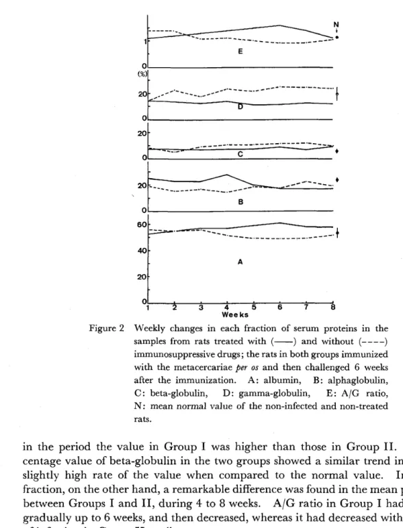
3 Wee ks 4samples from rats treated with ( ) and without (‑‑‑‑) immunosuppressive drugs ; the rats in both groups immunized with the metacercariae per os and then challenged 6 weeks after the immunization. A: albumin. B : alphaglobulin, C : beta‑globulin, D : gamma‑globulin, E : AjG ratio, N: mean normal value of the non‑infected and non‑treated
rats .
in the period the value in Group I was higher than those in Group II. The per‑
centage value of beta‑globulin in the two groups showed a similar trend indicating a slightly high rate of the value when compared to the normal value. In albumin fraction, on the other hand, a remarkable difference was found in the mean percentage between Groups I and II, during 4 to 8 weeks. A/G ratio in Group I had increased gradually up to 6 weeks, and then decreased, whereas it had decreased with the elapse of infection in Group 11 until necropsy.
DISCUSSION
Kondo and Hashiguchi (1979) have observed, for the first time, on the influence of primary infection with Japanese species of Fasciola metacercariae to superinfection in rats. They demonstrated that the recovery rates of Japanese Fasciola from chal‑
lenge infection were very low when compared to the natural‑immunity control.
The similar tendency, again, was also ascertained in the present study in rats, by