はじめに 脳はそのホメオスタシスを維持するために関門を持ち,神 経機能に必要なイオンやアミノ酸濃度を厳密にコントロール している.その中で中心的な役割を担っているのが脳毛細血 管に存在する血液-脳関門(blood-brain barrier; BBB)であり, この BBB によって血管内の血液と脳実質との間の高分子量 物質の交通は制限されている.その構造学的特徴としては, 脳毛細血管内皮細胞には窓(fenestration)がなく,小胞輸送 (vesicular transport)が高度に減少しているため,これによる 細胞質内の通過が制限される.さらに,内皮細胞間には密着 結合(tight junction)が存在し,細胞間の通過も制限されてい る1)~3).内皮細胞の基底側には内部に周皮細胞を含む基底膜 が存在し,さらにこの周囲をアストロサイトの終足が取り巻 くことでバリアの構造を特徴付けている(Fig. 1A).また,内 皮細胞の表面(管腔側)には陰性荷電が存在し,電気的な障 壁も形成されている.内皮細胞間の密着結合部位は claudin-5, occludin,junction adhesion molecule(JAM)などの物質を介 して細胞内蛋白である zonula occludens-1(ZO-1)同士で結合 しており,接着結合(adherens junction)間は膜貫通蛋白の cadherinで接着していて(Fig. 1B),高分子量物質の細胞間通 過を制限する役割を担っている. 一方で,脳内の細胞にとって必須な物質は,脳血管の内皮 細胞に存在する輸送体によって選択的に血管内から脳内へ取 り込まれている(BBB のキャリア機能).その代表的な選択 的輸送体としては,ブドウ糖などの糖を脳内に運ぶ glucose transporters,乳酸を運ぶ monocarboxylic acid transporters,amino
acid transporters,peptides transporters,Ion transporters,ATP-binding cassette(ABC)transporters,organic anion/cation transporters などが知られている4)~6).
次に,BBB とともによく知られているのが,脈絡叢に存 在する血液-脳脊髄液関門(blood-cerebrospinal fluid barrier; BCSFB)である(Fig. 2).脈絡叢内の毛細血管は有窓性であ り,血管内物質は毛細血管壁を通過して脈絡叢内の間質に移 行しうるが,その血管周囲にある上皮細胞には脳室側に密着 結合が存在している.この上皮細胞によって脈絡叢内と脳室 内との間における高分子量物質の移行は制限されており,こ の機能が BCSFB と捉えられている7)8).尚,脳室内の物質が エンドサイトーシスによって上皮細胞に取り込まれ,密着結 合を迂回して,細胞間腔あるいは基底側へと輸送される経路 は残る.さらに,脈絡叢上皮にも物質の移動を制限するだけ でなく,選択的に取り込むための輸送体が備わっており,特 定の物質の移行を可能にしている. 脳室内の脳脊髄液と脳実質との間には脳室上衣細胞が存在 するが,この上衣細胞には陰性電荷の存在,種々の接着因子 の発現,酵素活性の存在などが指摘されており,上衣細胞が 脳脊髄液-脳関門(cerebrospinal fluid-brain barrier; CSFBB) を形成することによって,物質の移動にある程度の制限をか けていると推測される(Fig. 2)7)8).しかしながら,上衣細胞 間は主として密着性の低いギャップ結合(gap junction)と接 着結合で結合されているとされており,高分子量物質は脳室 壁を介して移動しうる7)8).ラット上衣細胞間における部分的 な密着結合に関する報告9)もあるが,上衣細胞層でのバリア 機能は不完全なものと考えられる.
上野 正樹
*
要旨: 血管内物質が脳機能へ与える影響を考えるに際し,血液脳関門の存在が問題となる.また,脳内の組織 間液や脳脊髄液の脳外への排出に関しては,最近,二つの代表的な排出路の存在が確立されつつある.一つは脳組 織間液が毛細血管基底膜から細動脈の中膜平滑筋層の中を通る経路で,他の一つは脳脊髄液が細動脈周囲腔から アクアポリン 4 依存性アストロサイト細胞質内を経て細静脈周囲腔から脳脊髄液にはいる経路であり,いずれも 最終的に頸部リンパ節や静脈内に排出されるとされ,脳全体のバリア機能にも寄与していると考えられる.今回, 血液脳関門を含む脳のバリア機能の障害と血管性認知症の病態との関係に焦点を当て,最近の知見を紹介する. (臨床神経 2017;57:95-109) Key words: 血液脳関門,血液脳脊髄液関門,血管周囲排出路,グリアリンパ排出路,血管性認知症 *Corresponding author: 香川大学医学部・病理病態生体防御医学講座・炎症病理学〔〒 761-0793 香川県木田郡三木町池戸 1750-1〕 1)香川大学医学部・病理病態生体防御医学講座・炎症病理学(Received January 19, 2017; Accepted February 3, 2017; Published online in J-STAGE on February 22, 2017) doi: 10.5692/clinicalneurol.cn-001004
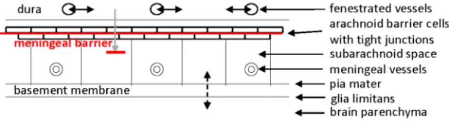
臨床神経学 57 巻 3 号(2017:3) 57:96 古くから,血管内に投与された色素がくも膜下腔に検出で きないことが指摘されていたが,硬膜とくも膜下腔の CSF の 間にもバリア機能が想定されており,髄膜関門(the meningeal barrier)として7)10),あるいは,血液とくも膜下腔 CSF の間 のバリアとして11)紹介されている(Fig. 3).硬膜内の血管は 有窓性毛細血管であるが,くも膜外側に存在する 2~3 層の扁 平な細胞(くも膜細胞)間は密着結合とギャップ結合で閉め られており7)12),硬膜とくも膜下腔との間の物質の移動は制 限されて関門となっている.また,脳実質とくも膜下腔との 間にある軟膜はギャップと窓を有する疎性結合組織であり, そこでは液体は通過できるとされているが,赤血球などの細 胞の通過は制限されている13). 脳室周囲には脈絡叢の他に,正中隆起(median eminence; ME),下垂体後葉(neurohypophysis; NH),脳弓下器官(subfornical Fig. 1 Blood-brain barrier and the tight junction.
(A) Schematic outline of a capillary of the blood-brain barrier in a transverse section showing not only endothelial cells with a tight junction, basement membrane, and few vesicles and no fenestration in the cytoplasm, but also a pericyte and the end feet of astrocytes. The localization of transporters is conceptually shown in the endothelial cell. The tightness of the blood-brain barrier (heavy solid red line: B-B, B) is structurally characterized by these structures. (B) Schematic representation of the protein interaction associated with tight and adherens junctions at the blood-brain barrier and endothelial glycocalyx at the surface. Claudin-5 (a major constituent of tight junctions in brain endothelial cells), occludin, and junction adhesion molecules (JAMs) are the transmembrane proteins in the tight junction, while zonula occludens-1 (ZO-1) are the cytoplasmic proteins. Cadherin is shown in the adherens junction, while catenins are shown in the cytoplasm. Transporters are shown in the cytoplasmic membrane of the endothelial cell.
Fig. 2 Blood-cerebrospinal fluid barrier and cerebrospinal fluid-brain barrier.
Schematic representation of the blood-cerebrospinal fluid barrier (solid red line: B-CSF, B) and cerebrospinal fluid-brain barrier (dashed red line: CSF-B, B). Fenestrated capillaries are located in the stroma of the choroid plexus, which is separated from the ventricle with epithelial cells bound by tight junctions. Ependymal cells bound by complex junctions (#: see text) are located between the ventricle and the brain parenchyma. Transporters are localized in the cytoplasm of epithelial cells of the choroid plexus and in the cytoplasm of the ependymal cells.
organ; SFO),終板器官(organum vasculosum of the lamina terminalis; OVLT),最後野(area postrema; AP),交連下器官 (subcommisural organ; SCO),松果体(pineal body; PI)という 脳室周囲器官(circumventricular organs)が存在している7)14). SCOを除くこれらの脳室周囲器官に存在する毛細血管は有 窓性であり,循環血液と器官内組織間液との間で交通がある. しかしながら,これらの器官の脳室側では密着結合で結合さ れた特殊脳室上衣細胞が脳室壁を裏打ちし,脳室内 CSF と接 している(Fig. 4). 以上のように,脳内には BBB と BCSFB を中心に複数のバ リアが存在し,必要な物質を取り込みながらも毒性を持つ物 質の侵入は防いで脳内環境を一定に保っていると考えられ る.それ故,これらのバリアが障害されれば,様々な脳機能 異常が生じると考えるのは自然である.特に,最も重要な鍵を 握っている BBB においては,脳血管が障害されている血管性 認知症とBBB機能障害との関係がすでに指摘されている15)16). 血液脳関門を迂回する経路 脈絡叢や脳室周囲器官に存在する不完全なバリア機能しか 持たない血管がその組織内や周囲の領域にどのような影響を 及ぼすかについては,よく知られていない.以前は,脳室周 囲器官及びその境界部では血管周囲の上衣細胞間に密着結合 の存在することが示され,器官内から周辺組織への拡散は防 がれているであろうとの報告もあった17)18).しかしながら, 内因性タンパク質の脳内移行を検討した実験19)や,horseradish peroxidase(HRP)をマウスの尾静脈から注入しその HRP を テトラメチルベンチジンで反応させた実験20)~22)では,血管 内物質が ME からその周辺の視床下部へ移動していると考え られる所見19)や,SFO の血管から組織間腔を伝ってその背 側部の脳梁19)20)や尾側部の海馬内側部20)21)へ移動していると 考えられる所見を認めた.また,静注された HRP は脈絡叢付 着部付近の白質を含む脳室周囲領域21)や扁桃体内側部22)に も流入していた(Fig. 5).興味深いことに,脈絡叢付着部付 Fig. 3 The meningeal barrier.
Arachnoid barrier cells with tight junctions, separating the subarachnoid space from the dura mater with fenestrated capillaries, constitute the meningeal barrier (solid red line). Glia limitans and pia mater separate the brain parenchyma from the subarachnoid space.
Fig. 4 Circumventricular organs.
A section through the brain in the median sagittal plane is shown. Located around the ventricles are the following circumventricular organs (enclosed by dashed or solid red lines): subfornical organ (SFO), median eminence (ME), neurohypophysis (NH), organum vasculosum of the lamina terminalis (OVLT), pineal body (PI), subcommissural organ (SCO), and area postrema (AP). Two portions of the choroid plexus (CP) are also drawn in the ventricles. Dashed or solid red lines indicate slightly weaker or tighter barrier functions, respectively. Broadwell et al.19) reported that
endog-enous serum proteins spread extracellularly from some of the circumventricular organs, such as the median eminence, area postrema, and subfornical organ, into the adjacent brain parenchyma. It is described that the vessels of the sub-commissural organ have no fenestrated walls and that the fenestrated aspect of capillaries with a lack of the BBB in the pineal gland is not accepted by all researchers14).
臨床神経学 57 巻 3 号(2017:3) 57:98 近の脳室周囲領域には HRP の流入は明らかであるが,脈絡叢 付着部から離れた脳室周囲領域には HRP は全く流入してい なかった(Fig. 5D, E).この結果からは,同じ脳室周囲領域と 言えども,脈絡叢付着部付近の脳室周囲領域が特殊であるこ とが推察される.尚,血管内物質が BBB を迂回して脳室周囲 の特定の領域に運ばれる役割はいまだ明らかでない. 組織間液および脳脊髄液の排出路
アルツハイマー病(Alzheimerʼs disease; AD)脳で沈着する アミロイドベータ蛋白(Aβ)などの脳組織間液内物質が脳内 から排除されるのか,あるいは沈着するのかを規程する因子 として,脳内での合成および脳外からの流入と脳からのクリ アランスが考えられる.脳からのクリアランスの具体的機序 として,(A)血管壁に存在する輸送体を介した直接的な血管 内への排出,(B)種々の酵素による分解,(C)グリア細胞な どの細胞質内への取り込み,そして,(D)血管壁を伝わる (頸部リンパ節への)排出が想定されている(Fig. 6).この合 成・流入と分解・排出のバランスが崩れると,その物質の脳 内沈着に繋がっていくと推測することは難くなく,AD では この行程のどこかに異常をきたして Aβ 蛋白が沈着してしま うのではないかと考えられる23). (A)BBB や BCSFB を介する Aβ の脳外への排出機構とし て,いくつか想定されているが,報告論文によって必ずしも 一致していない.最近,総説を含む様々な論文でその排出機 構は報告されており23)~32),その一部を紹介する.
(1)LRP-1(low-density lipoprotein receptor-related protein-1): Aβ は BBB に発現する LRP-1 に直接結合して経細胞的に脳 内から血液中に排出されることが提唱されているが24)33), その一方で,BBB を介した Aβ の排出輸送に対する寄与は 小さいとの報告もある34). (2)ABCB1(P-glycoprotein; P-gp):脳毛細血管における ABCB1 発現量と脳内および脳血管に蓄積した Aβ 量に負の相関関 係があることが報告されている35)が,これまでに ABCB1 が直接 Aβ 輸送を担っている事を示す報告は明確ではない とも考えられている.
(3)ABCG2(breast cancer resistance protein; BCRP):ABCG2
の発現量が AD 脳の毛細血管にて健常人より高いこと36)
Fig. 5 Leakage of intravascular HRP in the brain.
The staining reaction for horseradish peroxidase (HRP) with tetramethyl benzidine incubation is shown in the sections obtained from perfusion-fixed mice at 90 min after HRP was intravenously injected (A–E). The sections cut through the subfornical organ (SFO) (A) and two portions of the hippocampus (B, C) are shown. Strong staining for HRP appears in the SFO (long arrowhead in (A)), the medial portion of the hippocampus (HIP) (long arrowhead in (B)) and the amygdala (AMY) (short arrowhead in (B)), the dorsal portion of the thalamus (THL) (small arrow in (B)), the hypothalamus (HYP) (large arrows in (B, C)) around the median eminence, and the periventricular area (PV) (large arrowhead in (C)). Very strong staining for HRP appears in the choroid plexus (CP) (E) and the PV (long arrow in (D)) including the hippocampal fimbria (HF) (D, E) and the optic tract (OT) (small arrow in (D) and large arrowhead in (E)). Small arrowheads in (D, E) indicate vessels in the PV showing increased vascular permeability. IC: internal capsule. Modified from the paper reported by Ueno et al.21)
や,ABCG2 が Aβ 輸送に関与している可能性についての報 告がある31).
(4)ABCC1(multidrug resistance-associated protein; MRP1):
ABCC1は Aβ 排出輸送に間接的に寄与している可能性が報 告されている29). (5)ABCA1:ABCA1 は Aβ の排出に間接的に関与していると の報告31)がある一方で,ABCA1 欠失が Aβ の排出を有意に 減少させなかったことから,ABCA1 の BBB を介した排出 輸送に与える影響は小さいとも報告されている37).
(6)IDE(insulin degrading enzyme):Aβ 分解酵素の一つであ る IDE が Aβ のクリアランスに関与していることが指摘さ れている27)38). (7)ABCG4:ABCG4 は ABCG2 と協調してコレステロール 代謝と関連した BBB を介する Aβ の脳外への排出に関与し ている可能性が報告されている39). (8)LRP2:LRP2 が脳血管内皮細胞や脈絡叢上皮細胞での Aβ とアポ J の複合体の排出に関わっていることが指摘され ている26)40).
(9)LDLR(low-density lipoprotein receptor):LDL 受容体を 過剰発現させると,組織間液中の Aβ のクリアランスを増 加させ,Aβ 蛋白の沈着を阻害したとする報告41)42)や,LDL 受容体を欠失させると脳内 Aβ 蛋白の沈着が増加したとす る報告がある43). (10)CD36:ミクログリアや脳血管内皮細胞に発現している CD36が Aβ 線維の受容体として働き活性酸素の産生に関与 していることが報告されている44). (11)FPRL1(formylpeptide receptor-like-1):Aβ が FPRL1 と 結合しミクログリアなどの細胞内に取り込まれることが報 告されている45). (12)VLDL 受容体:Aβ とアポ E4 との複合体は VLDL 受容 体を介してゆっくり脳外へ排出されると報告されてい る46).一方,Aβ とアポ E2 やアポ E3 との複合体はより早 く排出される LRP1 と VLDL 受容体を介してアポ E4 より 早く排出されると報告されている46). 上記のような輸送体や受容体が Aβ のクリアランスに関 わっている可能性が指摘されている.しかしながら,BBB を 介する経細胞的かつ直接的な Aβ の輸送が実際に行われてい るのか,あるいはどのような機構で行われているのかといっ たことは,現時点では完全には解明されていないと言える. これは,脳血管内皮細胞における BBB と脈絡叢上皮細胞にお ける BCSFB が区別されずに評価されていることも一因であ るのかも知れない.一方,Aβ の BBB を介する血管内から脳 内への流入輸送体に関しては,RAGE(receptor for advanced glycation end products)が良く知られている47)~49).Aβ の輸送 に関与している可能性が指摘されている輸送体の脈絡叢上皮 細胞,脳室上衣細胞,血管内皮細胞における局在に関するヒ ト剖検脳での免疫染色結果を Fig. 7 に示すが50)51),mRNA レ ベルでの発現検討や実際の輸送機能を有しているかを検討す る in vitro 実験結果が待たれる. (B)脳内における酵素による Aβ の分解もクリアランス機 Fig. 6 Flow and clearance of interstitial solutes and substances.
Interstitial solutes or substances including Aβ are synthesized in the brain or flow in the brain parenchyma from the blood through specific influx transporters. Then, the intracerebral Aβ is eliminated via: (A) efflux through specific efflux transporters into the blood, (B) degradation, such as by neprilysin (NEP) or insulin-degrading enzyme (IDE), (C) cellular uptake by astrocytes or microglia, and (D) discharge through a perivascular drainage pathway possibly to the cervical lymph nodes.
臨床神経学 57 巻 3 号(2017:3) 57:100 構の一つに挙げられる.その代表的な酵素としてネプリライ シンが知られており,その発現量と Aβ 沈着との逆相関関係 が報告されている52).さらに,Aβ を分解する酵素として IDE も良く知られるようになってきている27)53)が,この酵素はま さしくインスリンを分解する酵素でもあり,高インスリン状 態とアミロイド沈着の関係を解明する際に鍵となる物質の一 つであることは言うまでもない. (C)脳内 Aβ は細胞内に取り込まれることによっても排除 されることが知られている.周皮細胞54),平滑筋細胞55),神 経細胞56),アストロサイト57)58)やミクログリア59)において, LRP-1や LDLR などの受容体を介して Aβ が取り込まれる可 能性が指摘されている. (D)さらには,脳内 Aβ のクリアランス機構の一つとし て,血管周囲排出路を介する排除が挙げられるが,詳細は後 述する. ところで,現在,脳組織間液や脳脊髄液の脳外への排液路 として複数の通路が想定されている60)~64)(Fig. 8)が,血管周 囲排出路(perivascular drainage pathway)とグリアリンパ排 出路(glymphatic pathway)と称されている経路に大別される. 血管周囲排出路では,脳組織間液は血管壁に到達すると脳毛 細血管基底膜から動脈平滑筋層内を血流とは逆行する方向に 移動し,最終的に頸部リンパ節に流入すると考えられている. 脳脊髄液のグリアリンパ排出路は,動脈周囲からアクアポリ ン 4 依存性にアストロサイト細胞質内,組織間,アストロサ イト細胞質内,そして,血流と順行性に静脈周囲を伝わって 脳脊髄液に入り,頸部リンパ節や静脈洞といった脳外へと排 出されるものである65)~69).尚,この際,脳脊髄液に含まれる 高分子量物質の脳組織内への移動に関しては制限されている ことも指摘されている67)68).そして,脳脊髄液は,従来から 言われているくも膜絨毛から上矢状洞内へ移行する経路,脳 内くも膜下腔から嗅神経周囲のくも膜下腔を伝わり嗅粘膜か ら頸部リンパ節内に移行する経路70),最近確立しつつあるく も膜下腔から硬膜内リンパ管に入り頸部リンパ節に流入する 経路65)66)を介して脳外へ排出されると想定されている(Fig. 8). このように,脳組織間液および脳脊髄液は複数の経路に よってリンパ管内や血管内(脳外)に戻されているのではな いかとの提唱がなされるようになった64).そして,これらの 排出経路が障害されれば,種々の脳疾患の病態増悪に繋がる ことは想像に難くない.実際,このクリアランス機構が破綻 して血管壁にタンパク質が蓄積するような病態が PEFA (protein-elimination failure arteriopathies)と称され,様々な疾
患での関与が推定されている62).AD 脳における血管周囲リ ンパ排液路に関しての知見として,初期の脳アミロイド血管 症における Aβ の沈着は,脳組織間液の排液経路である血管 中膜平滑筋基底膜から始まることが指摘されている61).アミ ロイド血管症などではこれらの排出路が障害を受けているこ とは十分に考えられ,この排出路対策が認知機能改善に結び つくものと考えられる62).
Fig. 7 Aβ transport through the choroid plexus, ependymal cells, and cerebral vessels.
Recent studies of the autopsied human brain on efflux transporters of Aβ50)51) have reported the
immuno-histochemical expressions of LDLR, LRP1, LRP2, FPRL1, ABCA1, ABCC1, ABCG4, and IDE in the choroid plexus epithelium. Immunohistochemical expressions of CD36 as well as LDLR, LRP1, LRP2, FPRL1, ABCA1, ABCC1, ABCG4, and IDE are observed in the ventricular ependymal cells. Clear expressions of transporters are indicated by the underlined bold type. Clear expressions of ABCB1 and ABCG2 (indicated by bold underlined type) are observed in the vessel wall, while the expression of LDLR is also seen in the vessel wall. Weak expression of RAGE, a representative influx transporter of Aβ, is occasionally observed in the vessel wall. In addition, the study by Daood et al.50) reported the immunohistochemical expression of ABCB1
バリアの破綻から血管性認知症への道筋は? BBBが脳虚血などの侵襲によって障害されると,脳実質は 障害性のある抗体や補体などの血管内物質にさらされやすく なり,さらに栄養物質が神経細胞に輸送されず,脳機能障害 をもたらすと考えられる.それ故,脳血管の BBB 機能障害が 様々な脳病態に関与している可能性が推定され,多くの論文で 血管病としての血管性認知症の病態が論じられている71)~75). 老化に伴い増悪しうる血管性認知症は,脳の虚血あるいは 出血を原因とし,多発梗塞性認知症と小血管性認知症に大き く分類され,後者は,病理学的にはラクナ梗塞の多発や白質 病変によって特徴づけられる.具体的には,高血圧に基づく 細動脈硬化の結果,血管壊死とフィブロヒアリノーシスがも たらされる.そして,血管壊死からは小血管閉塞やラクナ梗 塞が引き起こされ,血管周囲の線維化を特徴とするフィブロ ヒアリノーシスからは慢性低灌流状態や白質病変といったビ ンスワンガー型脳梗塞が引き起こされ,このような血管病変 に基づき血管性認知症が形成されるという仮説も提唱されて いる71).近年,この血管壊死とフィブロヒアリノーシスの形 成に BBB 障害の関与が想定されだしている15)71).血管性認知 症における BBB 障害の役割を考えるに際し,どのような侵襲 が BBB を障害しているのか,どの領域の BBB が障害を受け やすいのか,どのような機序で障害されるのかを明らかにす ることは治療を考える上でも有意義であると考えられる.こ Fig. 8 Perivascular drainage and glymphatic pathways.
Recent reports60)–69) have suggested that clearance of interstitial fluid (ISF) and cerebrospinal fluid (CSF) to the outside of the brain involves both
perivascular drainage and glymphatic pathways. (A) Through the perivascular drainage pathway (indicated by thin solid red lines), ISF flows through the basement membrane of the capillary walls, tunica media of arteries, and the vessel walls of the internal carotid artery, and then drains into the cervical lymph nodes. This may be affected by cellular uptake or degradation. (B) Through the glymphatic pathway (indicated by heavy solid red lines), CSF flows through the para-arterial routes, enters the interstitial space by aquaporin 4-dependent transport through the astroglial cytoplasm, drains into the paravenous routes, and may be dispersed into the subarachnoid CSF or enter the bloodstream across the vasculature. CSF in the subarachnoid space drains directly into the blood via the arachnoid villi of the dural sinus (B-1), and drains into the cervical lymph nodes via the subarachnoid spaces around the olfactory nerves and nasal lymphatics (B-2), or via the meningeal lymphatic vessels (B-3). (#): It have been reported that the mixing of CSF with ISF is dependent upon the presence of astrocytic aquaporin 467)68), while it
has been pointed out that the exact relationship between the perivascular drainage and glymphatic pathways requires further investigation69).
臨床神経学 57 巻 3 号(2017:3) 57:102 こでは,血管性認知症を引き起こす侵襲として,(1)老化(記 憶学習障害を伴うものと伴わないもの),(2)急性虚血後再灌 流,(3)慢性低灌流,(4)高血圧,(5)高血糖,(6)水頭症 状態の 6 因子(侵襲)に対する実験モデル動物における BBB 機能,及びヒト脳での BBB 機能のデータを紹介する.尚,実 験動物モデルでは初期変化を明らかにするため,できるだけ 軽微な侵襲かつ若齢での状態を検討することを試みた. (1)老化,記憶学習障害時の BBB 機能
老化促進モデルマウス(senescence-accelerated mouse; SAM), 中でも,加齢に伴い記憶学習障害を発症する SAMP8 の海馬 と嗅球の BBB 機能は加齢に伴い障害されていた76)~81).尚, コントロールの記憶学習障害を発症しない SAMR1 において は,ヨードラベルされたアルブミンの脳内移行に定量的には 加齢変化を認めるものの,形態学的な裏付けは得られなかっ た76)80).また,マウス脳室周囲領域はもともと BBB 機能が脆 弱であり,加齢変化も受けやすい事が示唆された21)82).一方, ヒト脳の BBB の加齢変化に関してはいままで意見の相違が みられてきたが,最近の大規模メタアナリシスの結果からは, 老化に伴う BBB 機能の障害が認められており,これは特に 認知症や白質障害のある患者で明らかであった16).さらに, BBB障害はヒト脳の初期の加齢変化であり,海馬から始まる との報告もある83). (2)急性虚血後再灌流時の BBB 機能 一過性に片側頸動脈を結紮した砂ネズミの BBB 機能は,結 紮側の海馬の hippocampal fissure に沿った血管において障害 されていた84).一方,BBB 障害はヒト急性虚血脳でも確認さ れており,再灌流状態によってその程度は変化することが報 告されている85). (3)慢性低灌流時の BBB 機能 脳血流が 30~40%にまで低下している両側総頸動脈結紮 ウイスターラットでは,結紮後 7 日目に脳梁の粗鬆化を来す が86),その BBB 機能を検討したところ,結紮後 3 日目の脳 梁ですでに BBB 障害が生じていた87).一方,ヒト虚血脳あ るいは AD 脳において,白質障害部位に BBB 障害が生じるこ とが報告されている88)89). (4)高血圧状態の BBB 機能 実験的高血圧モデル動物である脳卒中易発症系自然発症高 血圧ラット(SHRSP)では,3 ヶ月齢にて海馬の hippocampal fissureに沿った血管の BBB 機能が障害されていた90).さら に,4~5 ヶ月齢では浮腫を伴う白質障害が生じていた91)92). この 3 ヶ月齢 SHRSP 海馬の BBB 障害血管で発現が変化して いる物質を検討したところ,オステオポンチン93),matrix metalloproteinase-13(MMP-13)94),CD3695)などの物質の発 現が亢進し,エストロゲン受容体の発現が低下していた93). 障害後の結果をみている可能性も十分考えられるが,これら の物質が BBB 障害を引き起こす可能性も推測される.一方, ヒトでは高血圧状態が BBB 障害を伴う白質障害に先行して いたことが報告されており96)97),高血圧によって白質障害が 生じうると考えられる. (5)高血糖状態の BBB 機能 ストレプトゾトシンを負荷して実験的に高血糖状態にした マウスやラットでは,デキストランやスクロースといった糖 関連物質の脳血管透過性は亢進していた98)99).この際,高血 糖に加えて MMP 活性の亢進が必要であるとの指摘もある99). また,レプチン受容体遺伝子変異によって高血糖状態にある db/dbマウスでは,大脳皮質における HRP の血管透過性は変 化しなかったが100),脳室周囲領域におけるアルブミンの血管 透過性が亢進していた101).この db/db マウスではコントロー ルと比べて糖化アルブミンが増加していること102),および血 管内皮細胞表面の荷電を有する glycocalyx の密度が減少して いること100)が知られている.従って,db/db マウスでは糖化 アルブミンの増加と血管内皮細胞での電気的障壁の障害に基 づいて,糖化物質の血管透過性が亢進しているものと推測さ れた.電気的障壁の影響を受けないと考えられる HRP の血管 透過性は亢進していないにも関わらず,糖化が進んでいると 想定されるアルブミンなどの糖関連物質の血管透過性は亢進 しやすいことは興味深い.高血糖状態では荷電を有する毒性 物質の脳内移行が増加していることが推測され,このことか らも糖尿病患者における HbA1c のコントロールは重要であ ると考えられる.一方,ヒトではガドリニウム MRI を用いた 2型糖尿病患者脳の検討において,基底核での血管透過性が 亢進していることが報告されている103).この際,MRI にて 白質高信号を認める患者に血管透過性の亢進が認められてい る.また,認知症に罹患していない患者における糖化ヘモグ ロビンレベルが深部白質の MRI 高信号と相関することも指 摘されている104).これらの報告からは,脳室周囲白質領域に MRI異常信号を持つ糖尿病患者における血糖コントロール のさらなる重要性が示唆される. (6)水頭症状態の BBB 機能
低酸素誘導因子(hypoxia-inducible factor-1α; hif-1α)の中枢 神経系細胞特異的ノックアウトマウスは水頭症を発症する が105),この水頭症を発症している 10 週齢のマウスにおける BBB機能障害は明らかではなかった106). 以上の実験動物を主体とした実験結果からは,BBB のバリ ア機能は主として急性虚血後再灌流状態と高血圧状態の海 馬,及び,慢性低灌流状態の脳梁において障害されていた. また,高血圧は血管障害を伴う白質障害をもたらしていると 推測された.さらに,高血糖状態は血管壁での glycocalyx の 障害やプロテアーゼ活性の亢進を伴った脳室周囲領域のバリ ア障害に寄与しており,老化は海馬および脳室周囲白質領域 の両者のバリア障害を助長しているものと考えられた.これ らの因子が合わさることにより,血管性認知症の認知機能の 増悪が引き起こされている可能性が推測される(Fig. 9).尚, 脳機能障害にリンパ球や抗原提示細胞の関与も推測されるが,
それら血球系細胞の血管内から脳内,あるいは脳内から脳外 への移動経路に関して,最近,優れた総説がでている69).現状 では,免疫担当細胞は細動脈壁の狭い平滑筋中を経る血管周 囲排出路ではなく,脳脊髄液のグリアリンパ排出路を通過し て頸部リンパ節に移動するものと推測されている63)~65)69)107). 種々の認知症の病態解明のためには,この免疫担当細胞の役 割の解明も待たれる. バリア障害の具体的機序について 高血圧症を発症している 3 ヶ月齢 SHRSP 海馬の脳血管内 皮細胞では,HRP の小胞輸送が増加することによる血管透過 性の亢進を認めたが,HRP が内皮細胞同士の密着結合間を通 過している像は観察されなかった90).急性虚血後再灌流およ び低灌流状態においても小胞輸送の増加が示唆され,加えて 内皮細胞障害も認められた84)87).また,加齢による影響にお いても内皮細胞障害を認めている80).一方,高血糖状態では HRPの血管透過性は増加していなかったが100),血管内皮細 胞表面の glycocalyx の障害を伴ったアルブミン(高血糖に よって血管内で糖化が進んでいるとされている102))の血管透 過性が亢進していた101).このように,侵襲の種類によって血 管透過性亢進の機序は異なっており,特に,高血圧(小胞輸 送の増加)と糖尿病(糖化と glycocalyx の障害)で血管透過 性亢進の機序が異なることは,脳血管障害性疾患の治療を考 える上で配慮すべき点と考えられる. バリア障害と種々の物質の発現との関係について 高血圧に基づく BBB 障害血管の生化学的分析結果からは, BBB障害部位ではオステオポンチン93),MMP-1394),CD3695) などの発現が亢進していることが明らかになった.また,こ れらの物質は高血糖状態や糖尿病の実験モデル動物において も同様に発現が亢進していることが報告されている108)~110). オステオポンチンは AD の脳脊髄液においても増加してお り,その濃度と認知機能とは関連があることが指摘されてい る111).また,オステオポンチンは,動脈硬化性病変をはじめ とした様々な病的状態で発現が亢進していることが報告され ている112).一方で,マウスの脳梗塞モデルにおいてトロンビ ンで切断されたオステオポンチンの断片を脳室内あるいは鼻 粘膜内投与すると,脳梗塞巣の大きさの縮小効果や脳虚血障 害に対する保護効果がみられたとの報告がある113)114).また, マウスの脊髄では,オステオポンチンと白質障害との関連性 を指摘する論文もみられる115).これらの報告からは,オステ オポンチンの脳機能に対する作用は不確定であるが,トロン ビンで切断されたオステオポンチン断片なのか,あるいは MMPで切断されたオステオポンチン断片なのか116)で作用が 異なってくる可能性も推測され,病変部およびその周辺の環 境が重要であるのかもしれない. MMPの虚血性脳障害における役割はよく検討されてお り117),MMP-2 は BBB 障害を伴う血管性認知症やラクナー梗 塞患者脳で発現が亢進していることが知られている118).ま
Fig. 9 A hypothesis on relationships among insults, barrier damage, and dementia.
On the basis of the experimental results, the effects of acute ischemia followed by reperfusion, hypertension, senescence, hypoperfusion, and hyperglycemia, on barrier dysfunction in the hippocampus, white matter, and the cerebrum including periventricular areas are shown by solid arrows. Acute ischemia followed by reperfusion clearly induces barrier dysfunction in the hippocampus (A)84)
, while hypertension induce not only clear barrier dysfunction in the hippocampus (B)90)
but also that in the white matter (C)91)
. Senescence induces mild barrier dysfunction in the hippocampus (D)76)78)80)
and periventricular white matter (E)82)
. Hypoperfusion induces barrier dysfunction in the white matter (F)87)
. Hyperglycemia induces barrier dysfunction in the cerebrum including periventricular areas (G)98)99)101)
. It is likely that barrier dysfunction in the hippocampus related to increased expressions of osteopontin, MMP, CD36, and other molecules due to hypertensive insult (H)93)–95)
induces brain dysfunction, followed by the occurrence or aggravation of dementia, as indicated by solid arrows. In addition, barrier dysfunction in the cerebrum, especially the periventricular areas, due to hyperglycemic insult may also be related to the increased expression of these molecules (I)108)–110)
, shown by dashed arrows. The relationship between barrier dysfunction in the white matter and the increased expression of the molecules remains to be clarified, as shown by the dashed arrow with a question mark.
臨床神経学 57 巻 3 号(2017:3) 57:104 た,MMP-2 は慢性低灌流モデル動物における白質障害部位の BBB障害にも関連している119).さらに,MMP-2 のみならず MMP-3の発現も血管性認知症患者の白質障害部位で観察さ れている120).一方,MMP-9 は claudin-5 や occludin の発現抑 制を伴った BBB 障害をもたらし,虚血性脳障害に関係してい ることが報告されている117)121).我々の検討では,脳血管障 害の初期段階と考えられる血管透過性亢進状態の 3 ヶ月齢 SHRSP海馬血管においては,MMP-2,MMP-3,MMP-9 では なく MMP-13 の発現が亢進しており94),claudin-5 や occludin の 発現に影響を与えていなかった.MMP-13 に関しては,MMP-13の発現亢進そのものが血管障害を助長している可能性も 指摘されており122),高血圧に基づく MMP-13 発現亢進状態 が初期段階の血管障害になんらかの影響を与えているものと 考えられる. 高血圧,糖尿病,Aβ 刺激は酸化ストレスを誘導することが 知られており123)~125),この酸化ストレスには CD36 の関与が 指摘されている126).タンパク質の糖化により AGE が産生さ れて酸化ストレスを惹起させるだけでなく,タンパク質の β シート構造を誘導する127)ことで,Aβ の沈着促進に繋がる可 能性が推測されている.Aβ 刺激は上述の MMP-13 のみなら ず128),CD36 発現増加をももたらすことが報告されている129). そして,この CD36 の発現そのものが血管障害を助長すると の指摘もある130)131).また,CD36 は Aβ のクリアランスにも 関係している物質である44).このように,タンパク質の糖化 亢進を含む何らかの誘引によって脳内の Aβ が増加すると,そ のクリアランスのために CD36 などの物質の発現が亢進し, この CD36 の発現亢進そのものがさらなる血管障害を生じさ せ,血管性認知症の増悪に繋がるという機序が推測される. アルツハイマー病と血管性認知症が合併する際,この CD36 などの物質の発現亢進が両者を橋渡ししている可能性は検討 するに値するものと思われる.最近,ヒト剖検脳の脳室上衣 細胞および脳室下ビメンチン陽性グリア様細胞に CD36 の発 現があることが報告され51),脳室周囲領域のグリオーシスに おける CD36 の役割も推測されるが,これら CD36 陽性細胞 の存在意義の解明は今後の課題である.これまでの報告結果 に基づき,認知機能障害機序の一つの考え方(仮説)ではあ るが,バリア機能の障害と認知症との関連について Fig. 9 に まとめた.(海馬,白質,脳室周囲領域を含めた脳全体の BBB 機能に分け,関連物質についても記載している.) 総 括 これまでに述べた動物実験からは,血管性認知症を引き起 こす種々の侵襲に対するバリア機能の検討結果として,高血 圧,急性虚血後再灌流,老化が海馬血管のバリア障害を,慢 性低灌流と高血圧が白質血管のバリア障害を,糖尿病が脳室 周囲領域を中心とした脳全体のバリア障害をもたらしている と考えられた.これらは MRI や CT 画像所見から高齢者脳に 生じた病態を類推し,治療へ応用されうる結果である.また, 血管内での糖化を防止することや CD36 などの発現を低下さ せる作用のある薬剤投与が BBB 障害の軽減をもたらし血管 障害に基づく認知症の予防や治療薬になりうることも示唆し ている.さらに,ラクナ梗塞,脳室周囲白質粗鬆症,血管性 認知障害を引き起こす脳内微小血管病の脳において,トロン ビンの発現をコントロールすることにより抗血栓性を発揮す るトロンボモジュリンの発現が亢進していることも報告され ており132),抗凝固系のトロンビン・トロンボモジュリン・プ ロテイン C 系をコントロールすることも血管性認知症を含む 脳血管性疾患の治療に有効であるかもしれない.尚,今回の 検討では,種々の侵襲による実験動物脳の初期段階の変化を 中心にみており,また,ヒトの血管性認知症脳を用いた検討は 行っていないため,多方面からのさらなる検討が必要である. ※本論文に関連し,開示すべき COI 状態にある企業,組織,団体 はいずれも有りません. 文 献
1) Reese TS, Karnovsky MJ. Fine structural localization of a blood-brain barrier to exogenous peroxidase. J Cell Biol 1967;34:207-217.
2) Brightman MW, Reese TS. Junctions between intimately apposed cell membranes in the vertebrate brain. J Cell Biol 1969;40:648-677.
3) Brightman MW, Klatzo I, Olsson Y, et al. The blood-brain barrier to proteins under normal pathological conditions. J Neurol Sci 1970;10:215-239.
4) Zlokovic BV. The blood-brain barrier in health and chronic neurodegenerative disorders. Neuron 2008;57:178-201. 5) Redzic Z. Molecular biology of the brain and the
blood-cerebrospinal fluid barriers: similarities and differences. Fluids Barriers CNS 2011;8:3.
6) Abuznait AH, Kaddoumi A. Role of ABC transporters in the pathogenesis of Alzheimer’s disease. ACS Chemical Neurosci 2012;3:820-831.
7) Davson H, Welch K, Segal MB. Morphological aspects of the barriers. In: Davson H, Welch K, Segal MB, editors. Physiology and pathophysiology of the cerebrospinal fluid. Edinburgh, London, Melbourne, New York: Churchill Livingstone; 1987. p. 105-188. 8) Jimenez AJ, Dominguez-Pinos MD, Guerra MM, et al. Structure
and function of the ependymal barrier and diseases associated with ependymal disruption. Tissue Barriers 2014;2:e28426. 9) Brightman MW, Palay SL. The fine structure of ependyma in
the brain of the rat. J Cell Biol 1963;19:415-439.
10) Saunders NR, Habgood MD, Mollgard K, et al. The biological significance of brain barrier mechanisms: help or hindrance in drug delivery to the central nervous system? F1000Research 2016;5. pii: F1000 Faculty Rev-313.
11) Abbott NJ. Evidence for bulk flow of brain interstitial fluid: significance for physiology and pathology. Neurochem Int 2004;45:545-552.
12) Nabeshima S, Reese TS, Landis DMD, et al. Junctions in the meninges and marginal glia. J Comp Neurol 1975;164:127-170. 13) Hutchings M, Weller RO. Anatomical relationships of the pia
16) Farrall AJ, Wardlaw JM. Blood-brain barrier: Ageing and microvascular disease—systematic review and meta-analysis. Neurobiol Aging 2009;30:337-352.
17) Krisch B, Leonhardt H, Buchheim W. The functional and structural border of the neurohemal region of the median eminence. Cell Tiss Res 1978;192:327-339.
18) Krisch B, Leonhardt H. The functional and structural border between the CSF- and blood-milieu in the circumventricular organs (Organum vasculosum laminae terminalis, Subfornical organ, Area postrema) of the rat. Cell Tiss Res 1978;195:485-497.
19) Broadwell RD, Sofroniew MV. Serum proteins bypass the blood-brain fluid barriers for extracellular entry to the central nervous system. Exp Neurol 1993;120:245-263.
20) Ueno M, Akiguchi I, Hosokawa M, et al. Accumulation of blood-borne horseradish peroxidase in medial portions of the mouse hippocampus. Acta Neurol Scand 1994;90:400-404.
21) Ueno M, Akiguchi I, Hosokawa M, et al. Blood-brain barrier permeability in the periventricular areas of the normal mouse brain. Acta Neuropathol 2000;99:385-392.
22) Ueno M, Akiguchi I, Hosokawa M, et al. The passage of blood-brone horseradish peroxidase into the amygadaloid area of the mouse brain. Histochem Cell Biol 1999;11:265-270.
23) 伊藤慎悟,大槻純男,寺崎哲也.脳内アミロイド β クリアラ ンスにおける血液脳関門排出輸送分子機構とその破綻.血液 脳関門:BBB の概念提唱から百年の時を越えて捉えたその 実体.細胞工学 2013;32:965-970.
24) Shibata M, Yamada S, Kumar SR, et al. Clearance of Alzheimer’s amyloid-β1-40 peptide from brain by LDL-receptor-related
protein-1 at the blood-brain barrier. J Clin Invest 2000;106: 1489-1499.
25) Zlokovic BV. Cleaning amyloid through the blood-brain barrier. J Neurochem 2004;89:807-811.
26) Zlokovic BV. Neurovascular mechanisms of Alzheimer’s neuro-degeneration. Trends Neurosci 2005;28:202-208.
27) Shiiki T, Ohtsuki S, Kurihara A, et al. Brain insulin impairs amyloid-β(1-40) clearance from the brain. J Neurosci 2004;24: 9632-9637.
28) Cirrito JR, Deane R, Fagan AM, et al. P-glycoprotein deficiency at the blood-brain barrier increases amyloid-β deposition in an Alzheimer’s disease mouse models. J Clin Invest 2005;115: 3285-3290.
29) Krohn M, Lange C, Hoftichter J, et al. Cerebral amyloid-β proteostasis is regulated by the membrane transport protein ABCC1 in mice. J Clin Invest 2011;121:3924-3931.
30) Abuznait AH, Kaddoumi A. Role of ABC transporters in the pathogenesis of Alzheimer’s disease. ACS Chemical Neurosci 2012;3:820-831.
31) Gosselet F, Saint-Pol J, Candela P, et al. Amyloid-β peptides,
interaction mediates differential brain efflux of Aβ isoforms. Neuron 2004;43:333-344.
34) Ito S, Ueno T, Ohtsuki S, et al. Lack of brain-to-blood efflux transport activity of low-density lipoprotein receptor-related protein-1 (LRP-1) for amyloid-β peptide(1-40) in mouse: involvement of an LRP-1-independent pathway. J Neurochem 2010;113:1356-1363.
35) Volgelgesang S, Cascorbi I, Schroeder E, et al. Deposition of Alzheimer’s beta-amyloid is inversely correlated with P-glycoprotein expression in the brains of elderly non-demented humans. Pharmacogenetics 2002;23:535-541.
36) Xiong H, Callaghan D, Jones A, et al. ABCG2 is upregulated in Alzheimer’s brain with cerebral amyloid angiopathy and may act as a gatekeeper at the blood-brain barrier for Aβ1-40 peptides.
J Neurosci 2009;29:5463-5475.
37) Akanuma S, Ohtsuki S, Doi Y, et al. ATP-binding cassette transporter A1 (ABCA1) deficiency does not attenuate the brain-to-blood efflux transport of human amyloid-β peptide (1-40) at the blood-brain barrier. Neurochem Int 2008;52:956-961. 38) Behl M, Zhang Y, Zheng W. Involvement of insulin-degrading
enzyme in the clearance of beta-amyloid at the blood-CSF barrier: Consequences of lead exposure. Cerebrospinal Fluid Res 2009;6:11.
39) Do TM, Noel-Hudson MS, Ribes S, et al. ABCG2- and ABCG-4-mediated efflux of amyloid-β peptide 1-40 at the mouse blood-brain barrier. J Alzheimer’s Dis 2012;30:155-166.
40) Zlokovic BV, Martel CL, Matsubara E, et al. Glycoprotein 330/ megalin: probable role in receptor-mediated transport of apolipoprotein J alone and in a complex with Alzheimer disease amyloid β at the blood-brain and blood-cerebrospinal fluid barriers. Proc Natl Acad Sci U S A 1996;93:4229-4234.
41) Castellano JM, Deane R, Gottesdiener AJ, et al. Low-density lipoprotein receptor overexpression enhances the rate of brain-to-blood Aβ clearance in a mouse model of β-amyloidosis. Proc Natl Acad Sci U S A 2012;109:15502-15507.
42) Kim J, Castellano JM, Jiang H, et al. Overexpression of low-density lipoprotein receptor in the brain markedly inhibits amyloid deposition and increases extracellular Aβ clearance. Neuron 2009;64:632-644.
43) Cao D, Fukuchi K, Wan H, et al. Lack of LDL receptor aggravates learning deficits and amyloid deposits in Alzheimer transgenic mice. Neurobiol Aging 2006;27:1632-1643.
44) Coraci IS, Hu Coraci IS, Husemann J, et al. CD36, a class B scavenger receptor, is expressed on microglia in Alzheimer’s disease brains and can mediate production of reactive oxygen species in response to beta-amyloid fibrils. Am J Pathol 2002; 160:101-112.
45) Le Y, Gong W, Tiffany HL, et al. Amyloid β42 activates a G-protein-couples chemoattractant receptor, FPR-like-1. J Neurosci
臨床神経学 57 巻 3 号(2017:3) 57:106
2001;21:1-5.
46) Deane R, Sagare A, Hamm K, et al. apoE isoform-specific disruption of amyloid β peptide clearance from mouse brain. J Clin Invest 2008;118:4002-4013.
47) Yan SD, Chen X, Fu J, et al. RAGE and amyloid-β peptide neurotoxicity in Alzheimer’s disease. Nature 1996;382:685-691. 48) Deane R, Du YS, Submamaryan RK, et al. RAGE mediates
amyloid-β peptide transport across the blood-brain barrier and accumulation in brain. Nat Med 2003;9:907-913.
49) Deane R, Wu Z, Zlokovic BV. RAGE (yin) versus LRP (yang) balance regulates Alzheimer amyloid β-peptide clearance through transport across the blood-brain barrier. Stroke 2004;35:2628-2631.
50) Daood M, Tsai C, Ahdab-Barmada M, et al. ABC transporter (P-gp/ABCB1, MRP1/ABCC1, BCRP/ABCG2) expression in the developing human CNS. Neuropediatrics 2008;39:211-218. 51) Matsumoto K, Chiba Y, Fujihara R, et al. Immunohistochemical
analysis of transporters related to clearance of amyloid-β peptides through blood-cerebrospinal fluid barrier in human brain. Histochem Cell Biol 2015;144:597-611.
52) Fukami S, Watanabe K, Iwata N, et al. Aβ-degrading endo-peptidase, neprilysin, in mouse brain: synaptic and axonal localization inversely correlating with Aβ pathology. Neurosci Res 2002;43:39-56.
53) Leissring MA, Farris W, Chang AY, et al. Enhanced proteolysis of β-amyloid in APP transgenic mice prevents plaque formation, secondary pathology, and premature death. Neuron 2003;40: 1087-1093.
54) Wilhelmus MM, Otte-Holler I, van Triel JJ, et al. Lipoprotein receptor-related protein-1 mediates amyloid-β-mediated cell death of cerebrovascular cells. Am J Pathol 2007;171:1989-1999. 55) Kanekiyo T, Liu C-C, Shinohara M, et al. LRP1 in brain vascular
smooth muscle cells mediates local clearance of Alzheimer’s amyloid-β. J Neurosci 2012;32:16458-16465.
56) Kanekiyo T, Crrito JR, Liu C-C, et al. Neuronal clearance of amyloid-β by endocytic receptor LRP1. J Neurosci 2013;33: 19276-19283.
57) Koistinaho M, Lin S, Wu X, et al. Apolipoprotein E promotes astrocyte colocalization and degradation of deposited amyloid-β peptides. Nature Med 2014;10:719-726.
58) Basak JM, Verghese PB, Yoon H, et al. Low-density lipoprotein receptor represents an apolipoprotein E-independent pathway of Aβ uptake and degradation by astrocytes. J Biol Chem 2012;287:13959-13971.
59) Lee CY, Landreth GE. The role of microglia in amyloid clearance from the AD brain. J Neural Transm 2010;117:949-960. 60) Carare RO, Bernardes-Silva M, Newman TA, et al. Solutes, but
not cells, drain from the brain parenchyma along basement membranes of capillaries and arteries: significance for cerebral amyloid angiopathy and neuroimmunology. Neuropathol Appl Neurobiol 2008;34:131-144.
61) Weller RO, Djuanda E, Yow HY, et al. Lymphatic drainage of the brain and the pathophysiology of neurological disease. Acta Neuropathol 2009;117:1-14.
62) Carare RO, Hawkes CA, Jeffrey M, et al. Review: Cerebral amyloid angiopathy, prion angiopathy, CADASIL and the
spectrum of protein elimination failure angiopathies (PEFA) in neurodegenerative disease with a focus on therapy. Neuropathol Appl Neurobiol 2013;39:593-611.
63) Laman JD, Weller RO. Drainage of cells and soluble antigen from the CNS to regional lymph node. J Neuroimmune Pharmacol 2013;8:840-856.
64) Carare RO, Hawkes CA, Weller RO. Afferent and efferent immunological pathways of the brain. Anatomy, function and failure. Brain Behav Immun 2014;36:9-14.
65) Louveau A, Smirnov I, Keyes TJ, et al. Structural and functional features of central nervous system lymphatic vessels. Nature 2015;523:337-341.
66) Aspelund A, Antila S, Proulx ST, et al. A dural lymphatic vascular system that drains brain interstitial fluid and macro-molecules. J Exp Med 2015;212:991-999.
67) Iliff JJ, Wang M, Liao Y, et al. A paravascular pathway facilitates CSF flow through the brain parenchyma and the clearance of interstitial solutes, including amyloid β. Sci Transl Med 2012; 4:147ra111.
68) Tarasoff-Conway JM, Carare RO, Osorio RS, et al. Clearance systems in the brain—implications for Alzheimer disease. Nat Rev Neurol 2015;11:457-470.
69) Engelhardt B, Carare RO, Bechmann I, et al. Vascular, glial, and lymphatic immune gateways of the central nervous system. Acta Neuropathol 2016;132:317-338.
70) Bradbury MW, Cserr HF, Westrop RJ. Drainage of cerebral interstitial fluid into deep cervical lymph of the rabbit. Am J Physiol 1981;240:F329-F336.
71) Roman GC, Erkinjuntti T, Wallin A, et al. Subcortical ischemic vascular dementia. Lancet Neurol 2002;1:426-436.
72) Tomimoto H. Subcortical vascular dementia. Neurosci Res 2011;71:193-199.
73) van de Haar HJ, Burgmans S, Hofman PAM, et al. Blood-brain barrier impairment in dementia: current and future in vivo assessments. Neurosci Biobehav Rev 2015;49:71-81.
74) Popescu BO, Toescu EC, Popescu LM, et al. Blood-brain barrier alterations in ageing and dementia. J Neurol Sci 2009;283:99-106.
75) Ueno M, Chiba Y, Matsumoto K, et al. Blood-brain barrier damage in vascular dementia. Neuropathol 2016;36:115-124. 76) Ueno M, Akiguchi I, Yagi H, et al. Age-related changes in
barrier function in mouse brain. I. Accelerated age-related increase of brain transfer of serum albumin in accelerated senescence prone SAM-P/8 mice with deficits in learning and memory. Arch Gerontol Geriatr 1993;16:233-248.
77) Ueno M, Dobrogowska DH, Vorbrodt AW. Immunocytochemical evaluation of the blood-brain barrier to endogenous albumin in the olfactory bulb and pons of senescence-accelerated mice (SAM). Histochem Cell Biol 1996;105:203-212.
78) Ueno M, Akiguchi I, Hosokawa M, et al. Age-related changes in the brain transfer of blood-borne horseradish peroxidase in the hippocampus of senescence-accelerated mouse. Acta Neuropathol 1997;93:233-240.
79) Ueno M, Akiguchi I, Hosokawa M, et al. Ultrastructural and permeability features of microvessels in the olfactory bulbs of SAM mice. Acta Neuropathol 1998;96:261-270.
82) Ueno M, Sakamoto H, Kanenishi K, et al. Ultrastructural and permeability features of microvessels in the periventricular area of senescence-accelerated mice (SAM). Microsc Res Tech 2001;53:232-238.
83) Montagne A, Barnes SR, Sweeney MD, et al. Blood-brain barrier breakdown in the aging human hippocampus. Neuron 2015;85:296-302.
84) Shinnou M, Ueno M, Sakamoto H, et al. Blood-brain barrier damage in reperfusion following ischemia in the hippocampus of the Mongolian gerbil brain. Acta Neurol Scand 1998;98:406-411. 85) Simpkins AN, Dias C, Leigh R, et al. Identification of reversible
disruption of the human blood-brain barrier following acute ischemia. Stroke 2016;47:2405-2408.
86) Wakita H, Tomimoto H, Akiguchi I, et al. Axonal damage and demyelination in the white matter after chronic cerebral hypo-perfusion in the rat. Brain Res 2002;924:63-70.
87) Ueno M, Tomimoto H, Akiguchi I, et al. Blood-brain barrier disruption in white matter of chronic cerebral hypoperfusion. J Cereb Blood Flow Metab 2002;22:97-104.
88) Tomimoto H, Akiguchi I, Suenaga T, et al. Alterations of the blood-brain barrier and glial cells in white-matter lesions in cerebrovascular and Alzheimer’s disease patients. Stroke 1996; 27:2069-2074.
89) Hanyu H, Asano T, Tanaka Y, et al. Increased blood-brain barrier permeability in white matter lesions of Binswanger’s disease evaluated by contrast-enhanced MRI. Dement Geriatr Cogn Disord 2002;14:1-6.
90) Ueno M, Sakamoto H, Tomimoto H, et al. Blood-brain barrier is impaired in the hippocampus of young adult spontaneously hypertensive rats. Acta Neuropathol 2004;107:532-538. 91) Hazama F, Chue CH, Kataoka H, et al. Pathogenesis of
lacuna-like cyst formation and diffuse degeneration of the white matter in the brain of stroke-prone spontaneously hypertensive rats. Clin Exp Pharmacol Physiol 1995;22:S260-S261.
92) Lin JX, Tomimoto H, Akiguchi I, et al. White matter lesions and alteration of vascular cell composition in the brain of spontaneously hypertensive rats. Neuroreport 2001;12:1835-1839.
93) Iwanaga Y, Ueno M, Ueki M, et al. The expression of osteopontin is increased in vessels with blood-brain barrier impairment. Neuropathol Appl Neurobiol 2008;34:145-154.
94) Ueno M, Wu B, Nishiyama A, et al. The expression of matrix metalloprotease-13 is increased in vessels with blood-brain barrier impairment in a stroke-prone hypertensive model. Hypertens Res 2009;32:332-338.
95) Ueno M, Nakagawa T, Nagai Y, et al. The expression of CD36 in vessels with blood-brain barrier impairment in a stroke-prone hypertensive model. Neuropathol Appl Neurobiol 2011; 37:727-737.
96) Vaslievko V, Passos G, Quiring D, et al. Aging and
cerebro-endothelial barrier permeability changes in the cerebral tissue of streptozotocin-induced diabetic rats. Diabetes 2005;54:2977-2982.
99) Hawkins BT, Lundeen TF, Norwood KM, et al. Increased blood-brain barrier permeability and altered tight junctions in experi-mental diabetes in the rat: contribution of hyperglycaemia and matrix metalloproteinases. Diabetologia 2007;50:202-211. 100) Liao YJ, Ueno M, Nakagawa T, et al. Oxidative damage in
cerebral vessels of diabetic db/db mice. Diabetes Metab Res Rev 2005;21:554-559.
101) Fujihara R, Chiba Y, Nakagawa T, et al. Albumin microvascular leakage in brains with diabetes mellitus. Microsc Res Tech 2016;79:833-837.
102) Cohen MP, Hud E, Shea E, et al. Normalizaing glycated albumin reduces increased urinary collagen IV and prevents renal insufficiency in diabetic db/db mice. Metabolism 2002;51:901-905. 103) Starr JM, Wardlaw J, Ferguson K, et al. Increased blood-brain
barrier permeability in type II diabetes demonstrated by gadolinium magnetic resonance imaging. J Neurol Neurosurg Psychiatry 2003;74:70-76.
104) Murray AD, Staff RT, Shenkin SD, et al. Brain white matter hyperintensities: Relative importance of vascular risk factors in nondemented elderly people. Radiology 2005;237:251-257. 105) Tomita S, Ueno M, Sakamoto M, et al. Defective brain
development in mice lacking the Hif-1α gene in neural cells. Moll Cell Biol 2003;23:6739-6749.
106) Ueno M, Tomita S, Nakagawa T, et al. Effects of aging and HIF-1α deficiency on permeability of hippocampal vessels. Microsc Res Tech 2006;69:29-35.
107) Kaminski M, Bechmann I, Pohland M, et al. Migration of monocytes after intracerebral injection at entorhinal cortex lesion site. J Leukoc Biol 2012;92:31-39.
108) Cai M, Bompada P, Atac D, et al. Epigenetic regulation of glucose-stimulated osteopontin (OPN) expression in diabetic kidney. Biochem Biophys Res Comm 2016;469:108-113. 109) Chen YJ, Chan DC, Lan KC, et al. PPARγ is involved in the
hyperglycemia-induced inflammatory responses and collagen degeneration in human chondrocytes and diabetic mouse cartilages. J Orthop Res 2015;33:373-381.
110) Lu H, Yao K, Huang D, et al. High glucose induces upregulation of scavenger receptors and promotes maturation of dendritic cells. Cardiovasc Diabetol 2013;12:80.
111) Comi C, Carecchio M, Chiocchtti A, et al., Osteopontin is increased in the cerebrospinal fluid of patients with Alzheimer’s disease and its levels correlate with cognitive decline. J Alzheimer Dis 2010;19:1143-1148.
112) Giachelli CM, Bae N, Almeida M, et al. Osteopontin is elevated during neointima formation in rat arteries and is a novel component of human atherosclerotic plaques. J Clin Invest
臨床神経学 57 巻 3 号(2017:3) 57:108
1993;92:1686-1696.
113) Meller R, Stevens SL, Minami M, et al. Neuroprotection by osteopontin in stroke. J Cereb Blood Flow Metab 2005;25: 217-225.
114) Doyle KP, Yang T, Lessov NS, et al. Nasal administration of osteopontin peptide mimetics confers neuroprotection in stroke. J Cereb Blood Flow Metab 2008;28:1235-1248.
115) Hashimoto M, Sun D, Rittling SR, et al. Osteopontin-deficient mice exhibit less inflammation, greater tissue damage, and impaired locomotor recovery from spinal cord injury compared with wild-type controls. J Neurosci 2007;27:3603-3611. 116) Scatena M, Liaw L, Giachelli CM. Osteopontin, a multifunctional
molecule regulating chronic inflammation and vascular disease. Arterioscler Thromb Vasc Biol 2007;27:2302-2309.
117) Chang JJ, Stanfill A, Pourmotabbed T. The role of matrix metalloproteinase polymorphisms in ischemic stroke. Int J Mol Sci 2016;17,pil: E1323.
118) Batra A, Latour LL, Ruetzler CA, et al. Increased plasma and tissue MMP levels are associated with BCSFB and BBB disruption evident on post-contrast flair after experimental stroke. J Cereb Blood Flow Metab 2010;30:1188-1199.
119) Nakaji K, Ihara M, Takahashi C, et al. Matrix metalloproteinase-2 plays a critical role in the pathogenesis of white matter lesions after chronic cerebral hypoperfusion in rodents. Stroke 2006; 37:2816-2823.
120) Rosenberg GA, Sullivan N, Esiri MM. White matter damage is associated with matrix metalloproteinases in vascular dementia. Stroke 2001;32:1162-1168.
121) Barr TL, Latour LL, Lee KY, et al. Blood-brain barrier disruption in humans is independently associated with increased matrix metalloproteinase-9. Stroke 2010;41:e123-e128.
122) Lenglet S, Montecucco F, Mach F, et al. Analysis of the expression of nine secreted matrix metalloproteinases and their endogenous inhibitors in the brain of mice subjected to ischaemic stroke. Thromb Haemost 2014;112:363-378.
123) Butterfield DA. Amyloid beta-peptide(1-42)-induced oxidative
stress and neurotoxicity: implications for neurodegeneration in Alzheimer’s disease brain. A review. Free Radic Res 2002;36: 1307-1313.
124) Sinha N, Dabla PK. Oxidative stress and antioxidants in hypertension-A current review. Curr Hypertens Rev 2015;11: 132-142.
125) Asmat U, Abad K, Iamail K. Diabetes mellitus and oxidative stress-A concise review. Saudi Pharm J 2016;24:547-553. 126) Cho S, Park EM, Febbraio M, et al. The class B scavenger
receptor CD36 mediates free radical production and tissue injury in cerebral ischemia. J Neurosci 2005;25:2504-2512. 127) Bouma B, Kroon-Batenburg LMJ, Wu YP, et al. Glycation
induces formation of amyloid cross-β structures in albumin. J Biol Chem 2003;278:41810-41819.
128) Ito S, Kimura K, Haneda M, et al. Induction of matrix metalloproteinases (MMP3, MMP12 and MMP13) expression in the microglia by amyloid-β stimulation via the PI3K/Akt pathway. Exp Gerntol 2007;42:532-537.
129) Pellicano M, Bulati M, Buffa S, et al. Systemic immune responses in Alzheimer’s disease: in vitro mononuclear cell activation and cytokine production. J Alzheimers Dis 2010;21: 181-192.
130) Cho S, Szeto HH, Kim E, et al. A novel cell-permeable antioxidant peptides, SS31, attenuates ischemic brain injury by down-regulating CD36. J Biol Chem 2007;282:4634-4642. 131) Park L, Wang G, Zhou P, et al. Scavenger receptor CD36 is
essential for the cerebrovascular oxidative stress and neuro-vascular dysfunction induced by amyloid-β. Proc Natl Acad Sci U S A 2011;108:5063-5068.
132) Giwa MO, Williams J, Elderfield K, et al. Neuropathologic evidence of endothelial changes in cerebral small vessels disease. Neurology 2012;78:167-174.
133) Ueno M, Chiba Y, Murakami R, et al. Blood-brain barrier and blood-cerebrospinal fluid barrier in normal and pathological conditions. Brain Tumor Pathol 2016;33:89-96.
1)Inflammation Pathology, Department of Pathology and Host Defense, Faculty of Medicine, Kagawa University
It is well-known that the blood-brain barrier (BBB) plays significant roles in transporting intravascular substances
into the brain. The BBB in cerebral capillaries essentially impedes the influx of intravascular compounds from the blood
to the brain, while nutritive substances, such as glucose, can be selectively transported through several types of influx
transporters in endothelial cells. In the choroid plexus, intravascular substances can invade the parenchyma as
fenestrations exist in endothelial cells of capillaries. However, the substances cannot invade the ventricles easily as
there are tight junctions between epithelial cells in the choroid plexus. This restricted movement of the substances
across the cytoplasm of the epithelial cells constitutes a blood-cerebrospinal fluid barrier (BCSFB). In the brain, there
are circumventricular organs, in which the barrier function is imperfect in capillaries. Accordingly, it is reasonable to
consider that intravascular substances can move in and around the parenchyma of the organs. Actually, it was reported in
mice that intravascular substances moved in the corpus callosum, medial portions of the hippocampus, and
periventricular areas via the subfornical organs or the choroid plexus. Regarding pathways of intracerebral interstitial
and cerebrospinal fluids to the outside of the brain, two representative drainage pathways, or perivascular drainage and
glymphatic pathways, are being established. The first is the pathway in a retrograde direction to the blood flow through
the basement membrane in walls of cerebral capillaries, the tunica media of arteries, and the vessels walls of the internal
carotid artery. The second is in an anterograde direction to blood flow through the para-arterial routes, aquaporin
4-dependent transport through the astroglial cytoplasm, and para-venous routes, and then the fluids drain into the
subarachnoid CSF. These fluids are finally considered to drain into the cervical lymph nodes or veins. These clearance
pathways may play a role in maintenance of the barrier in the entire brain. Obstruction of the passage of fluids through
the perivascular drainage and glymphatic pathways as well as damage of the BBB and BCSFB may induce several kinds
of brain disorders, such as vascular dementia. In this review, we focus on the relationship between damage of the barriers
and the pathogenesis of vascular dementia and introduce recent findings including our experimental data using animal
models.
(Rinsho Shinkeigaku (Clin Neurol) 2017;57:95-109)
Key words: blood-brain barrier, blood-cerebrospinal fluid barrier, perivascular drainage pathway, glymphatic pathway, vascular dementia