気管支結核治療終了後に paradoxical response によって
新たにリンパ節穿孔型気管支結核を生じた 1 例
斎藤美和子 鈴木 朋子 新妻 一直
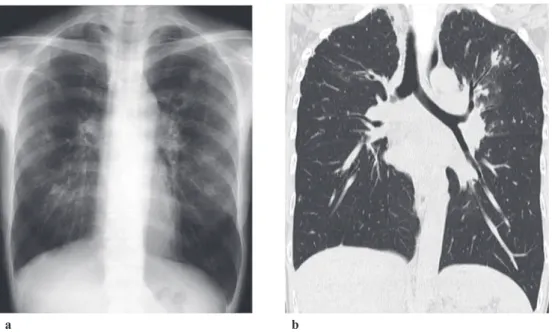
は じ め に 気管支結核の明確な定義は,定まっていないが,倉澤1) は,区域気管支より中枢側の気管・気管支の結核性粘膜 病変と定義している。また気管支結核の発症機序とし て,末梢気道からの直接浸潤と結核性リンパ節炎の気管 支内への波及および気管内穿孔をあげている。 小児結核の既往はあるが,体重減少と低栄養以外の基 礎疾患のない成人女性が,肺結核・気管支結核にて加療 を受けた。経過は良好であったが,結核治療終了後に新 たに,左主気管支の閉塞を伴う paradoxical response(PR) によると思われるリンパ節穿孔型の病変を呈したと考え られた症例を経験したので報告する。 症 例 症 例:49 歳,女性。 主 訴:咳嗽,呼吸困難。 既往歴:小児結核(時期と治療内容等の詳細不明),38 歳に子宮頸癌の手術を施行された。 家族歴:娘が肺結核に罹患した。 職業・生活歴:縫製会社で 10 年間従事した。喫煙歴 はない。機会飲酒。 現病歴:20X 年 3 月から咳嗽と微熱が出現した。同時 期,娘が肺結核と診断されている。その接触者検診にて 抗原特異的インターフェロン-γ遊離検査が陽性だった ので 7 月に紹介受診した。胸部画像所見では左上葉陳旧 性所見(Fig. 1-a, b)と傍大動脈リンパ節,左肺門部リン パ節の腫脹(Fig. 3-a)を指摘されたが,喀痰を喀出でき ず,8 月に気管支内視鏡検査(BF)を施行された。気管 支鏡所見(Fig. 2-d)は,左主気管支前壁側に潰瘍性病変 があり,この気管支洗浄液から結核菌群増殖同定検査陽 性〔TB-PCR(+)〕だったので気管支結核と肺結核(結 福島県立医科大学会津医療センター感染症呼吸器内科 連絡先 : 斎藤美和子,福島県立医科大学会津医療センター感染 症呼吸器内科,〒 969 _ 3492 福島県会津若松市河東町谷沢字前 田 21 _ 2(E-mail: aizuanes@fmu.ac.jp)(Received 19 May 2017 / Accepted 31 Jan. 2018)
要旨:症例は 49 歳の女性。既往歴は,小児結核と子宮癌術後。基礎疾患はなし。娘が結核を発症し たころから,咳嗽・微熱が出現し,娘の接触者検診で受けた抗原特異的インターフェロン-γ遊離検 査が陽性となったため当院を紹介された。この間体重が 7 kg 減少して BMI 15.2 であった。胸部異常 陰影を指摘され肺結核を疑われたが,排菌なく症状出現 5 カ月後に気管支鏡検査(以下 BF)を施行 された。BF では,左主気管支に潰瘍性病変あり,洗浄液の TB-PCR(+)にて気管支・肺結核と診断 され,入院にて標準結核治療を開始された。経過良好にて 3 カ月で退院となり治療は 6 カ月で終了し た。退院 2 カ月後に呼吸困難と喘鳴が出現した。胸部 CT にて左主気管支に腫瘤性病変が描出されて いたため,BF を行ったところ,同部は,大量の白苔で閉塞されていた。洗浄液の抗酸菌検査は陰性で あった。その後のステロイド剤投与にて閉塞所見が改善したことから結果的には paradoxical response (PR)による縦隔リンパ節の穿孔に伴う新たな気管支潰瘍性病変と考えられた。結核治療開始 14 カ月 後に BF を再検したところ,気管支前壁は瘢痕治癒していたが縦隔側は白苔を伴う肉芽腫病変が残っ ていた。粘膜型気管支結核の治療後に PR によって新たなリンパ節穿孔型気管支結核を生じたと思わ れる稀有な症例と考察した。 キーワーズ:気管支結核,奇異性反応,paradoxical response,低栄養,リンパ節穿孔
Fig. 1 a: Chest radiograph image at the time of diagnosis of pulmonary tuberculosis showed multiple
spotty infi ltrations in both lung fi elds. b: Chest CT image at the time of diagnosis of pulmonary tuberculosis showed multiple spotty infi ltrations in both lung fi elds and swelling of hilar lymph node.
91.0%,FEV1は 2.07 リットル,FEV1/FEV は 79.6% と改善
していた。胸部単純 CT(Fig. 2-c)を施行したところで は,左主気管支腔内の腫瘤陰影は縮小し,左肺門リンパ 節(Fig. 3-c)はさらに縮小傾向にあった。BF(Fig. 2-f) にては,左主気管支の縦隔側に白苔を伴う潰瘍性病変と 左上下葉支分岐部にポリープ病変を認めた。ポリープ生 検では,壊死を伴う肉芽腫性病変であり,ラングハンス 型巨細胞が散見されたが,チール・ネールゼン染色で抗 酸菌塗抹検査は陰性であった。同部位の洗浄液と組織の 抗酸菌塗抹・培養検査は陰性であった。その後治療終了 2 年後も,結核の再燃や喘鳴等の症状はみられない。 考 察 気管・気管支結核は,年代とともに減少傾向にあり,肺 結核の数パーセントにみられる程度となってきている2)。 気管支結核の臨床的特徴は,若年女性に多く,咳嗽,嗄 声,喘鳴などを主症状とすると報告されている2)。本症 院した。20X+ 1 年 1 月(治療 5 カ月後)から仰臥位に なると喘鳴と呼吸困難が出現するようになり,当院を緊 急受診した。呼吸困難の日内変動や労作時の呼吸困難は なかったが,聴診上左下肺野に強い wheezing を聴取し た。気管支喘息の診断で再入院し,ステロイドの点滴に て喘鳴は消失した。退院後,抗結核療法は予定どおり 6 カ月で終了したが,再び喘鳴と呼吸困難が増悪し,仰臥 位で寝られなくなった。3 月に撮影した胸部単純 CT (Fig. 2-b)で左主気管支内に腫瘤性病変を指摘され精査 のため 3 度目の入院となった。体重は 43.5 kg と増加し て BMI 17,PNI 53.4 であった。呼吸機能検査で,肺活量 (VC)は 2.2l,%VC は 81.8%,一秒量(FEV1)は 1.72l,一 秒率(FEV1/ FEV)は 79.6% であった。気管支拡張剤吸 入後の FEV1は,1.81l で,その改善率は 5.2% と気道可逆 性は証明されなかった。気管支鏡所見(Fig. 2-e)は,左 主気管支前壁の輪状軟骨構造は消失し肉芽を残さずに治 癒しているのに対し,左上下葉支分岐部は,大量の白苔 a b
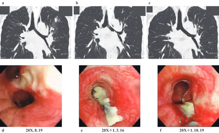
Fig. 2 a: CT image when pulmonary tuberculosis was diagnosed: no abnormal fi ndings in the left main bronchus. b:
CT image 7 months after the beginning of anti-tuberculous therapy: tumorous shadows in the left main bronchus. c: CT image 14 months after the beginning of anti-tuberculous therapy: smaller tumorous shadows in the left main bronchus. d: BF image when pulmonary tuberculosis was diagnosed: ulcerative lesions in the left main bronchus. e: BF image 7 months after the beginning of anti-tuberculous therapy: obstruction with fi brin formation in the left main bronchus. f: BF image 14 months after the beginning of anti-tuberculous therapy: granulation and scar formation with ulcer in the left main bronchus.
Fig. 3 a: Mediastinal condition CT images when pulmonary tuberculosis was diagnosed: swelling of left hilar lymph
node and bronchial bifurcation lymph node was shown. Arrow showed swollen mediastinal lymph node. b: Mediastinal condition CT images 7 months after the beginning of anti-tuberculous therapy: both lymph nodes swelling was getting smaller than the previous. Arrow showed swollen mediastinal lymph node. c: Mediastinal condition CT images 14 months after the beginning of anti-tuberculous therapy: both lymph nodes swelling was even smaller than the previous.
a
d 20X. 8. 19 e 20X+1. 3. 16 f 20X+1. 10. 19
a 20X. 8. 10 b 20X+1. 3. 7 c 20X+1. 10. 19
Ⅲa・隆起性潰瘍型Ⅲb),肉芽型(結節隆起性肉芽型Ⅳa・ ポリープ状肉芽型Ⅳb),瘢痕型(瘢痕非狭窄型Ⅴa・瘢 痕狭窄型Ⅴb),リンパ節穿孔型 LN とされおおむね病態 と病進行過程,病期を表したものになっている4)。未治 療例は潰瘍型が 80% と多く,治療の過程で瘢痕型に移行 する。 本症例は,診断時には荒井の分類Ⅲb だったが,治療 開始後 7 カ月の内視鏡所見は,左主気管支前壁の輪状軟 骨構造は消失して狭窄せず分類Ⅴa に移行していた。一 方で,左上下葉支分岐部は,多量な白苔により閉塞され ていた。ステロイド投与にて喘鳴,呼吸困難の症状は 徐々に軽快した。治療終了 8 カ月後の気管支鏡検査で は,縦隔側に一部白苔を伴う瘢痕性病変と肉芽腫性ポリ ープによる病変に変化していた。 気管支結核は,診断される前に喘鳴があるため気管支 喘息と誤診されて診断が遅れる症例報告が珍しくない4) 5)。 本症例は,当初から気管支・肺結核として診断され標準 治療も順調に経過したが,治療終了間近に新たな喘鳴と いう症状が出現し,結核治療終了後には呼吸困難もみら れた。これらの新たな症状や内視鏡所見の変化は全て PR によるものと考えられた。 PR とは,抗結核薬による治療によって臨床的症状や 画像的に以前から存在していた病変が悪化したり,通常 の経過では起こらないような新たな病変が出現したりす る現象である。治療中の PR はリンパ節腫脹や肺病変の ン血症と低 BMI が危険因子として当てはまっていた。 著者の COI(confl icts of interest)開示:本論文発表内 容に関して特になし。 文 献 1 ) 倉澤卓也:もう一つの結核:Endobronchial Tuberculosis. 結核. 2010 ; 85 : 805 808. 2 ) 田村厚久, 蛇沢 晶, 益田公彦, 他:気管支結核の現 状―103例の解析. 結核. 2007 ; 82 : 647 654. 3 ) 荒井他嘉司:気管支結核の新しい気管支鏡所見分類の 有用性について. 気管支学. 2001 ; 23 : 352 360. 4 ) Williams DJ, York EL, Nobert EJ, et al.: Endobronchial
tuberculosis presenting as asthma. Chest. 1988 ; 93 (4) : 836 838.
5 ) Argun Baris S, Onyilmaz T, Basyigit I, et al.: Endobronchial Tuberculosis Mimicking Asthma. Tuberculosis research and treatment. 2015 ; 2015 : 781842.
6 ) Cheng SL, Wang HC, Yang PC: Paradoxical response during anti-tuberculosis treatment in HIV-negative patients with pulmonary tuberculosis. Int J Tuberc Lung Dis. 2007 ; 11 : 1290 1295.
7 ) Bloch S, Wickremasinghe M, Wright A, et al.: Paradoxical reactions in non-HIV tuberculosis presenting as endobron-chial obstruction. Eur Respir Rev. 2009 ; 18 : 295 299. 8 ) 友田義崇, 内藤圭祐, 小川知洋, 他:初期悪化による気
管支ポリープで無気肺を生じた結核性縦隔リンパ節炎 の 1 例. 気管支学. 2017 ; 39 : 241 245.
Abstract A 49-year-old woman was admitted to our
hospi-tal due to positive result for interferon-gamma release assay, persistent cough, and body weight loss of 7 kg which had started when her daughter had been diagnosed with tubercu-losis (TB) 9 months before. Her body mass index (BMI) was 15.2 and her prognostic nutritional index (PNI) was also low as 36.3. She had no underlying diseases. Since ulcerative lesion at the left main bronchus was found by bronchoscopy (BF) and Mycobacterium tuberculosis was detected, she was diagnosed with pulmonary and endobronchial TB. Anti-TB therapy was started and went successfully, and since her complaints were all disappeared after 3 months, she was discharged from the hospital. However, in 2 months since then, she started to have cough and dyspnea at supine posi-tion again. Tumorous lesion in the left main bronchus was revealed by CT and obstruction of left main bronchus with
white mass was found by BF. It was considered as a case of bronchial tuberculosis due to mediastinal lymph node perforation to left main bronchus caused by paradoxical response after completion of tuberculosis treatment.
Key words : Endobronchial tuberculosis, Paradoxical
re-sponse, Poor nutrition, Lymph node perforation
Department of Infectious Disease and Pulmonary Medicine, Aizu Medical Center, Fukushima Medical University Correspondence to: Miwako Saitou, Department of Infectious Disease and Pulmonary Medicine, Aizu Medical Center, Fukushima Medical University, 21_2, Maeda, Tanisawa, Kawahigashi, Aizuwakamatsu-shi, Fukushima 969_3492 Japan. (E-mail: aizuanes@fmu.ac.jp)
−−−−−−−−Case Report−−−−−−−−