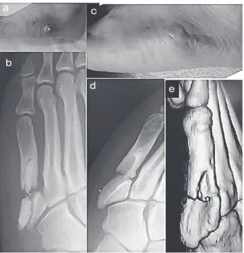
P
roximal fifth metatarsal fracture is classified into 3 categories. The first is avulsion fracture of tuber-osity (zone 1), the second is Jones fracture in the metaphyseal-diaphyseal junction (zone 2), and the third is diaphyseal stress fracture (zone 3) (Fig.1) [1,2].Jones fracture was first described by Sir Robert Jones in 1902. Although his patients had a proximal fifth metatarsal fracture in the metaphyseal-diaphyseal
junc-tion due to trauma [3], many subsequent authors have defined stress fracture of the proximal fifth metatarsal diaphysis as a Jones fracture. In the cases reported in this study, a proximal fifth metatarsal diaphyseal stress fracture was considered a Jones fracture.
Jones fracture is often seen in athletes, particularly soccer and basketball players [4]. Conservative treat-ment is an option for Jones fracture because it is a stress fracture, but pseudarthrosis or delayed union occurs in 25% to 28% of patients [5-7]. Ongoing participation in athletic activities, moreover, caries the risk of re-frac-ture, since running creates forces of 270% to 350% of body weight on the outer foot. Surgical treatment immediately after the diagnosis is often selected for ath-letes to shorten the duration of treatment and rehabili-tation. Most patients achieve bone union with surgery and are able to return to athletic activity, but re-fracture and other complications occasionally occur. Other complications include infections, peroneal tendon rup-ture, peroneal nerve damage, sural nerve damage [8], screw head impingement [9], looseness of proximal CopyrightⒸ 2020 by Okayama University Medical School.
http ://escholarship.lib.okayama-u.ac.jp/amo/
Case Report
Four Cases with Rare Complications of Intramedullary Screw Fixation for
Jones Fracture
Yusuke Morimoto
*, Taichi Komatsu, and Yasuaki Tokuhashi
Department of Orthopaedic Surgery, Nihon University School of Medicine, Itabashi-ku, Tokyo 173-8610, Japan
Active treatment with intramedullary screw fixation is now common for athletes with Jones fracture. Outcomes are generally good, but complications can occur. We report 4 rare complications of intramedullary screw fixa-tion. Two cases developed osteomyelitis and pseudarthrosis caused by thermal necrosis. In the other two cases, screw-related complications occurred during the insertion of the tapered headless screw. Although thermal necrosis and screw insertion failures are considered rare complications and not widely reported in the litera-ture, they do occur occasionally. Knowing the mechanisms underlying these complications could help prevent them, and knowing their course could lead caregivers to appropriate interventions when they do occur.
Key words: Jones fracture, thermal necrosis, tapered headless screw
Received October 24, 2019 ; accepted July 20, 2020.
*Corresponding author. Phone : +81-3-3792-8111; Fax : +81-3-3792-4824
E-mail : morimoto.yusuke@nihon-u.ac.jp (Y. Morimoto) Conflict of Interest Disclosures: No potential conflict of interest relevant to this article was reported. Fig. 1 Zone 1, Avulsion fracture of tuberosity [1]; Zone 2,
Jones fracture in the metaphyseal-diaphyseal junction; Zone 3, Diaphyseal stress fracture.
screws, iatrogenic fracture, metatarsal pain, distal metatarsal stress fracture [10], and thermal necrosis [8,11]. Although the presence of these other complica-tions has been noted in the literature, none have been described in detail.
At our institute, we actively perform intramedullary screw fixation for the treatment of Jones fracture in ath-letes. We herein present 4 cases involving rare, diffi-cult-to-treat complications.
Case Reports
Case 1. The patient was a 20-year-old male col-lege basketball player. He developed sudden pain in the left foot while pivoting during a basketball game. A diagnosis of Jones fracture was made based on cortical bone thickening and a fracture line in zone 3 of the left proximal fifth metatarsal observed in the X-ray (Fig.2 a,b). Nineteen days after the injury, intramedullary screw fixation was performed with a tourniquet via a small skin incision with a 4.5 mm-diameter cannulated compression screw (Japan Medical Dynamic Marketing, Tokyo) (Fig.2 c,d). The process of insert-ing the guide wire into the intramedullary fifth metatar-sal while drilling is made more difficult in cases such as this due to the large amount of sclerosis present at the fracture site.
Twenty-one days postoperatively, blisters formed on the skin at the fracture site, followed by skin necrosis and ulceration. Bone exposure and continual exudation
a b c d
Fig. 2 (Case 1) A-P (a) and oblique (b) preoperative foot X-ray images, Jones fracture was observed in zone 3 of the left proximal fifth
metatarsal; A-P (c) and oblique (d) postoperative foot X-ray images, Intramedullary screw fixation was performed using a 4.5 mm-diameter cannulated cancellous screw.
a
b c d e
Fig. 3 (Case 1) A photograph of the left foot is shown in (a),
Bone exposure was seen at the ulcerated area; oblique (b) and A-P (c) of foot X-ray 5 weeks after operation; A-P (d) and lateral (e) of foot X-ray 8 weeks after operation, the screw was removed and a synthetic bone graft with an antibiotic was performed.
were seen in the ulcerated area (Fig.3 a,b,c).
Osteomyelitis occurred 8 weeks postoperatively. The screw was removed, curettage was administered, and a synthetic bone graft was performed under antibiotic prophylaxis (Fig.3 d,e). Antibiotic treatment and hyperbaric oxygen therapy were administered postop-eratively. The infection subsided, but bone union was not achieved. The patient, however, had no pain and was able to return to playing basketball 9 months after the initial operation. The bone graft became exposed (Fig.4 a,b) and had to be removed at 5 years postoper-atively. Pseudarthrosis was present at the fracture site (Fig.4 c,d, e), but the patient was able to continue playing basketball without pain.
Case 2. The patient was a 19-year-old, non-pro-fessional, male rugby player. He developed pain in the right foot while standing on his toes during a rugby match. X-ray and computed tomography (CT) showed Jones fracture in zone 3 (Fig.5 a,b,c). Twenty-seven days after the injury, intramedullary screw fixation was performed using a tourniquet via a small skin incision with an Acutrak Plus Screw (Japan Medical Next, Osaka, Japan) (Fig.5 d,e). The guide pin did not read-ily pass through the fracture site during the operation. Fourteen days postoperatively, blisters formed on the skin at the fracture site, followed by skin necrosis. Wound healing was delayed, and osteomyelitis
occurred 2 months postoperatively (Fig.6 a,b,c). The screw was removed, and curettage was performed (Fig.6 d,e). Hyperbaric oxygen therapy and
vacu-a
b
c
d e
Fig. 4 photograph of left foot (a), A-P (a) and oblique (b) of
foot X-ray, The bone graft became exposed; photograph of left foot (c) oblique (d) of X-ray and CT of foot (e), The bone graft had to be removed 5 years postoperatively.
a b c d e
Fig. 5 A-P (a) and oblique (b) of preoperative foot X-ray and CT, Jones fracture in zone 3 of the Right proximal fifth metatarsal was
observed; A-P (d) and oblique (e) of postoperative foot X-ray, intramedullary screw fixation was performed using an Acutrak Screw Plus (Japan Medicalnext, Osaka, Japan)
um-assisted closure were administered postoperatively. The infection subsided, and the wound closed 6 months postoperatively (Fig.6 f,g,h). One year post-operatively, the patient had not achieved bone union (Fig.6 i), but he was able to continue playing rugby without pain.
Case 3. The patient was a 16-year-old, female, high-school soccer player. She was injured while play-ing soccer. She underwent conservative therapy at
another medical institution, resuming soccer after a month, but suffered re-fracture in zone 3. Intramedullary screw fixation was performed, at the same medical institution, 2 months after the initial fracture. She resumed soccer at 2 months postoperatively, but 2 months thereafter she suffered a recurrence of left foot pain when dribbling the ball. An X-ray taken after this recurrence showed re-fracture associated with screw breakage (Fig.7). At this point, the patient came
a
b c d e g
f
h i
Fig. 6 photograph of right foot (a), osteomyelitis occurred; A-P (b) and oblique (c) of foot X-ray 2 months after operation; A-P (d) and
lateral (e) of foot X-ray, The screw was removed, and curettage was performed 3 months after operation; photograph of right foot (f) and A-P (g) and oblique (h) of foot X-ray, Infection subsided, and the wound closed 6 months postoperatively; A-P (i) of foot X-ray 2 months after operation; A-P (d) and lateral (e) of foot X-ray, The patient had not achieved bone union one year after operation.
a b c d e f
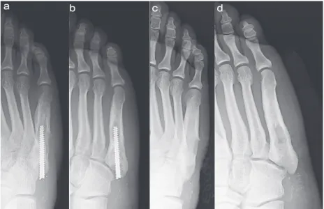
to our institution seeking treatment. The broken screw was removed 1 month after re-injury, and intramedul-lary screw re-fixation was performed with an Acutrak 4/5 screw (Japan Medical Next, Osaka, Japan) (Fig.8 a,b). The patient achieved bone union (Fig.8 c,d) and resumed soccer 5 months after the re-fixation. Two years and 9 months postoperatively (Fig.8 e,f), she continues to play soccer without complications.
Case 4. The patient was a 17-year-old, male, high- school soccer player. He was injured while playing soc-cer. He presented to our department with persistent pain. X-ray and CT imaging showed Jones fracture in zone 3 of the right foot (Fig.9 a,b,c). Intramedullary screw fixation was performed with an Acutrak 4/5 screw 1 month after the injury. The tip of the screw-driver broke in the screw head during screw insertion,
a b c d e f
Fig. 8 A-P (a) and oblique (b) of foot X-ray, The broken screw was removed 1 month after re-injury, and intramedullary screw re-fixation
was performed with an Acutrak 4/5; A-P (c) and oblique (d) foot X-ray images, The patient achieved bone union at 5 months after this re-operation; A-P (e) and oblique (f) foot X-ray images, Two years and 9 months after re-operation.
a b c d e
Fig. 9 (Case 4) A-P (a) and oblique (b) preoperative foot X-ray and CT images (c), The Jones fracture of the right foot is visible; A-P
(d) and oblique (e) X-ray images of the foot, Intramedullary screw fixation was performed with an Acutrak 4/5 screw at 1 month after injury. Circumstances necessitated the excision of the protruding proximal end of the screw.
preventing full insertion. Removal was not possible because the screw was locked into the surrounding bone. This circumstance necessitated the excision of the protruding proximal end of the screw (Fig.9 d,e).
Exudate continued to flow from the wound postop-eratively. X-ray imaging showed loosening around the screw (Fig.10 a,b). Although the exudate persisted, the patient achieved bone union and his pain disap-peared, which allowed him to resume playing soccer. Exudate continued to flow from the wound. A
Pseudomonas aeruginosa infection was present for a
time but improved with oral antibiotic treatment. Immediate screw removal and curettage were recom-mended to the patient, but, because his symptoms were mild, he decided to forgo such treatment in order to continue playing soccer and to take his college entrance exams.
The screw was removed 1 year and 1 month postop-eratively (Fig.10 c,d). The wound healed soon after removal.
Discussion
Many reports have noted that pseudarthrosis (25-28%), delayed union (60.7%) and re-fracture (61.1%) are common complications of intramedullary screw fixation performed to treat Jones fracture [12,13]. Although many other complications can occur, these have not been described in detail in the literature. In
particular, as far as we are aware, there has been only one report of Jones fracture involving thermal necrosis [10]. The overall frequency of occurrence is unclear because the number of cases is small, even in this report. At the time of the present report, 25 cases of Jones fracture had been treated at our institution, 2 of which involved thermal necrosis and 2 of which involved screw-related complications. The number of cases of Jones fracture treated at our institution is not large, and thus we cannot describe the incidence of complications associated with Jones fracture with accu-racy. However, the complications discussed in this paper are rarely reported, and our description of the treatment of such cases may be of clinical value.
Thermal necrosis. Thermal necrosis is a compli-cation characterized by necrosis of the bone and sur-rounding soft tissue due to the heat of friction occurring during intramedullary drilling or reaming. Most of the 15 cases of thermal necrosis identified in the literature involve the use of intramedullary nails to repair tibial fracture. Leuning et al. reported thermal necrosis fol-lowing intramedullary nail fixation for a tibial shaft fracture in 3 patients [14]. All 3 patients required flap coverage for the soft tissue defect. Giannonds et al. compared fibula reaming temperatures in 16 patients in whom a tourniquet was used and 18 patients in whom a tourniquet was not used. They found an increase in temperature associated with tourniquet use, although this association did not reach the level of statistical
sig-a b c d
Fig. 10 (Case 4) A-P (d) and oblique (e) foot X-ray images, Loosening around the screw was observed; A-P (c) and oblique (d) foot
nificance [15]. Saldua et al., noting that thermal necro-sis is a rare complication, emphasized the need for caution because of the serious consequences of the condition [16]. There is little information available regarding thermal necrosis following intramedullary fixation for Jones fracture. Tsukada et al. reported that thermal necrosis developed in 1 of 15 patients who underwent the operation [10]. They noted that heat may be generated during the drilling of hard sclerotic bone or from friction between a bent guide wire and the drill. They recommend releasing the tourniquet during drilling to prevent heat accumulation due to the lack of blood flow and propose cooling the skin with running water.
Thermal necrosis may have occurred in the two patients in our report due to the extra time needed to perform the drilling of hard sclerotic bone because the guide wire was inserted at an angle. In the two present cases, the guide pin insertion position was poor and the pin was inserted obliquely, causing friction to occur between the bone cortex and pin or the bone cortex and drill during pin insertion and reaming, which resulted in thermal necrosis. In addition, the use of tourniquets during surgery resulted in poor local circulation and increased the temperature at the fracture site. To avoid thermal necrosis, it is necessary to insert the guide pin from the correct position in order to reduce the friction between the drill and the cortex during reaming. Also during reaming, it is necessary to open the tourniquet, maintain local circulation, and perform a cooling treat-ment such as saline application.
Screw selection. Intramedullary screw fixation is a widely used surgical procedure [17]. However, re-fracture following intramedullary screw fixation is occasionally reported [12,13].
Because Jones fracture is a type of stress fracture, the goal of treatment should be to prevent recurrence [18]. Surgery should be performed with a focus on dis-tributing stress rather than compressing the fracture site or maintaining repositioning. Because Jones fracture occurs predominantly in athletes, minimally invasive surgery is preferable. In an experiment comparing intramedullary screw fixation with low-profile plate fixation, Huh et al. found that screw fixation provided superior biomechanical performance [19]. Collectively, the above findings suggest that intramedullary screw fixation is a useful surgical option for Jones fracture.
Cannulated cancellous screws may be used, but they
are associated with screw head damage [9] as a compli-cation. Thus, cannulated screws, whose headless design simplifies surgery, are a better option [18].
Headless cannulated screws come in tapered and Herbert varieties. Tapered screws are threaded along the entire axis, robustly compressing the fracture. However, Jones fractures require only a small distance to be pulled by the screw because the gap between the fragments is small. Also, the amount of cancellous bone in the metatarsal bones is small, and if the screw becomes stuck in the cortical bone, excessive torque will be generated and further insertion will not be possible.
In Patients 3 and 4, the screw was inserted at an angle and could not be inserted further after catching on cortical bone. A gap formed between the proximal screw and cortical bone in Patient 3, who experienced loosening and consequent re-fracture from screw breakage following resumption of activity. In Patient 4, the screw entered out of position and was locked into the cortical bone. It is likely that the resulting high torque caused the tip of the screwdriver to break. Surgeons are advised to ensure that they insert the screw at the correct angle and, in patients with a small medullary space, use a Herbert screw rather than a fully threaded tapered screw so that excessive torque is not generated. To prevent complications related to screw insertion, the guide pin must be inserted from the cor-rect position, and a screw must be selected which does not generate excessive torque during screw insertion.
The complications presented in this paper are rare; most patients achieve bone union and return to athletic activity without complication. However, complica-tions, when they arise, may keep the patient off the playing field and are psychologically stressful for patient and surgeon alike. By being aware of these complica-tions, surgeons can often prevent them and can better inform their patients to facilitate treatment.
References
1. Hartog BDD: Fracture of the proximal fifth metatarsal. J Am Acad Orthop Surg (2009) 17: 458-464.
2. Lawrence SJ and Botte MJ: Joneʼs fracture and related fracture of the proximal fifth metatarsal. Foot Ankle (1993) 14: 358-365. 3. Jones R: Fracture of the base of the fifth metatarsal bone by
indi-rect violence. Ann Surg (1902) 35: 697-700.
4. Saita Y, Nagao M, Kawasaki T, Kobayashi Y, Kobayashi K, Nakajima H, Takazawa Y, Kaneko K and Ikeda H: Range limita-tion in hip internal rotalimita-tion and fifth metatarsal stress fractures (Jones fracture) in professional football players. Knee Surgery
Sport Traumatol Arthrosc (2017) 26: 1943-1949.
5. Mologne TS, Lundeen JM, Clapper MF and OʼBrien TJ: Early screw fixation versus casting in the treatment of acute Jones frac-tures. Am J Sports Med (2005) 33: 970-975.
6. Dameron TB Jr: Fractures and anatomical variations of the proxi-mal portion of the fifth metatarsal Fractures and Anatomical Variations of the Proximal Portion of the Fifth. J Bone Joint Surg Am (1975) 57: 788-792.
7. Clapper MF, OʼBrien TJ and Lyons PM: Fractures of the fifth metatarsal. Analysis of a fracture registry. Clin Orthop Relat Res (1995) 315: 238-241.
8. Donley BG, McCollum MJ, Murphy GA and Richardson EG: Risk of sural nerve injury with intramedullary screw fixation of fifth meta-tarsal fractures: a cadaver study. Foot Ankle Int (1999) 20: 182-184.
9. DeLee JC, Evans JP and Julian J: Stress fracture of the fifth metatarsal. Am J Sports Med (1983) 11: 349-353.
10. Tsukada S, Ikeda H, Seki Y, Shimaya M, Hoshino A and Niga S: Intramedullary screw fixation with bone autografting to treat proxi-mal fifth metatarsal metaphyseal-diaphyseal fracture in athletes: a case series. Sport Med Arthrosc Rehabil Ther Technol (2012) 4: 25.
11. Cheung CN and Lui TH: Proximal Fifth Metatarsal Fractures: Anatomy, Classification, Treatment and Complications. Arch Trauma Res (2016) 5: e33298.
12. Wright RW, Fischer DA, Shively RA, Heidt RS and Nuber GW:
Refracture of proximal fifth metatarsal (Jones) fracture after intra-medullary screw fixation in athletes. Am J Sports Med (2000) 28: 732-736.
13. Larson CM, Almekinders LC, Taft TN and Garrett WE: Intramedullary screw fixation of Jones fractures. Am J Sports Med (2002) 30: 55-60.
14. Leunig M and Hertel R: Thermal Necrosis After Tibial Reaming for Intramedullary Nail Fixation a Report of Three Cases. J Bone Jt Surg Br (1996) 78: 584-587.
15. Giannoudis PV, Snowden S, Matthews SJ, Smye SW and Smith RM: Friction burns within the tibia during reaming. Are they affected by the use of a tourniquet? J Bone Joint Surg Br (2002) 84: 492-496. 16. Saldua NS, Kuhn KM and Mazurek MT: Thermal necrosis compli-cating reamed intramedullary nailing of a closed tibial diaphysis fracture: A case report. J Orthop Trauma (2008) 22: 737-741. 17. Porter DA: Fifth Metatarsal Jones Fractures in the Athlete. Foot
Ankle Int (2018) 39: 250-258.
18. Tanaka J: [Efforts to prevent re-injury and re-injury after surgery- The mechanism and practice of rehabilitation] Elucidation of re-in-jury / re-damage mechanism after surgery- Fifth metatarsal fatigue fracture in football. The Journal of Clinical Sports Medicine. (2011) 28: 387-394 (in Japanese).
19. Huh J, Glisson RR, Matsumoto T and Easley ME: Biomechanical Comparison of Intramedullary Screw Versus Low-Profile Plate Fixation of a Jones Fracture. Foot Ankle Int (2016) 37: 411-418.
![Fig. 1 Zone 1, Avulsion fracture of tuberosity [1]; Zone 2, Jones fracture in the metaphyseal-diaphyseal junction; Zone 3, Diaphyseal stress fracture.](https://thumb-ap.123doks.com/thumbv2/123deta/5830763.1036566/1.901.94.441.819.945/avulsion-fracture-tuberosity-fracture-metaphyseal-diaphyseal-junction-diaphyseal.webp)