Title
オステオポンチンの多機能性と臨床応用への戦略 (第
59回日本泌尿器科学会中部総会)
Author(s)
辻, 秀憲; 梅川, 徹; 植村, 天受
Citation
泌尿器科紀要 (2011), 57(1): 49-54
Issue Date
2011-01
URL
http://hdl.handle.net/2433/135432
Right
許諾条件により本文は2012-02-01に公開
Type
Departmental Bulletin Paper
Textversion
publisher
オステオポンチンの多機能性と臨床応用への戦略
辻
秀憲,梅川
徹,植村 天受
近畿大学医学部泌尿器科学教室
MULTIFUNCTIONAL CHARACTER OF OSTEOPONTIN AND
STRATEGY FOR CLINICAL APPLICATIONS
Hidenori Tsuji, Tohru Umekawa and Hirotsugu Uemura The Department of Urology, Kinki University School of Medicine
Osteopontin (OPN) is the major constituent of calcium-containingurinary stones and is involved in the inhibition of nucleation and aggregation of calcium oxalate (CaOx) crystals, promotion of the adherence of CaOx crystals to cultured renal epithelial cells, and regulation of inflammatory cells as chemokine. OPN has different effects (inhibitor and promoter) at each stage of stone formation in vitro and these multifunctional actions of OPN have not been fully elucidated. We developed a modified crystal method using collagen granules (CG) and immobilized OPN. OPN had strong inhibitory activity on the aggregation/growth of CaOx crystals, but the inhibitory activity decreased by use of OPN-immobilized CG. OPN is also a critical promoter of adherence for CaOx crystals to cultured renal epithelial cells in an in vitro experimental system. We examined the effect of OPN in vivo, by OPN siRNA transfection in rats. Hydrodynamic intravenous and renal subcapsular injections with lipofection were performed on days 1 and 8. The calcium concentration in the kidney was significantly lower and the frequency of CaOx crystal deposits in the tubules was lower in the OPN siRNA transfection group (drinking 1.5% ethylene glycol (EG)), than in the EG drinkinggroup (sham operation) at day 15. We examined the effect of candesartan, an angiotensin II (Ang II) type 1 receptor blockers (ARB) in hyperoxaluric rats. ARB reduced crystal formation and calcium concentrations in the whole kidney. Hyperoxaluria leads to CaOx crystallization and the development of tubulointerstitial lesions in the kidney. AngII mediates OPN synthesis, which is involved in both macrophage recruitment and CaOx crystallization. OPN synthesis and production increased with hyperoxaluria but to a lesser extent in ARB-treated hyperoxaluric rats. These results show that oxalate can activate the renal renin-angiotensin system and that oxalate-induced upregulation of OPN is in part mediated via the renal renin-angiotensin system.
(Hinyokika Kiyo 57 : 49-54, 2011) Key words : Osteopontin, Urolithiasis
緒 言 尿路結石の形成機序には様々な因子が関与している が,尿中に排泄される高分子蛋白もその1つである. そのうちオステオポンチン (OPN) は尿路結石の形成 に重要な役割を有していることが諸家の報告からも明 らかにされている1~3).OPNは尿路結石のマトリック スを構成する蛋白の1つでもあり,その機能は in vitroにて相反する作用も報告されており,尿路結石 形成の全体,各段階にどのように関与するのか,その 詳細は明らかとなっていない部分も多い.OPNの作 用として, 1)結晶成長・凝集の抑制,2)結晶の 尿細管上皮細胞への付着の促進,3)炎症細胞の遊走 などのchemical mediatorとしての作用,があげられ る.第59回日本泌尿器科学会中部総会シンポジウムで はOPNの多機能性という視点でわれわれがこれまで 集積してきたデータを示し,臨床に応用できる分野に つ い て 考 察 し た.1,2)を in vitro の,3)は in vivoのOPNに関する実験系として分けて提示する. 方 法
・
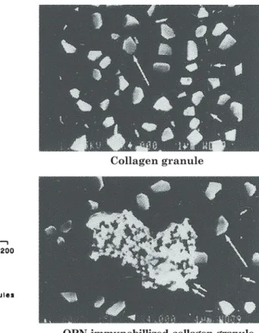
結 果 1)In vitro Fig. 1に Isotopes法に準じたシードクリスタル法の 概略を示す4~6).人工尿のmetastable solusionに蓚酸カ ルシウムのseed crystalを添加し,蓚酸カルシウムの結 晶凝集・成長阻止能 (inhibitory activity) を測定した. OPNを単独で添加した場合とコラーゲン顆粒を加え たもの,さらにこのコラーゲン顆粒の表面にOPNを コーティングしたものを添加して比較した.OPNを 単独で加えると,37.5μ/mlでおよそ90%のinhibitory activityを示した (Fig. 2).一方,コラーゲン顆粒の表 面にOPNをコーティングするとinhibitory activityは逆に低下した7,8).電顕像でもコラーゲン顆粒への結
泌57,01,11-1
Fig. 1. The modified seed crystal method using collagen granules. とで明らかに増加した (Fig. 3).以上よりOPNは人 工尿中にフリーで存在すると結晶の凝集抑制効果 (inhibitor) を持ち,OPNが何らか基盤に接着した状 態では逆に結晶析出が促進 (promoter) されることが 示された.OPNの作用のうち,2)結晶の尿細管上 皮細胞への付着の促進についての知見の1つとして
Madin Darby canine kidney cellnmembrane(MDCK) 培
養細胞にてFig. 4に示すように,COM細胞表面への
付着はOPN を添加すると有意に増加する9)ことが
泌57,01,11-3
Fig. 3. Effect of collagen granules and OPN-immobilized collagen granules in the seed crystal method. Inhibitory activity of collagen granules with a surface area of 180 cm2/5ml was 90%. Inhibitory activity of
OPN-immobilized collagen granules (180 cm2/5 ml) was 60%.
泌57,01,11-2
Fig. 2. Effect of osteopontin (OPN) in the seed crystal method. The inhibitory activity of OPN at a concentration of 37.5μg/ml was 90%.
ある.COM 結晶が曝露されるとbikunin,heparanx
sulfate (HS), prostaglandin E2などと同時にOPNの
発現も増加する.細胞表面への結晶の付着はOPNを 添加した状態では有意に増加する (Fig. 4).さらに ラット遠位尿細管細胞 (NRK-52E) を用い,蓚酸を暴 露するとOPN発現の亢進とともにCOM結晶の細胞 への付着も有意に増加する.OPN siRNAを尿細管細 胞に導入すると,結晶の細胞への付着は有意に減少し 泌尿紀要 57巻 1 号 2011年 50
泌57,01,11-4
Fig. 4. Difference in the adhesion of crystals between OPN and thrombin by SEM image and comparison of45Ca
concentrations after the addition of negative control, OPN(10μg/ml), thrombin (10 U/ml), RGD (Arg-Gly-Asp peptide, 10μg/ml) and RGE (Arg-Gly-Glu peptide, 10μg/ml) by scintillation counting.
泌57,01,11-5
*: P<0.05 vs control, N=5
Fig. 5. COM crystal adherence was increased by exposure of the cells to high oxalate concentration and reduced significantly by OPN siRNA transfection to NRK-52E cultured cells. た (Fig. 5).つまり培養細胞の実験系では OPN が promoter(COM結晶と細胞の付着を亢進する)とし て働くことが示唆された. 2)In vivo OPNは3)間質への炎症細胞の遊走などの chem-ical mediatorとしての作用をもち,前述した作用も合 わせた多機能を有するが故,尿路結石形成に対して inhibitorか promoterかも一元的には説明できない. そ こ で OPN の in vivo で の 作 用 に 着 目 し,OPN siRNA導入によるin vivoの実験系を作成した.導入
試薬であるAtelogeneTMを用いて腎被膜下注入とリポ
フェクションを用いた hydrodynamic injection をラッ
ト尾静脈より行い OPN siRNA を導入した.Target
sequence : 5′AAGGCGCATTACAGCAAACAC 3′(167
-187,87 base downstream from the start codon) Sense Sequence : 5′GGCGCAUUACAGCAAACACUU 3′, An-tisense Sequence : 5′GUGUUUGCUGUAAUGCGCCUU 3′(Ambion○R siRNA Target Finder) を用い,サイレン
シングを評価した10).作成したラットモデルはA 群 : コントロール (n=2),B群 : 1.5% エチレング リコール (EG) 自由摂取 (n=6),C群 : リポフェク ショ ン を 用 い た hydrodynamic injection に よ る 導 入 (n=6),D群 :腎被膜下注入による導入 (n=6) の4 群である.C,D群はいずれも1.5% EGの自由摂取 とした.ラットは24週齢雌Sprague-Dawley(SD) を用 いた.In vivo transfectionの処置はday 1,8に行い,
day 15にsacrificeした.Fig. 6に各群の原子吸光法に
よる腎組織カルシウム含有量を示す.EG投与による
カルシウム含有量の増加が,OPN siRNA 導入を行っ
泌57,01,11-6
*: P<0.05 vs group B
Fig. 6. Hyperoxaluria was induced in 6-week-old male Sprague-Dawley rats by administering 1.5% ethylene glycol in drinking water for 3 weeks. Four groups of 6 rats each were studied : group A, untreated control animals ; group B, hyperoxaluria without treatment ; group C, hyperoxaluria treated hydrodynamic administration of OPN siRNA by lipofection ; group D, hyperoxaluria treated renal capsular injection of OPN siRNA usingatelocollagen. The renal calcium concentration was higher in groups B, C and D than in group A. The increase of renal calcium concentration induced by hyperoxaluria (group B) was significantly reduced by OPN siRNA transfection (group C and D).
て作成した結石形成モデル11)ではあるが,OPNはin
vivoで全体として結石形成にpromoterとして作用し
ていることが示唆された.
このOPNの発現を抑制することで,臨床応用する
ことができないか? その1つとして,Angiotensin II
type I receptor blocker(ARB) であるカンデサルタンを
泌57,01,11-7
Group B : 1% EG for 4 weeks, group C : 1% EG+CS (20 mg/ml) for 4 weeks
Fig. 7. (Left) : Sections of a kidney from the rat in group B and C. Calcium oxalate crystals in the tubular lumen were decreased in group C. H & E staining, reduced from×200. (Right) : Kidney sections immunostained for ED-1, reduced from×200. ED-1-positive cells, correspondingto monocytes/macrophare, are present in the renal interstitum. In group C, ED-1-positive cells were reduced.
用いてのラット腎結石抑制効果について検討した.作 成したラットモデル(10週齢雄SD) はA群 : 水道水 (n=10),B群 : 1.0%EG(n=10),C群: 1.0%EG とカンデサルタン20μg/ml EG(n=10),D群 : カン デサルタン20μg/ml EG(n=10) を4週間投与した4 群である.まず腎組織像のEG投与群 (group B) と 泌尿紀要 57巻 1 号 2011年 52
泌57,01,8-8A
(A)
泌57,01,8-8B
(B)
Cont : control. EG : 0.1% EG for 4 weeks, CS : 20μg/ml for 4 weeks, N=10 mean±SD
Fig. 8. (A) renal calcium concentration. (B) calcium oxalate crystal deposits (%nephron/×200 fields). Group A : control, group B : EG, group C : EG+CS (Candesartan), group D : CS. High levels of renal calcium concentrations and calcium oxalate crystal deposits in group B were significantly decreased in group C.
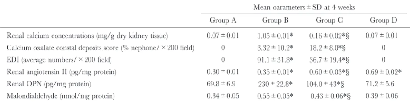
Table 1. Urinary and serum concentrations of various substances
Mean oarameters±SD at 4 weeks
Group A Group B Group C Group D
Renal calcium concentrations (mg/g dry kidney tissue) 0.07±0.01 1.05±0.01* 0.16±0.02*§ 0.07±0.01 Calcium oxalate constal deposits score (% nephone/×200 field) 0 3.32±10.2* 18.2±8.0*§ 0
EDI (average numbers/×200 field) 0 91.1±31.8* 36.7±19.4*§ 0
Renal angiotensin II (pg/mg protein) 0.30±0.01 0.35±0.01* 0.60±0.03*§ 0.69±0.02*
Renal OPN (pg/mg protein) 69.8±6.9 230±22.8* 104.0±43*§ 71.2±5.6 Malondialdehyde (nmol/mgprotein) 0.34±0.05 0.55±0.05* 0.43±0.06*§ 0.39±0.06
*: P<0.05 vs group A,§P<0.05 vs group C. Group A : control, group B : l% EG for 4 weeks, group C : 1%EG+CS (20 mg/ml) for 4weks, group D : CS (20 mg/ml) for 4 weeks.
EG+ARB投与群 (group C) の比較をFig. 7に示す.
Group Cでは尿細管でのクリスタルの沈着が減少し, 間質のED1陽性細胞もgroup Cで明らかな減少をみ た.腎組織カルシウム含有量および石灰化係数 (H & E stain標本200倍視野でネフロンの総数のうち管腔内 に結晶が付着しているネフロンの割合)をみると,と もにEG投与にて著明に増加したが,ARB投与にて 有 意 に 減 少 し た (Fig. 8).そ の 他 の 結 果 も 含 め て Table 1に示す10). 腎組織内OPN,Malondialdehyde濃度は有意に減少 した. 考 察 Angiotensin II は酸化ストレスにも関与していて NADPH oxidaseの活性化から酸素活性ラジカルを産 生させる12).このNADPH oxidaseは蓚酸の曝露など により活性化され,スーパーオキシド (・O2−)の産 生,さらにOPNの亢進につながる.このOPNの亢 進にはレニンアンギオテンシン系の関与もある. 結石形成ラットでは腎尿細管に様々なストレスが影 響し13),活性酸素の増加,レニンアンギオテンシン 系の亢進よりOPNの発現が増加する.ARBの投与 によりAngiotensin IIを介する経路がブロックされる ため,その分OPNの発現が抑制され,結晶の沈着の 減少と腎カルシウム含有量が低下したのではないかと 泌57,01,11-9
Fig. 9. Hypothetical schema depictingthe multiple actions of OPN that may be required to adequately inhibit the intrarenal deposition of calcium oxalate crystals.
推察された (Fig. 9).
Angiotensin II は血管に作用する他に,腎障害およ び腎臓病の進行に影響を与える免疫調節因子も活性化 させると報告されている14).さらにAngiotensin IIは
RANTES(regulated on activation normal T-cell expres-sion and secreted),monocyte chemoattactant protein-1
(MCP-1) およびOPNを up-regulation させ,サイト カイン, ケモカインの誘導より, 腎内でのmonocytes/ macrophagesの浸潤が促進される.これらは腎尿細管 間 質 の 機 能,構 築 に も 影 響 を も つ.ARB に よ る Angiotensin IIの抑制は腎尿細管間質の障害を緩和し, サイトカインとマトリックス蛋白質の発現を減少させ る15). 臨床応用について考えてみるとARB(カンデサル タン)はすでに市販されている薬剤であるが,今回の 結果をうけて実際にヒトに投与した場合,尿路結石の 予防効果として期待できるのか? それは今後まだま だデータの蓄積が必要である.またNADPH oxidase が産生するスーパーオキシドは,second messengerの 役割を担ってOPNの発現を亢進させることから,さ らに2種の薬剤もinhibitorとして導き出される.フ リー ラ ジ カ ル ス カ ベ ン ジャー で あ る Nicaraven,
Edaravone と Rho-kinase inhibitor で あ る.後 者 は フ リーラジカルスカベンジャーとしての活性はないが, 好中球凝集,遊走,活性酸素産生の抑制作用をもち, 尿路結石形成のinhibitorとしての効果も期待できる かも知れない. 過剰な蓚酸イオン (Ox) や蓚酸カルシウム結晶の尿 細管上皮への曝露は多様なストレス性の反応を生み出 す.この代表がOPNやMCP-1の発現の亢進であり, 尿路結石の初期段階である尿細管腔での結晶形成に寄 与している16).このように尿細管上皮から間質に遊 走された炎症細胞より分泌された様々な酵素やサイト カイン,ケモカインにより結晶にマトリックス成分が 取り込まれて成長し,結石形成に移行すると考えられ ている.尿路結石を予防するという観点からいうと, 尿細管上皮細胞から惹起される過剰な炎症を抑制する ことも重要な機序の1つと考えられた. 結 語 尿路結石の形成機序に関与する尿中高分子蛋白のう ちOPNに関するこれまでに得られた知見と臨床応用 への可能性について考察した. 文 献
1) Wesson JA, Johnson RJ, Mazzali M, et al. : Osteopontin is a critical inhibitor of calcium oxalate crystal formation and retention in renal tubules. J Am Soc Nephrol 14 : 139-147, 2003
2) Khan SR : Role of renal epithelial cells in the initiation of calcium oxalate stones. Nephron Exp Nephrol 98 : 55-60, 2004
3) Yasui T, Fujita K, Asai K, et al. : Osteopontin regu-lates adhesion of calcium oxalate crystals to renal epithelial cells. Int J Urol 9 : 100-109, 2002 4) Robertson WG, Peacock M and Nordin BE : Inhibitors
of the growth and aggregation of calcium oxalate crystals in vitro. Clin Chim Acta 43 : 31-37, 1973 5) Ligabue A, Fini M and Robertson WG : Influence of
urine on “in vitro” crystallization rate of calcium oxalate : determination of inhibitory activity by a [14C]
oxalate technique. Clin Chim Acta 98 : 39-46, 1979 6) Ryall RL, Ryall RG and Marshall VR : Interpretation of particle growth and aggregation patterns obtained from the Coulter counter : a simple theoretical model. Invest Urol 18 : 396-405, 1981
7) Umekawa T, Iguchi M and Kurita T : The effect of osteopontin immobilized collagen granules in seed crystal method. Urol Res 29 : 282-286, 2001 8) Konya E, Umekawa T, Iguchi M, et al. : The role of
osteopontin on calcium oxalate crystal formation. Eur Urol 43 : 564-571, 2003
9) Yamate T, Kohri K, Umekawa T, et al. : Interaction between osteopontin on Madin Darby canine kidney cellnmembrane and calcium oxalate crystal. Urol Int 62 : 82-86, 1999
10) Umekawa T, Hatanaka Y, Kurita T, et al. : Effect of Angiotensin receptor blockage on osteopontin expres-sion and calcium oxalate crystal deposition in rat kidneys. J Am Soc Nephrol 15 : 635-644, 2004 11) Kharn SR, Johnson JM, Peck AB, et al. : Expression of
osteopontin in rat kidneys : induction duringethyrene glycol induced calcium oxalate nephrolithiasis. J Urol 168 : 1173-1181, 2002
12) Ito H, Mukoyama M, Pratt RE, et al. : Multiple autocrine growth factors modulate vascular smooth muscle cell growth response to angiotensin II. J Clin Invest 91 : 2268-2274, 1993
13) Khan SR : Hyperoxaluria-induced oxidative stress and antioxidants for renal protection. Urol Res 33 : 349-357, 2005
14) Antus B, Exton MS and Rosivall L : AngiotensinⅡ : a regulator of inflammation during renal disease ? Int J Immunopathol Pharmacol 14 : 25-30, 2001
15) Cao Z and Cooper ME : Role of angiotensin II in tubulointerstitial injury. Semin Nephrol 21 : 554-562, 2001
16) Umekawa T, Tsuji H, Uemura H, et al. : Superoxide from NADPH oxidase as second messenger for the expression of osteopontin and monocyte chemoat-tractant protein-1 in renal epithelial cells exposed to calcium oxalate crystals. BJU Int 104 : 115-120, 2009