ORIGINAL ARTICLE
THE RELATIONSHIP BETWEEN MUSCLE DAMAGE AND REACTIVE OXYGEN SPECIES PRODUCTION CAPABILITY AFTER JUDO EXERCISE
Hisashi Kudoh
1),Makoto Yaegaki
2),Ippei Takahashi
1),Takashi Umeda
1), Kaori Sawada
1),Noriyuki Okubo
1),Yousuke Yamamoto
1,3)and Shigeyuki Nakaji
1)Abstract The relationship between neutrophil function and the muscles damage before and after exercise in judoists was examined. The changes in the body composition, biochemical parameters (neutrophil counts and myogenic enzymes such as aspartate aminotransferase (AST), alanine aminotransferase (ALT) etc. and the major neutrophil immune functions such as oxidative burst activity (OBA) and phagocytic activity (PA) were measured in 39 male university judoists before (pre value) and after 2 hours of unified exercise loading. Increases in myogenic enzymes were confirmed after the exercise, which suggested that the muscular tissue damage and degeneration was caused by the exercise itself. Neutrophil count and OBA increased and PA decreased after exercise. The amount of change (post value – pre value) of OBA showed a negative correlation with LDH and AST (P<0.01 all) and that of PA displayed a positive relationship with LDH and AST (P<0.05 all). Following the muscle damage, neutrophil PA may have increased due to increased biological detritus by phagocytosis such as muscle fragments. The reason for OBA movement against muscle damage (negatively correlated) may be due to the results of compensating OBA and PA e.g. increased OBA and decreased PA, or decreased OBA and increased PA.
Hirosaki Med.J. 64:176―185,2014 Key words: muscle damage; judo; neutrophil function; reactive oxygen species(ROS); exercise.
原 著
柔道稽古後の筋損傷と好中球活性酸素種産生能の関連について
工 藤 久
1)八重垣 誠
2)高 橋 一 平
1)梅 田 孝
1)沢 田 かほり
1)大久保 礼 由
1)山 本 洋 祐
1,3)中 路 重 之
1)抄録 柔道選手を対象として稽古前後の好中球機能と筋損傷の関係を調査した.
39人の男子大学柔道選手において,普段の 2 時間の稽古前後に体組成と生化学指標(好中球数,好中球機能(活性酸素 産生能(OBA),貪食能(PA)),筋逸脱酵素(LDH(乳酸脱水素酵素),アスパラギン酸アミノトランスフェラーゼ(AST),
アラニンアミノトランスフェラーゼ(ALT)など)を測定した.
稽古後に筋逸脱酵素は増加し,稽古に伴う筋組織の損傷と変性によると考えられた.また,稽古後に好中球数と OBA は増加し,逆にPAは低下した.一方,OBA の変化量は LDH および AST の変化量と負の相関関係を示し(p<0.01),
PA の変化量は LDH,AST の変化量と正の相関関係を示した(p<0.05).
筋損傷に対する PA の変動(正の相関)の要因としては,筋損傷に伴う筋肉の断裂片のような生理学的異物に対して好 中球の異物反応が亢進した可能性が考えられた.また,筋損傷に対する OBA の変動(負の相関)の要因としては,好中 球の各機能間における補完機構が OBA と PA の間で生じた可能性が考えられた.
弘前医学 64:176―185,2014 キーワード:筋損傷;柔道;好中球機能;活性酸素種:運動.
1)
Department of Social Medicine, Hirosaki University Graduate School of Medicine
2)
Department of Orthopaedic Surgery, Hirosaki University Graduate School of Medicine
3)
Department of Physical Education, Nippon Sport Science University
Correspondence: I. Takahashi
Received for publication, May 21, 2013 Accepted for publication, June 19, 2013
1)
弘前大学大学院医学研究科社会医学講座
2)
弘前大学大学院医学研究科整形外科学講座
3)
日本体育大学体育学部
別刷請求先:高橋一平
平成25年 5 月21日受付
平成25年 6 月19日受理
Introduction
Judo is a martial art originated from Japan and has become an international competitive sport since the Tokyo Olympics, and is a competitive sport that can also be classified as a contact sport
1).
Although the duration of each judo match is short, the burden on the body and the risk of injury are both high
2, 3). It is also thought that judo practice has a large effect on the body, especially the muscle tissue.
Exercise has been reported to elevate the levels of serum enzymes such as creatine kinase
(CK), aspartate aminotransferase (AST), alanine aminotransferase (ALT) and lactate dehydrogenase (LDH) through the mechanisms of muscular inflammation, increased permeability of muscle cell membranes
4-6), anoxic or hypoxic damage to muscles
7, 8)or production of toxic free radicals
9).
On the other hand, the number of neutrophils and the neutrophil function, i.e. neutrophil reactive oxygen species (ROS) production capability, has been reported to increase after high intensity exercise
10-15). Neutrophils represent one of the cellular factors that play an important role in the first line of defense against invading pathogens, including microorganisms.
Neutrophils engulf microorganisms through phagocytic activity (PA) and destroy them through the production of cytotoxic reactive oxygen species (ROS)
16, 17). However, the meaning behind the changes in these neutrophil functions remains unclear. For example, ROS can destroy invading microorganisms
18, 19), however, they can cause oxidative damage to normal body tissues and organs under different circumstances
20, 21). Unfortunately, the borderline between the beneficial and detrimental effects of ROS production remains unclear.
In recent studies, modified muscle use such as exercise or injury can produce an
inflammatory response in which neutrophils rapidly invade muscle tissue, followed by macrophages, which coincide with muscle repair, regeneration, and growth, and which involve the activation and proliferation of satellite cells, followed by their terminal differentiation
22, 23). Furthermore, recent studies have contributed to painting a complex picture in which inflammatory cells including neutrophils and macrophages promote both muscle injury and repair, through the combined actions of free radicals such as ROS, growth factors and chemokines
22). It has become clear that a role has emerged for neutrophils in promoting muscle damage soon after muscle injury or modified use
24, 25). Unfortunately, no direct evidence is yet available to show that neutrophils play a beneficial role in muscle repair or regeneration, although few studies have evaluated the amount of neutrophil ROS production and its relationship with muscular tissue damage
26).
We focused our attention on the relationship between muscular tissue damage and neutrophil ROS production following judo exercise in this study. The characteristics of this study used two neutrophil function such as ROS and phagocytic activity (PA) (which generally show opposite movement following exercise).
Subjects and Methods
1. Subjects
The 39 male subjects of this study belonged to the Nippon Sport Science University judo team that participated in a summer training camp. The athletes’ demographics and anthropometrics are shown in Table 1.
This study was approved by the Ethics
Review Committee of Hirosaki University
School of Medicine. Each participant was
adequately informed of the goal, methods, and
potential risks, as well as the right to abstain
from participating in this study. Freely given
informed consent was obtained in writing from all participants.
2. Methods
We measured the changes in the body composition, biochemical parameters and the major neutrophil immune functions in 39 male university judoists before (pre value) and after a 2-hour unified exercise loading (UEL) (post value).
1. Training program
The 2-hour UEL consisted of a warm-up for 15 min; “uchicomi” (the same technique practiced repeatedly, such as throw down, push down and hook down) for 20 min;“randori”
(exercise training in the form of a match) for 70 min; and cool-down for 15 min.This training was classified as irregular-interval training, and is a model of acute exercise. The mean heart rate during the training was 128.8 ± 12.0 bpm and the maximum heart rate was 180.5 ± 14.0 bpm, recorded in the randori phase. The heart rate was measured and recorded at intervals of 15 seconds using VANTAGE XL (POLAR ELECTLIC Inc.,Finland)
27), and was analyzed at intervals of 1 minute after the end of the measurement using POLAR HR ANALYSIS SOFTWARE VER. 4 (POLAR ELECTLIC Inc., Finland).
2. Anthropometry
The anthropometric parameters included body weight, relative body fat, and fat-free mass.
An apparatus employing the impedance method
(TBF-110, TANITA, Tokyo, Japan) was used in their measurement.
3. Blood chemistry parameters
Twenty milliliter samples of blood were collected from the forearm vein before and after the training. The collected blood samples were split up as described and allocated for
measurement with the following methods. Five milliliters were used to analyze the hemocyte elements, 10 ml were used to analyze the serum elements and 5 ml were used to analyze the neutrophil function. Leukocyte and neutrophil counts, hemoglobin (Hb), hematocrit (Ht)
were determined from venous blood treated with ethylenediaminetetraacetic acid using an automatic blood cell counter (Sysmex K-2000, Kobe, Japan). Serum samples were separated from blood samples by centrifugation for 10 min at 3,000 rpm and kept frozen at -30°C until used for measurement at a later date. The measurement items of the serum elements were AST, ALT, CK and LDH which were measured with the ultraviolet method. These values after the exercise were adjusted by the plasma volume calculated by the levels of hematocrit
(Ht) and hemoglobin before and after the exercise
28)because dehydration was observed in the changes of weight and Ht before and after the training.
4. Neutrophil oxidative burst activity (OBA)
and phagocytic activity (PA)
These activities were measured with a FACScan system (Becton Dickinson, San Jose, CA) using two-color flow cytometry.
Hydroethidine (HE: 44.4 µM, Polysience Inc., Warrington, PA) was used as an indicator for OBA (representing the ROS production capability), and opsonized zymosan particles
(Sigma Chemical Co., St. Louis, MO) labeled with fluorescein isothiocyanate (FITC, Sigma)
were used as indicators for PA.
In brief, 100 µl of heparinized whole blood was mixed with 22 µl of HE (final concentration;
f.c. 8 µM), and incubated at 37°C for 5 min.
After the addition of 25 µl of FITC-labeled
opsonized zymosan (FITC-OZ, f.c. 5 mg/ml),
the samples were incubated at 37°C for 35
min. To measure basal oxidative burst activity,
neutrophils labeled with only HE served as
controls. After the incubation, Lyse and Fix
(IMMUNOTECH, Marseille, France) were added to the red blood cells to fix the samples.
The samples were washed twice in phosphate buffered saline with sodium azide (PBS+), and the fluorescence intensity (FI) in the activated neutrophils was measured with FACScan. Just before the assay, 30 µl of trypan blue (0.25 mg/ml, pH 4.5) was added to differentiate the attached from the ingested FITC-OZ in the samples following measurement by fluorescence quenching
29, 30). The intensity of neutrophils expressing FITC-OZ and HE fluorescence was computed. Neutrophil OBA and PA were estimated as the mean channel number of the FI of activated neutrophils.
5. Statistical analysis
Data are presented as the mean ± standard deviation. The differences between before and after exercise were tested with the paired t-test.
The correlation between muscle tissue and ROS was tested with Spearman’s coefficient. The differences were considered to be statistically significant at p<0.05. All parameters were tested
for normal distribution with the Shapiro-Wilkes test for normality before further analysis. The statistical analysis was carried out using the PC version of the statistical package SPSS/PC.
Results
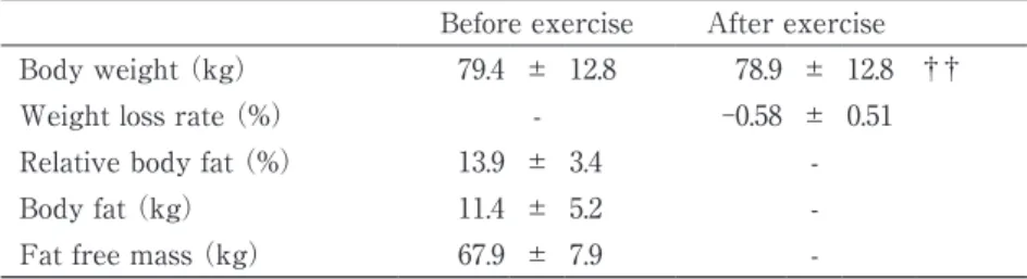
Body weight significantly decreased after the exercise (P<0.01) (Table 1).
Leukocyte counts did not show any significant change after the exercise. On the other hand, neutrophil counts showed a significant rise after the exercise (P<0.01) (Table 2).
As for myogenic enzymes, LDH and AST increased significantly after the exercise (P<0.01 for both) (Table 3).
As for neutrophil function, OBA increased significantly after the exercise (P<0.01). On the other hand, PA significantly decreased after the exercise (p<0.01) (Figure 1).
The amount of change (post value – pre value) of OBA showed a negative correlation with LDH and AST (P<0.01 for both) and that of PA displayed a positive relationship with LDH and AST (P<0.05 for both) (Figure 2).
Table 1 Physical characteristics and changes in anthropometric parameters
Before exercise After exercise
Body weight (kg) 79.4 ± 12.8 78.9 ± 12.8 ††
Weight loss rate (%) - -0.58 ± 0.51
Relative body fat (%) 13.9 ± 3.4 -
Body fat (kg) 11.4 ± 5.2 -
Fat free mass (kg) 67.9 ± 7.9 -
Results were expressed as mean ± standard deviation .
††: p<0.01 compared with pre value.
Table 2 Changes in blood leukocyte/neutrophil counts
Before exercise After exercise
*Leukocyte (cells/mm
3) 6469.2 ± 1268.9 6634.4 ± 1420.2 Neutrophil (cells/mm
3) 3455.4 ± 1089.4 4119.3 ± 1370.3 ††
Results were expressed as mean ± standard deviation.
††: p<0.01 compared with pre value.
*
: The post values were adjusted for dehydration by plasma volume.
Discussion
Exercise has been reported to elevate the levels of serum myogenic enzymes including those mentioned in the Introduction section.
Therefore changes in these enzyme levels may be effective markers for monitoring overtraining in athletes
31). Evans et al. have reported that CK levels increased more significantly in non- exercising subjects than in habitually exercising subjects under the same exercise load, and that habitual exercise might increase the capability of muscular function and tissue structure to perform more intense exercise without any
exercise-related muscle injury
4).
Koutedakis Y. et al. have also reported that intense and high quality physical training in Olympic rowers failed to produce any significant change in the above-stated enzyme levels, leading to the speculation that an inevitable adaptation occurs in such elite competitors, i.e., a blunted incremental response to exercise
33). This study confirmed the previous findings
1, 2, 10,30, 33)
that exercise increases the levels of serum CK, AST, ALT, and LDH.
The relationship between leukocytes and exercise has been well-reported: leukocyte and neutrophil counts increase with transient exercise, the degree of increase being
Table 3 Changes in serum myogenic enzymes
Before exercise After exercise
*CK (IU/l) 539.1 ± 352.3 556.5 ± 343.7 LDH (IU/l) 231.9 ± 33.9 246.6 ± 34.6 ††
AST (IU/l) 26.6 ± 8.2 27.9 ± 8.8 ††
ALT (IU/l) 21.6 ± 9.3 22.0 ± 9.5
Results were expressed as mean ± standard deviation.
††: p<0.01 compared with pre value.
*
: The post values were adjusted for dehydration by plasma volume.
Figure 1 Change in OBA and PA.
Results were expressed as mean ± standard deviation. OBA: oxidative burst activity, PA:
phagocytic activity. ††: p<0.01 compared with pre value.
Figure 2 Correlation between the change rates of neutrophil PA and OBA, and serum myogenic enzymes.
OBA: oxidative burst activity, PA: phagocytic activity. n.s.: not significant.
dependent on the intensity of the exercise
33). This study confirmed the previous findings that exercise increases the neutrophil count
8, 11, 12, 13, 15). Furthermore, the increases in the levels of these components are recognized as an anti- inflammatory reaction to the degeneration of and injury to muscle tissue caused by exercise, and it is possible that inflammatory cytokines participate in this reaction
6, 7). On the other hand, another study has suggested that the elevated white blood cell counts are not only due to the anti-inflammatory reaction, but possibly exercise itself may become the stressor which stimulates the production and release of growth hormones, adrenaline, and noradrenaline
6).
Eccentric exercise commonly results in muscle damage. The primary sequence of events leading to exercise-induced muscle damage is believed to involve initial mechanical disruption of sarcomeres, followed by impaired excitation- contraction coupling and calcium signaling, and finally, activation of calcium-sensitive degradation pathways
28, 34, 35). Muscle damage is characterized by ultrastructural changes to muscle architecture, increased muscle proteins and enzymes in the bloodstream, loss of muscular strength and range of motion, and muscle soreness. The inflammatory response to exercise-induced muscle damage is characterized by leukocyte infiltration and production of pro-inflammatory cytokines within damaged muscle tissue, systemic release of leukocytes and cytokines, in addition to alterations in leukocyte receptor expression and functional activity. Current evidence suggests that inflammatory responses to muscle damage are dependent on the type of eccentric exercise, previous eccentric loading (repeated bouts), age and gender
36-38). Circulating neutrophil counts and systemic cytokine responses are greater after eccentric exercise using a large muscle mass than after other types of eccentric exercise involving a smaller muscle mass
39,40)