ウイロイド感染で生じる
RNA
サイレンシング の病理学的及び農学的意義の解析
15580030
平成
15
年度~平成17
年度科学研究費補助金(基盤研究(C))研究成果報告書
平成
18
年3
月研究代表者 佐野 輝男 弘前大学農学生命科学部教授
< は し が き >
Potato spindle tuber viroid を ト マ ト に 感 染 さ せ る と 2 - 3 週 間 後 に 激 し い 葉 巻 と 矮 化 症 状 が 現 れ る が 、 や が て 病 徴 は 軽 減 し 、 ほ と ん ど 健 全 と 変 わ ら な い 葉 を 生 じ る 病 徴 回 復 現 象 が 見 ら れ る 。 本 研 究 は 、 高 等 植 物 に の み 見 ら れ る ユ ニ ー ク な RNA病 原 体 で あ る ウ イ ロ イ ド の 病 徴 発 現 と ウ イ ロ イ ド 感 染 植 物 に 誘 導 さ れ る RNA サ イ レ ン シ ン グ に 関 し て 、そ の 分 子 機 構 と 病 理 学 的 及 び 農 学 的 意 義 を 明 ら か に す る た め に 実 施 し た も の で あ る 。
研 究 組 織
研 究 代 表 者 : 佐 野 輝 男 ( 弘 前 大 学 農 学 生 命 科 学 部 教 授 )
交 付 決 定 額 ( 配 分 額 ) ( 金 額 単 位 : 円 )
直 接 経 費 間 接 経 費 合 計
平 成 15 年 度 1,800,000 0 1,800,000
平 成 16 年 度 800,000 0 800,000
平 成 17 年 度 1,100,000 0 1,100,000
総 計 3,700,000 0 3,700,000
研究発表
(1)学会誌等
Owens, R.A., Sano, T., Feldstein, P.A., Hu, Y. and Steger, G. (2003) Identification of a novel structural interaction in Columnea latent viroid. Virology 313(2): 604-614.
Sano, T. and Matsuura, Y. (2004) Accumulation of short interfering RNAs characteristic of RNA silencing precedes recovery of tomato plants from severe symptom of potato spindle tuber viroid infection. J. Gen. Pl. Pathol. 70(1):50-53.
Sano, T., Yoshida, H., Goshono, M., Monma, T., Kawasaki, H. and Ishizaki, K. (2004) Characterization of a new viroid strain from hops: evidence for viroid speciation by isolation in different host species. J. Gen. Pl. Pathol. 70(3):181-187.
Senda, M., Masuta, C., Ohnishi, K., Goto, K., Kasai, A., Sano, T., Hong, J. S. and Macfarlane, S.
(2004) Patterning of virus-infected soybean seed coat is associated with suppression of endogenous silencing of chalcone synthase genes. Plant Cell 16:807-818.
Yamamoto, H. and Sano, T. (2005) Occurrence of Chrysanthemum chlorotic mottle viroid in Japan.
J. Gen. Plant Pathol. 71(2): 156-157.
Takaki, F., Sano, T., Yamashita, K., Fujita, T., Ueda, K., and Kato, T. (2005) Complete nucleotide sequences of attenuated and severe isolates of Leek yellow stripe virus from garlic in northern Japan: Identification of three distinct virus types in garlic and leek world-wide. Arch. Virol.
150: 1135-1149, 2005.
Ogawa, T., Toguri, T., Kudoh, H., Okamura, M., Momma, T., Yoshioka, M., Kato, K., Hagiwara, Y. and Sano, T. (2005) Double-stranded RNA-specific ribonuclease confers tolerance against Chrysanthemum stunt viroid and Tomato spotted wilt virus in transgenic chrysanthemum plants. Breeding Science 55: 49-55.
Shi-fang Li, Qian-fu Su, Rui Guo, Tsuji, M. and Sano, T. (2006) The first report of Coleus blumei viroid from coleus in China. Plant Pathology (in Press : Accepted for publication 10/Nov/05) Shi-fang Li, Rui Guo, Tsuji, M. and Sano, T. (2006) First reports of two grapevine viroids in China
and the possible detection of a third. Plant Pathology (in Press : Accepted for publication 08/Dec/05)
(2)口頭発表
Sano,T.: Techniques for detection and diagnosis of viroids from fruit trees. Invited speaker in 12th Annual Meeting and 2nd Korea-Japan joint sympodium on plant virology. Kangwon National University, Chunchon, Korea, August 8, 2003.
佐野輝男・松浦陽子(2003)ウイロイド感染で生じるsmall RNAsによるウイロイド濃度 の低下と病徴の軽減. 第50回日本ウイルス学会学術集会(札幌)
吉田泰・山端直樹・荒木浩行佐野輝男・他3人(2003)ホップとブドウ・リンゴから検出
されるApscaviroidの塩基配列の比較. 日植病報69 (1) : 35-36(日本植物病理学会東北
部会)
山端直樹・佐野輝男・他 3 人(2003)ホップ矮化ウイロイド(HSVd)感染ホップきゅう 花のα酸含量と遺伝子発現の解析. 日植病報69 (3) : 315(日本植物病理学会)
Sano, T.(2003)Techniques for detection and diagnosis of viroids from fruit trees. 2nd Korea-Japan Joint Symposium on Plant Virology, Kangwon National University (Chunchon) Korea
(Invited speaker)
山端直樹・綿貫宏美・佐野輝男(2003)Potato spindle tuber viroid(PSTVd)特異的siRNAs
の蓄積とPSTVd感染トマトに見られる病徴回復の経時的分析. 第26回日本分子生物学
会年会.神戸ポートピア
佐野輝男:ウイロイドの病原性と宿主適応. 第7回植物ウイルス研究会 「ウイルス・ウ イロイドの遺伝的多様性と宿主適応」九州大学国際ホール 福岡市2004年3月31日 荒木浩行・吉田泰・辻雅晴・佐野輝男・李世訪(2004)日本及び中国の栽培ブドウから検
出されるホップ矮化ウイロイド、ブドウイエロースペックルウイロイド1、オースト ラリアブドウウイロイド. 日植病報70 (1) :52-53(日本植物病理学会東北部会)
山端直樹・佐野輝男・松浦陽子・森谷良平(2004)PSTVd感染で発現量が変化するトマト 13遺伝子の同定. 日植病報 70 (1) : 53(日本植物病理学会東北部会)
Sano,T. Matsuura,Y., Yamahata. Watanuki,H(2004)Correlation between PSTVd-specific short interfering RNAs characteristic of RNA silencing and recovery of tomato plants from severe infection. XVth International Plant Protection Congress. Beijing, China.
辻 雅晴・佐野輝男:トマト品種“Micro-Tom”と“Rutgers”のウイロイド感染に対する 感受性の違い.日本植物病理学会東北部会第41回大会 2005年9月.弘前市
佐野輝男:「ウイロイドとは」新潟市民大学「人類とウイルスの共存を考える」2005 年 11月12日 新潟市生涯学習センター
(3)出版物
T. Sano (2003) Hop stunt viroid in cucumber. in Viroids, (Eds. Hadidi, A. et al.) CSIRO Publishing (Australia), Part IV: 16, pp.134-136. ISBN 0 643 06789 2
T. Sano (2003) Hop stunt viroid in cucumber. in Viroids, (Eds. Hadidi, A. et al.) CSIRO Publishing (Australia), Part IV: 16, pp.134-136.
T. Sano (2003) Hop stunt viroid in plum and peach. in Viroids, (Eds. Hadidi, A. et al.) CSIRO Publishing (Australia), Part IV: 24, pp. 165-167.
T. Sano (2003) Hop stunt viroid. in Viroids, (Eds. Hadidi, A. et al.) CSIRO Publishing (Australia), Part IV: 29, pp. 207-212.
T. Sano (2003) Viroids in Japan. in Viroids, (Eds. Hadidi, A. et al.) CSIRO Publishing (Australia), Part V: 44, pp. 286-289.
T. Sano, R.W.Hammond and R.A.Owens (2003) Biotechnological approaches for controlling viroid diseases. in Viroids, (Eds. Hadidi, A. et al.) CSIRO Publishing (Australia), Part VIII:
55, pp. 343-349.
佐野輝男 (2004) ウイロイド-自律複製するノンコーディングRNA. 化学と生物 42 巻:508-513
研究成果による工業所有権の出願・取得状況 該当なし
Chapter 1
Accumulation of short interfering RNAs characteristic of RNA silencing precedes recovery of tomato plants from severe symptoms of Potato spindle
tuber viroid infection
I. Abstract
Tomato plants infected with Potato spindle tuber viroid (PSTVd) had severe leaf curling and vein necrosis. The disease symptoms began to diminish during the late stages of infection, however, and almost healthy-looking leaves began to appear on the upper portion of the plants.
PSTVd concentrations reached their highest levels in leaves with severe symptoms and decreased in upper leaves recovering from severe symptoms. PSTVd-specific short interfering RNAs (siRNAs), characteristic of RNA silencing, accumulated in all leaves in which PSTVd reached a detectable level, suggesting that recovery from severe disease was induced by RNA silencing via sequence-specific degradation of PSTVd.
II. Introduction
Viroids are small RNA pathogens that consist solely of a single-stranded circular RNA that contains 246–402 nucleotides. Viroids replicate from RNA to RNA via a rolling circle mechanism in infected cells and depend completely on host metabolism. Infected host plants are often stunted and epinastic. Potato spindle tuber viroid (PSTVd), the first viroid to be discovered and the type species of the family Pospiviroidae, replicates and accumulates in the nuclei. PSTVd infects tomato systemically, and infected plants develop mild to severe stunting, leaf curling, and veinal necrosis. PSTVd pathogenicity is regulated by a portion of the molecule known as the “pathogenicity domain” (Keese and Symons 1985; Schnölzer et al.
1985). Recently, small RNAs that had either 22 and 23 (Papaefthimiou et al. 2001) or 25 (Itaya et al. 2001) nucleotides and were homologous to PSTVd RNAs of both polarities were detected in tomato plants infected with PSTVd. A similar phenomenon has also been described for two other viroids that replicate and accumulate in the chloroplast of infected cells (Martinez de Alba et al. 2002). The finding of such small RNA species in viroid-infected plants indicates that, like many RNA plant viruses (Baulcombe 1999; Lindbo et al. 2001;
Marathe et al. 2000), viroids can induce RNA silencing (or posttranscriptional gene silencing).
However, detailed analysis using severe and mild PSTVd strains revealed that the quantity of small RNA and the degree of the viroid pathogenicity had no correlation, suggesting that RNA silencing was unlikely to be responsible for the symptoms observed (Itaya et al. 2001;
Papaefthimiou et al. 2001). While these studies were going on, we often observed amelioration of symptoms during the later stage of PSTVd infection. For example, PSTVd infected tomato plants had severe leaf curling accompanied by veinal necrosis and stunting within 4–5 weeks after inoculation. By 1–2 months after inoculation, however, they began to produce leaves with extremely mild or no symptoms (Sano et al. 1992); and although once seriously diseased, the plants recovered to an almost healthy condition. The phenomenon may be a general one for plants infected with members of the Pospiviroidae because a similar amelioration of symptom expression during the late stage of infection can be observed on tomato plants infected with Citrus exocortis viroid and cucumbers infected with Hop stunt viroid (Chapter 2). In this chapter we compare the progress of symptom expression and induction of short interfering RNAs (siRNA) homologous to PSTVd in infected tomato plants.
We then discuss the contribution of gene silencing to plant resistance to viroid replication and symptom expression.
III. Materials and Methods
In the first experiment on disease symptom development in PSTVd-infected plants and accompanying changes in PSTVd concentration, we inoculated five tomato seedlings at the cotyledon stage with PSTVd (ca. 2ng/10μl/plant) and observed symptom expression for 10 weeks in a greenhouse maintained at 20°–30°C, with supplementary lighting to provide a 16-h day. The intermediate strain of PSTVd [kindly provided by R.A. Owens, USDA-ARS, MPPL and imported with permission of Plant Quarantine (permission no. 6 Yokoshoku 2312)] was used for the inoculum. Ten weeks after inoculation a leaf disk (1cm diameter) was collected from each of the 1st to 14th leaves of each plant; the disks were used for extracting low-molecular-weight RNAs to compare the PSTVd concentration in each leaf (Li et al.
1995).
IV. Results
Leaf curling began to appear on the 3rd or 4th leaf 11–12 days after inoculation. The severity of leaf curling increased rapidly between the 4th and 6th leaves, and the plants finally became severely stunted 4–5 weeks after inoculation (Fig. I-1A,B). Veinal necrosis was also observed usually on the 3rd to 6th leaves. Beginning with the 10th or 11th leaf, the leaf
symptoms began to decrease, and the upper part of the infected plant began to grow at almost the same rate as the uninfected ones (Fig. I-1C,D).
Concentrations of PSTVd in the infected plant were consistent with the progress of symptom expression. That is, with both Northern analysis and microplate hybridization (Sano and Ishiguro 1996), the PSTVd concentration reached the highest level in the 3rd to 9th leaves and then gradually decreased in the upper leaves, showing a remarkable recovery from the disease (Fig. I-2). Because similar results were obtained from all five plants examined, this amelioration of disease symptoms accompanied by a decrease in PSTVd concentration during the late infection seemed to be a general phenomenon in tomato plants infected with PSTVd.
For the second experiment, we examined the accumulation of PSTVd-specific siRNA in infected plants and its possible relation with symptom expression. Three tomato seedlings were infected with PSTVd on the cotyledon and first true leaf stage and then incubated for 10 weeks under greenhouse conditions as already described. Pooled samples containing the corresponding leaves from all three plants (5–10g) were then used for extraction of low molecular-weight RNAs to examine PSTVd and PSTVd-specific siRNA concentrations in each leaf. Low-molecular weight RNA (10μg) was dissolved in 10μl of loading buffer containing 25% urea, 10% glycerol, and 0.01% each of bromophenol blue and xylene cyanol FF. After heat denaturation at 68°C for 15min, the samples were electrophoresed in a 12%
polyacrylamide (acrylamide: bisacrylamide 19 : 1) gel containing 8M urea. The RNA was then transferred to a nylon membrane (Biodyne plus, Pall) and hybridized with a DIG-labeled full-length PSTVd cRNA probe as described by Papaefthimiou et al. (2001).
In this experiment, primary leaf curling appeared on the 3rd or 4th leaf of each plant 14 days after inoculation. The severity of leaf curling increased rapidly toward the 5th to 7th leaves, and all the plants became severely stunted by 4 weeks after inoculation. Veinal necrosis was also observed on the 3rd to 7th leaves. Symptoms decreased beginning with the 11th leaf, and the upper part of the plants resembled the mock-inoculated controls. Although delayed by 2–3 days compared to the first experiment, the progress of symptom expression in each plant was similar and otherwise consistent with those examined in the previous experiment (Fig. I-3).
Low-molecular-weight RNAs extracted from each of the 16 expanded leaves at 10 weeks after inoculation were subjected to 12% polyacrylamide gel electrophoresis (PAGE) and transferred to a nylon membrane. Two replicates were prepared and used for Northern analysis: one to determine the PSTVd concentration and the other to examine PSTVd-specific siRNA accumulation. These analyses revealed that PSTVd concentrations were highest in the 3rd to 9th leaves and gradually decreased in the upper leaves (Fig. I-3, PSTVd). This reduction of PSTVd concentration in the upper leaves is consistent with the remarkable
recovery from disease in the upper part of the infected plants. PSTVd-specific siRNA, in contrast, was first detected faintly in the 2nd leaf, reached its highest level in the 3rd leaf, and was maintained at a steady level through the 16th leaf (Fig. I-3, PSTVd-siRNA). Because siRNA accumulation was detectable in the 3rd leaf, in which the PSTVd concentration also reached a detectable level, production of the siRNA in the tomato plant seems to be triggered whenever the PSTVd concentration exceeded a certain threshold.
V. Discussion
In summary, when tomato plants were infected with PSTVd at the cotyledon stage, primary symptoms appeared on the 3rd or 4th leaf 11–14 days after inoculation. More severe symptoms, consisting of leaf curling with occasional veinal necrosis, appeared soon thereafter on the 5th to 9th leaves, and the plants then became severely stunted. The severity of disease symptoms, however, decreased dramatically during the later stages of the infection, and almost healthy-looking leaves began to appear on the upper portion of infected plants.
Because the concentration of PSTVd reached its highest level in the leaves with severe leaf curling (i.e., 3rd to 9th or 10th leaf) and decreased in those above the 10th to 11th leaves that had remarkably recovered from severe infection, the recovery of tomato plants from severe infection may be dependent on a decrease in the PSTVd concentration in the upper leaves.
Furthermore, as PSTVd-specific siRNA accumulated in the 2nd leaf to the upper leaves, RNA silencing seemed to be activated when the PSTVd concentration exceeded a certain threshold level. Recent advances in RNA-silencing research have revealed that siRNA derived from dsRNA hybridizes to the complementary target sequence and guides a multisubunit ribonuclease, called RISC, that specifically degrades the target sequence (Hamilton et al.
2002; Hannon 2002; Plasterk 2002; Vance and Vaucheret 2001). Taking this RNA silencing process into consideration, the accumulation of PSTVd-specific siRNAs strongly suggests that sequence-specific degradation of PSTVd is taking place in the infected tomato cells.
Consequently, the recovery of PSTVd-infected tomato plants from primary severe infection seems to be a general phenomenon, which was induced by RNA silencing via sequence-specific degradation of the PSTVd molecule and the resulting decrease in the PSTVd concentration in the infected plant. However, when explaining the recovery phenomenon from the viewpoint of RNA silencing, we must raise the following question. As described before, recovery of tomato plants from severe infection was highly consistent with a decrease in PSTVd concentration; however, a considerable time lag was observed between the induction of PSTVd-specific siRNAs and the decrease in PSTVd concentration or the recovery. Concretely, PSTVd-specific siRNAs was first observed as early as in the 2nd leaf,
but lowering of the PSTVd concentration was not apparent until the 10th or 11th leaf expanded during the late stage of infection. Expression of the target gene, in general, immediately downshifts to a considerably lower level once the RNA silencing mechanism is activated. Why is the decrease in PSTVd concentration or the recovery from severe symptoms not observed immediately after the induction of PSTVd-specific siRNA in the PSTVd infected tomato plants? First, we have to consider the nature of a viroid that replicates directly from RNA to RNA, depending on the host enzyme. In particular, we must note that models proposed for the replication of viroids (Branch and Robertson 1984; Ishikawa et al. 1984) and the generation/accumulation of siRNAs by the RNA silencing mechanism (Nishimura 2001;
Plasterk 2002) share a somewhat similar process, although viroid replication seems to be dependent mainly on host RNA polymerase II (Diener 2001; Mühlbach and Sänger 1979).
The process during RNA silencing that produces double-stranded RNA from target RNA using target-specific siRNA primers may contribute to viroid accumulation, which could offset the rapid degradation of viroid during the subsequent process. As a result, rapid lowering of the viroid concentration might not be observed immediately after the induction of viroid specific siRNA. In addition, considering that disease symptoms develop during the process in which the leaf primordium develops into the leaf, it should have a critical effect on the severity of symptoms if RNA silencing symptoms have been induced in the differentiating primordium. Because siRNA or the signal to induce RNA silencing spreads systemically in the plant immediately after induction, any leaf primordium that develops after the induction of siRNA is subject to RNA silencing from the beginning of the differentiation. If the recovery starts in such a differentiated leaf after the establishment of RNA silencing, we cannot recognize it until the leaf fully expands. We are now conducting more detailed time-course analyses on viroid and viroid-specific siRNA accumulation to have a clearer understanding of the relation between the timing of recovery and viroid-specific siRNA accumulation. This phenomenon is interesting from the perspective of viroid–host interaction. That is, recovery of plants from severe infection can be considered a phenomenon in which the viroid-infected plant obtains acquired tolerance to viroids by an RNA-silencing mechanism. In other words, RNA silencing seems to have an important role in conferring acquired viroid tolerance on the viroid-infected plant itself.
Fig. I-1. Disease symptoms in Potato spindle tuber viroid (PSTVd)-infected tomato plants. A Healthy control plants 5 weeks after inoculation (wpi). B PSTV dinfected plants 5 wpi. C Healthy plants 10 wpi. D PSTVd-infected plants 10 wpi. Plants infected with PSTVd had severe leaf curling and stunting at 5 wpi (B). Note, however, that the upper portion of these plants (L11 - L16) later resembled the corresponding portions on uninfected plants (D). L11 - L16, leaves 11 to 16. Arrows indicate the 10th leaf of each plant.
Fig. I-2. Average PSTVd concentrations in tomato plants at 10 weeks after inoculation. PSTVd concentrations reached the highest level in the 3rd through 9th leaves and decreased in the upper portion, the 10th to 16th leaves. PSTVd concentrations were measured by microplate hybridization using in vitro PSTVd transcript as a standard. Bars are the standard deviation of five leaves collected from the same position on different plants
Fig. I-3. Relation between symptom expression (Symptoms), PSTVd concentration (PSTVd), and PSTVd-specific siRNA accumulation (PSTVd-siRNA) in PSTVd-infected tomato plants. PSTVd concentration reached the highest level when the plant had severe leaf curling, veinal necrosis (3rd to 9th or 10th leaf), or both. PSTVd-specific siRNA reached a detectable level in the 2nd leaf when PSTVd concentration also reached a detectable level and kept accumulating up to the 16th leaf. 5.8S RNA was included as an internal control.
VI. References
Baulcombe DC (1999) Viruses and gene silencing in plants. Arch Virol 15:S189–S201
Branch AD, Robertson HD (1984) A replication cycle for viroids and other small infectious RNAs. Science 223:450–455
Diener TO (2001) The viroid: biological oddity of evolutionary fossil. Adv Virus Res 57:137–184
Hamilton A, Voinnet O, Chappell L, Baulcombe D (2002) Two classes of short interfering RNA in RNA silencing. EMBO J 21:4671–4679
Hannon GJ (2002) RNA interference. Nature 418:224–251
Ishikawa M, Meshi T, Ohno T, Okada Y, Sano T, Ueda I, Shikata E (1984) A revised replication cycle for viroids: the role of longer than unit length RNA in viroid replication.
Mol Gen Genet 196:421–428
Itaya A, Folimonov A, Matsuda Y, Nelson RS, Ding B (2001) Potato spindle tuber viroid as inducer of RNA silencing in infected tomato. Mol Plant Microbe Interact 14:1332–1334 Keese P, Symons RH (1985) Domains in viroids: evidence of intermolecular RNA
rearrangements and their contribution to viroid evolution. Proc Natl Acad Sci USA 82:4582–4586
Li S-F, Onodera S, Sano T, Yoshida K, Wang G, Shikata E (1995) Gene diagnosis of viroids:
comparisons of return-PAGE and hybridization using DIG-labeled DNA and RNA probes for practical diagnosis of hop stunt, citrus exocortis and apple scar skin viroids in their natural host plants. Ann Phytopathol Soc Jpn 61:93–102
Lindbo JA, Fitzmaurice WP, Della-Cioppa G (2001) Virus-mediated reprogramming of gene expression in plants. Curr Opin Plant Biol 4:181–185
Marathe R, Anandalakshmi R, Smith TH, Pruss GJ, Vance VB (2000) RNA viruses as inducers, suppressors and targets of post-transcriptional gene silencing. Plant Mol Biol 43:295–306
Martinez de Alba AE, Flores R, Hernandez D (2002) Two chloroplastic viroids induce the accumulation of small RNAs associated with posttranscriptional gene silencing. J Virol 76:13094–13096
Mühlbach HP, Sänger HL (1979) Viroid replication is inhibited by alpha-amanitin. Nature 278:185–188
Nishimura K (2001) A short primer on RNAi: RNA-directed RNA polymerase acts as a key catalyst. Cell 107:415–418
Papaefthimiou I, Hamilton AJ, Denti MA, Baulcombe DC, Tsagris M, Tabler M (2001) Replicating potato spindle tuber viroid RNA is accompanied by short RNA fragments that
are characteristic of posttranscriptional gene silencing. Nucleic Acids Res 29:2395–2400 Plasterk RA (2002) RNA silencing: the genome’s immune system. Science 296:1263–1265 Sano T, Candresse T, Hammond RW, Diener TO, Owens RA (1992) Identification of
multiple structural domains regulating viroid pathogenicity. Proc Natl Acad Sci USA 89:10104–10108
Sano T, Ishiguro A (1996) A simple and sensitive non-radioactive microplate hybridization for the detection and quantification of picograms of viroid and viral RNA. Arch Phytopathol Plant Protect 30:303–312
Schnölzer M, Haas B, Ramm K, Hofmann H, Sänger HL (1985) Correlation between structure and pathogenicity of potato spindle tuber viroid (PSTV). EMBO J 4:2181–2190 Vance V, Vaucheret H (2001) RNA silencing in plants: defense and counterdefense. Science
292:2277–2280
Chapter 2
Time course analysis of viroid-specific small interfering RNAs in viroid-infected plants
I.Introduction
Viroids are small RNA pathogens, consisting of highly base-paired, covalently closed, single-stranded circular RNA. Since Potato spindle tuber viroid (PSTVd) was first discovered in 1971 (Diener, 1971, 2001) as a novel class of pathogen from potato suffering from spindle tuber disease, more than 30 species have been reported in the nature (Hadidi et al., 2003).
Viroids replicate autonomously via rolling circle mechanism in their host plants depending on host transcriptional machinery. Since viroids do not encode any genetic information translatable to protein, we now consider the unique RNA pathogen as a member of non-coding RNAs.
As it was expected from the unique highly base-paired secondary structure and the replication via RNA to RNA, viroids were shown to have a potential to induce RNA silencing in their host plants. In other words, replication of viroids in the cell is a target for RNA silencing; a host defense system. First, accumulation of PSTVd specific small-interfering RNA (siRNA) was detected in tomato plants infected with PSTVd (Itaya et al., 2001;
Papaefthimiou et al., 2001). Then, three types of viroids belonging to Avocado sunblotch viroid (ASBVd) group were also reported to induce RNA silencing (Martinez de Alba et al., 2002; Markarian et al., 2004). These results revealed that both of the viroids replicating in nuclei or in chloroplast can be inducers for RNA silencing, as well as RNA viruses and transgene transcripts in cytoplasm. Viroids target nuclei or chloroplast as the site for reproduction after invading in cytoplasm (Woo et al., 1999). The viroids accumulating in the nucleus or chloroplasts, then, return to cytoplasm for spreading to adjacent cells through plasmodesmata (Ding and Owens, 2003). Since RNA-silencing, in general, is triggered when the amount of RNA exceeded a certain level in the cytoplasm, it awaits further analysis whether viroids induce RNA silencing in nucleus or in chloroplast in the course of proliferation, or in the cytoplasm when they migrate from the proliferation site (i.e., nuclei or chloroplast) to the adjacent cells (Flores, 2005).
RNA silencing is potent in symptom expression of viroid-infected plants. Concentration of viroid-specific siRNA influenced the severity of leaf curling in Gynura aurantiaca infected with Citrus exocortis viroid (CEVd), and ASBVd-specific siRNA was detectable only in the
bleached parts of avocado leaves suffering from partial bleaching caused by ASBVd infection (Markatrian et al., 2004).
In the Chapter 1, we have reported a possible involvement of RNA silencing in recovery phenomena of PSTVd-infected tomato plants in the later stage of infection (Sano and Matsuura, 2004). Tomato seedlings infected with PSTVd showed dwarfing accompanied by severe leaf curling and necrosis by four weeks after infection. But another two – three weeks later, the plants started to develop leaves without curling and necrosis and the termination of dwarfing marked its amelioration or recovery. Accumulation of PSTVd-specific siRNA was evident in the infected tomato leaves, and the concentration of PSTVd decreased in the recovered leaves.
In this Chapter, with the aim to examine the relationship between RNA silencing and the recovery phenomena of the viroid-infected plants in the latter infection, we have carried out a time-course analysis of viroid-specific siRNA accumulation using several viroids-hosts combinations.
II. Results II-1.RNA Silencing in Tomato Infected with PSTVd.
II-1-1.Time course analysis of PSTVd and PSTVd-specific siRNA accumulation in tomato.
Materials and Methods
We infected 42 tomato (Lycopersicon esculentum, cv. Rutgers) seedlings with PSTVd, and reaped three each randomly on 2, 4, 6, 8, 10, 12, 14, 16, 18, 25, 30, and 45 days post inoculation (dpi). Leaves at the same position, from the 1st to the uppermost primordium, and the meristem of the three were combined and processed for extraction of 2M LiCl soluble low molecular weight RNA (LMW-RNA). Two ug of LMW-RNA was electrophoresed on 12%
polyacrylamide gel containing 8M urea and on 1.5% agarose gel containing formaldehyde for the subsequent northern hybridization to detect PSTVd-specific siRNA or PSTVd. But the meristem and primordial leaves of the uppermost, 2nd, 3rd, and 4th were so tiny to extract enough amount of LMW-RNA. Only 0.1 to 0.5 ug was charged for the analysis.
Results
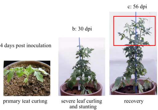
[Symptom expression] Infection of PSTVd in tomato seedlings of cotyledon stage caused primary leaf curling accompanied by vein necrosis on the 3rd to 4th leaves 2-3 weeks later (Fig. II-1a) and resulted in severe stunting of the plants 4-6 weeks later (Fig. II-1b). However, the 9th and 10th leaves newly expanded in 6-8 weeks later recovered from the symptoms, and the subsequently developing leaves showed neither leaf curling nor necrosis. Finally, the upper part of the plants higher than the 9th and 10th leaves recovered dramatically (Fig. II-1c).
c: 56 dpi
b: 30 dpi
a: 14 days post inoculation
primary leaf curling severe leaf curling recovery and stunting
Fig. II-1. Symptoms of tomato infected with PSTVd, 14 days post inoculation (dpi) (a), 30 dpi (b), and 56 dpi. Plant once stunted severely at 30 dpi, and then the upper portion (red box) recovered and started to grow like healthy at 56 dpi.
[Overall features of PSTVd and PSTVd-specific siRNA accumulation in tomato two to 45 days after inoculation]
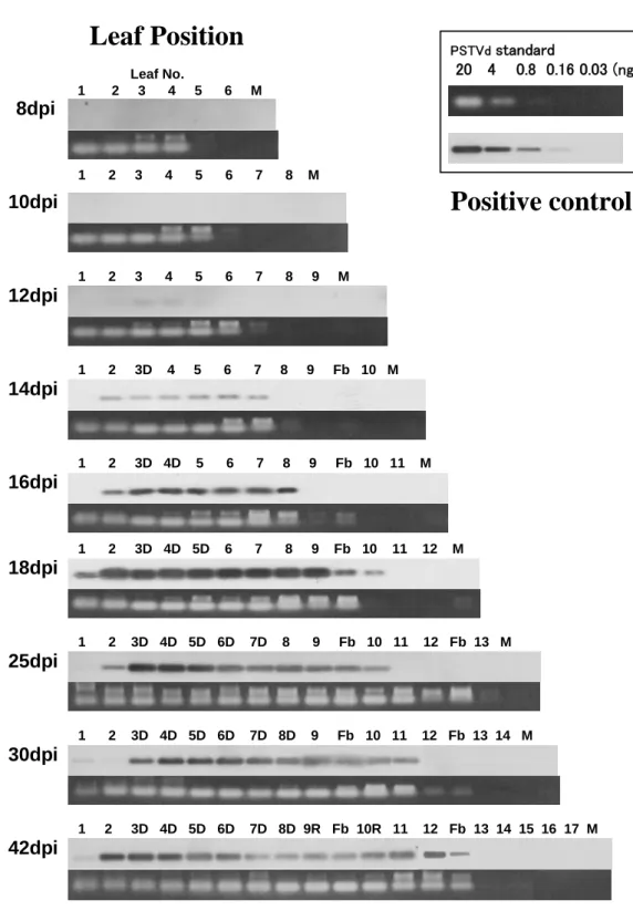
Neither PSTVd nor PSTVd-specific siRNA was detectable in any of the leaves up to 10 dpi (Fig.II-2). On 12 dpi, PSTVd reached the detectable level for the first time in the 3rd, 4th, and 5th leaves (Fig. II-2), however, PSTVd-specific siRNA was still under the detectable level (Fig.II-3). Two days later (14 dpi), when primary leaf curling appeared on some of the 3rd leaves (Fig.II-2, 14 dpi, 3D), PSTVd-specific siRNA first reached the detectable level in the 2nd, 3rd, 4th, 5th, and 6th leaves (Fig.II-3). Since the amount of LMW-RNA charged for the 7th, 8th, flower bud, 9th, 10th and the meristem was only 0.1 - 0.3 ug, further analysis was necessary for PSTVd or PSTVd-specific siRNA accumulation in these tissues (see Section II-1-2).
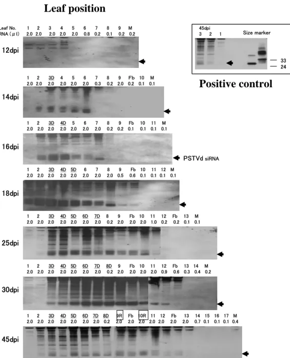
After 16 dpi, as the symptoms (leaf curl and stunt) were getting severer, PSTVd accumulation gradually increased and reached the maximum levels in the 3rd – 7th leaves on 25 dpi, 4th – 7th on 30 dpi, and 2nd – 6th on 42 dpi (Fig.II-2). Recovery began at the 9th and 10th leaves on 42 dpi when they fully expanded. It should be noteworthy that PSTVd accumulation in the recovered leaves was apparently lower than the maximum levels (Fig.II-2, 42 dpi, 9R, 10R). Meanwhile, PSTVd-specific siRNA kept the detectable level up to 42 dpi, the end of examination, from almost all the leaves that we charged at least 1.0 ug of
LMW-RNA. It should be noteworthy that two sizes (ca. 21 nucleotides and ca. 25 nucleotides) of PSTVd-specific siRNAs were clearly visible in the lower parts of the plants;
i.e., 2nd – 4th on 14 dpi, 2nd – 6th on 16 dpi, 1st – 7th on 18 dpi and 3rd – 10th on 25 dpi (Fig.II-3, 14 – 25 dpi, Fig.II-4). On the contrary, only short siRNA (ca. 21 nucleotides) was visible in the upper parts of the plants; i.e., 5th – 6th on 14 dpi, 7th on 16 dpi, 8th – flower bud on 18 dpi, and 11th on 25 dpi (Fig.II-3, 14 – 25 dpi, Fig.II-4). The double or single feature of siRNA accumulation was obscure in the latter stage of infection (Fig.II-2, 30 dpi, 42 dpi).
II-1-2.Detail analysis of PSTVd-specific siRNA accumulation on 15 days after inoculation, just after the induction of RNA silencing.
Materials and Methods
In the first experiment described above, the shoot meristem and the uppermost primordial leaves down to the 4th were so tiny that we could not get enough amounts of nucleic acids, 1.0 ug at least, which was necessary to detect viroid-specific siRNAs in our experiment conditions. Then, we infected 50 tomato seedlings with PSTVd under the same condition, and reaped all on 14 dpi when some of the plants started to show primary leaf curling in their 3rd true leaves. As in the first experiment, all the 50 leaves or primordial leaves at the same position, from the 1st to the uppermost (10th) primordial leaf and the meristem, were combined and processed for LMW-RNA extraction. Enough amount of nucleic acids were successfully obtained from the meristem as well as the tiny primordial leaves; i.e., the meristem (3 ug), 10th (3 ug), 9th (4 ug), 8th (5 ug) and 7th (5 ug). Two or 1.5 ug of the nucleic acids were electrophoresed on 12% PAGE containing 8M urea and on 1.5% AGE for the subsequent northern hybridization to detect PSTVd-specific siRNA or PSTVd.
Results
As the result, PSTVd-specific siRNA was detectable from all the primordial leaves higher than 7th but for the meristem. To our interest, only short siRNA of ca. 21-nucleotide was clearly visible in the upper primordial leaves; i.e., 7th – 10th (Fig.II-5c), and again two siRNAs (ca. 21 and 25 nucleotides) were visible in the lower expanded leaves; i.e., 2nd – 4th (data not shown, since the result was the same to Fig.II-3, 14 dpi). The result indicated that once RNA silencing was triggered in PSTVd-infected tomato plant, short PSTVd-specific siRNA of ca. 21 nucleotide accumulated quickly to the detectable level throughout the plant except for the meristem. Since long siRNA of ca. 25 nucleotides was also detected in the leaves up to 6th on 16 dpi, 7th on 18 dpi, and 10th on 25 dpi in the first experiment (Fig.II-3, 16 - 25 dpi), accumulation of long siRNA behinds two – several days of short siRNA.
Fig. II-2. Time course analysis of PSTVd accumulation in tomato infected with PSTVd.
Low molecular weight RNAs extracted from PSTVd-infected tomato, 8 – 45 days post inoculation (dpi) were loaded on 1.5% AGE, transferred to nylon membrane, and hybridized with DIG-labeled PSTVd cRNA. The upper column of each dpi indicates lumigraph obtained by northern hybridization, and the lower column indicates AGE image stained with ethidium bromide. Numbers and abbreviations are all the same to Fig. II-1. Positive control on the upper right of the figure, indicate intensity of PSTVd hybridization signals serially diluted by 5-folded from 20 – 0.03 ng per lane.
8dpi
10dpi
12dpi
14dpi
18dpi 16dpi
25dpi
30dpi
42dpi
1 2 3D 4D 5D 6D 7D 8D 9R Fb 10R 11 12 Fb 13 14 15 16 17 M PSTVdstandard
20 4 0.8 0.16 0.03 (ng)
1 2 3D 4D 5D 6D 7D 8D 9 Fb 10 11 12 Fb 13 14 M 1 2 3D 4D 5D 6D 7D 8 9 Fb 10 11 12 Fb 13 M 1 2 3D 4D 5D 6 7 8 9 Fb 10 11 12 M 1 2 3D 4D 5 6 7 8 9 Fb 10 11 M 1 2 3D 4 5 6 7 8 9 Fb 10 M 1 2 3 4 5 6 7 8 9 M 1 2 3 4 5 6 7 8 M
Leaf No.
1 2 3 4 5 6 M
Leaf Position
Positive control
Fig. II-3. Time course analysis of PSTVd-specific siRNA accumulation in tomato infected with PSTVd. Low molecular weight RNAs extracted from PSTVd-infected tomato, 12 – 45 days post inoculation (dpi) were loaded on 12% PAGE, transferred to nylon membrane, and hybridized with DIG-labeled PSTVd cRNA. Arrow head indicated the position of PSTVd-specific siRNA.
Numbers under the leaf position indicate amount of low molecular weight RNA loaded on the gels. Leaf number with D, for example 3D in 14 dpi, indicates the leaf showing disease symptoms such as leaf curl. Leaf number with R, for example 9R in 45 dpi, indicates the leaf showing recovery. ‘Fb’ means flower bud. ‘M’ means meristem. Positive control on the upper right of the figure, indicate samples from leaf 1 – 3 of PSTVd-infected tomato at 45 dpi, and PSTVd-sense oligo DNAs of 24 and 33 nucleotides.
14dpi
16dpi
18dpi
25dpi
30dpi
45dpi 12dpi
1 2 3D 4 5 6 7 8 9 Fb 10 M 2.0 2.0 2.0 2.0 2.0 2.0 0.3 0.2 2.0 0.2 0.1 0.1 Leaf No. 1 2 3 4 5 6 7 8 9 M RNA (μl) 2.0 2.0 2.0 2.0 2.0 0.8 0.2 0.1 0.2 0.2
33 24 Size marker
1 2 3D 4D 5 6 7 8 9 Fb 10 11 M 2.0 2.0 2.0 2.0 2.0 2.0 2.0 0.2 0.2 0.1 0.1 0.1 0.1
1 2 3D 4D 5D 6 7 8 9 Fb 10 11 12 M 2.0 2.0 2.0 2.0 2.0 2.0 2.0 2.0 0.5 0.6 0.1 0.1 0.1 0.1
1 2 3D 4D 5D 6D 7D 8 9 Fb 10 11 12 Fb 13 M 2.0 2.0 2.0 2.0 2.0 2.0 2.0 0.2 2.0 2.0 2.0 1.0 0.2 0.2 0.1 0.1
1 2 3D 4D 5D 6D 7D 8D 9 Fb 10 11 12 Fb 13 14 M 2.0 2.0 2.0 2.0 2.0 2.0 2.0 0.2 2.0 2.0 2.0 2.0 0.9 0.6 0.3 0.4 0.2
1 2 3D 4D 5D 6D 7D 8D 9R Fb 10R 11 12 Fb 13 14 15 16 17 M 2.0 2.0 2.0 2.0 2.0 2.0 2.0 0.2 2.0 2.0 2.0 2.0 2.0 2.0 2.0 0.7 0.1 0.1 0.1 0.4
45dpi 3 2 1
PSTVdsiRNA
Leaf position
Positive control
Leaf Number
4 5 6 7 8 9 Fb
siRNA (ca.25 nt) siRNA (ca.21 nt)
Fig. II-4. Accumulation of long and short PSTVd-specific siRNA in PSTVd-infected tomato leaves at 18 dpi. As described in the text 2.0 ug of low molecular weight RNAs from 4th to 9th leaf and primordial leaf, and a flower bud (Fb) were loaded on 12% PAGE, transferred to nylon membrane, and hybridized with DIG labeled PSTVd-cRNA. Double PSTVd-specific siRNA bands were visible in the leaf extract from 4th, 5th, 6th and 7th, but only a short PSTVd-specific siRNA band was visible in 8th, 9th and Fb.
CC
Fig. II-5. Accumulation of PSTVd-specific siRNA from PSTVd-infected tomato leaf primordia at 15 dpi. (A) and (B) indicate a representative PSTVd-infected tomato plant at 15 dpi, showing primary leaf curling. Numbers in (A) and (B) indicate leaf numbers from first to 9 and meristem (M). As described in the text 1.0 – 2.0 ug of low molecular weight RNAs from 6th to 9th leaf primordia and the meristem were loaded on 12% PAGE, transferred to nylon membrane, and hybridized with DIG labeled PSTVd-cRNA. A single PSTVd-specific siRNA band was visible except for meristem.
M
8 9 7
6
1
2
3 4 5
A B
6 7 8 9 M
PSTVd -siRNA
C
II-2.RNA Silencing in Cucumber Infected with Hop stunt viroid.
II-2-1.Recovery phenomena of cucumber plant infected with HSVd in the later stage of infection
Materials and Methods
Recovery of disease symptom in the later stage of infection was also examined in cucumber infected with Hop stunt viroid (HSVd). We infected cucumber (Cucmis sativus, cv.
Suyo) seedlings of cotyledon stage with HSVd, incubated in a grows-chamber conditioned at 20-25C, 16-h day length, and observed the symptom expression for 3 months. At the end of observation, LMW-RNA was extracted from selected leaves and 2 ug of the LMW-RNA was electrophoresed in 12% polyacrylamide gel containing 8M urea and on 1.5% agarose gel containing formaldehyde for the subsequent northern hybridization to detect HSVd-specific siRNA or HSVd.
Results [Symptom expression]
Infection of cucumber seedlings of the cotyledon stage with HSVd resulted in primary leaf curling in the 4th leaf 15 – 25 days post inoculation (dpi). The plants stunted severely and the leaves became small and curled about 6 weeks post inoculation at the 10-11th true-leaf stage.
However, the 30-40th leaf expanded in 10 – 12 weeks post inoculation recovered from curling and the upper stems and leaves (40-50th) started to grow more vigorously (Fig.II-6). The result indicated that the recovery phenomenon in the later stage of viroid-infection was also the case in cucumber infected with HSVd.
[HSVd and HSVd-specific siRNA accumulation in cucumber]
Three months, i.e., 12 weeks, post inoculation, we selected a plant which showed typical recovery, and extracted LMW-RNA from the 15 upper leaves, that means leaves of the 36-50th in Fig.II-6. Two ug of the LMW-RNA was electrophoresed in agarose gel containing folmaldehyde (Fig.II-7b), blotted onto nylon membrane, and hybridizaed with DIG-labeled HSVd cRNA probe (Fig.II-7a). HSVd concentration looked slightly lower in the 36-39th leaves which developed just after recovery from severe stunting and leaf curling (a).
LMW-RNA was also electrophoresed in 12% polyacrylamide gel containing 8M urea for northern hybridization to detect HSVd-specific siRNA. HSVd-specific siRNA was visible in all the leaves examined (data not shown).
Fig. II-6. Recovery of cucumber plant infected with HSVd in the late stage of infection, 12 weeks post inoculation. Lower part of the plant shows stunting and leaf curling, but the upper part (in the red box) recovered and started to grow vigorously.
the 30th leaf the 40th leaf The 50th leaf
Leaf Number
36 37 38 39 40 41 42 43 44 45 46 47 48 49 50
a
b
Fig.II-7. Lumigraph of HSVd northern hybridization. Two ug of LMW-RNA extracted from the 36 – 50th HSVd-infected cucumber leaves at 12 weeks post inoculation was electrophoresed in agarose gel containing folmaldehyde (b), blotted onto nylon membrane, and hybridized with DIG-labeled HSVd cRNA probe (a). HSVd concentration looked slightly lower in the 36-39th leaves which developed just after recovery from severe stunting and leaf curling (a).
II-2-2.Time course analysis of HSVd and HSVd-specific siRNA in cucumber.
Materials and Methods
We infected 15 cucumber seedlings of cotyledon stage with HSVd, and reaped 10 of them one month post inoculation. Leaves at the same position, from the 1st to the uppermost primordium, of the ten were combined and processed for extraction of 2M LiCl soluble low molecular weight RNA (LMW-RNA). Two ug of LMW-RNA was electrophoresed on 12%
polyacrylamide gel containing 8M urea and on 1.5% agarose gel containing formaldehyde for the subsequent northern hybridization to detect HSVd-specific siRNA or HSVd.
Results
[Accumulation of HSVd and HSVd-specific siRNA in cucumber]
As in the tomato infected with PSTVd, all the 10 leaves at the same position, from the 1st to 18th, were combined and processed for LMW-RNA extraction. Since the primordial leaves higher than 19th were so tiny and grew with bracts in cluster as to collect separately, we were unable to use them for further analysis. Two ug of the nucleic acids were electrophoresed on 12% PAGE containing 8M urea for the subsequent northern hybridization to detect
HSVd-specific siRNA.
As a result, two siRNAs consisting of ca.21 and ca.25 nucleotides were detected from the lower parts of the plants, i.e., 4-14th leaves, while only ca. 21 nucleotide siRNA was detectable in the younger primordial leaves; 15th and higher (Fig.II-8). The result confirmed that the accumulation pattern of long and short HSVd-specific siRNA in cucumber plant is quite similar to PSTVd-specific siRNA accumulation in tomato. However, the accumulation of HSVd-specific siRNA in cucumber was apparently lower than PSTVd-specific siRNA in tomato.
(a)
Leaf Number
M25 1 2 3 4 5 6 7 8 M25 9 10 11 12 13 14 15 16 17 18 M20
Leaf Number (b) Enlarged images of leaf number
4 5 6 7 17 18
Fig.II-8. Accumulation of long and short HSVd-specific siRNA in HSVd-infected cucumber leaves one month post inoculation. As described in the text 2.0 ug of LMW RNA from 1st to 18th leaf were loaded on 12% PAGE, transferred to nylon membrane, and hybridized with DIG labeled HSVd-cRNA. Double HSVd-specific siRNA bands were visible in the leaf extract from 4 to 14th, but only a short HSVd-specific siRNA band was visible in 17 and 18th (a). M25; 25 nucleotides size marker. M20; 20 nucleotide size marker. Arrow indicates the position of M20 faint band. Panel (b) is enlarged images of siRNA in leaf number 4,5,6,7, 17 and 18.
II-3.RNA Silencing in Cultivated Hops and Grapevines Harboring Viroids.
Once PSTVd and HSVd reached their detectable levels in tomato or cucumber, short viroid-specific siRNA of ca.21 nucleotide accumulated throughout the plant, which was followed by accumulation of long viroid-specific siRNA of ca. 23-25 nucleotides. To verify the pervasiveness of this phenomenon in the other viroid-infected host plants, analysis was carried out for viroid-specific siRNA accumulation in hops mixed-infected with HSVd and Hop latent viroid (HLVd) or with Apple fruit crinkle viroid (AFCVd) and HLVd.
II-3-1.Time course analysis of HSVd, HLVd, AFCVd and their siRNA accumulation in cultivated hops.
Materials and Methods
Five each of young shoots were collected from hops (Humulus lupulus, cv. Kirin II) cultivated in the Chitose Experiment Field of Faculty of Agriculture and Life Science, Hirosaki University, Aomori, Japan, on June 3 and 20, 2005. Mature hop leaves and cones were also collected on September 20. They were collected from hops infected with HSVd-citrus or AFCVd, and also from healthy as a control. All the hops, however, were latently infected with HLVd.
On June 3, shoots were ca.15 cm long and had two pairs of expanded leaves, but have not yet shown any disease symptoms (Fig.II-9a). On June 20, on the other hand, shoots grew ca.30cm long with four pairs of expanded leaves, and those infected with HSVd began to show typical leaf curling (Fig.II-9b).
For the samples collected on June 3 and 20, all the 5 pairs of leaves at the same position, from the 1st to the meristem, were combined and processed to extract 2M LiCl soluble nucleic acids (LMW-NA). Nevertheless, the primordial leaves higher than the 10th were mixed and extracted together with the meristem, because they were so tiny and grew with bracts in cluster as to collect separately. For the mature leaves and cones collected on September 20, all the leaves and cones of ca. 60g were combined and used for LMW-RNA extraction. Twenty ug of LMW-NA from young shoots and of LMW-RNA from matured leaves, were electrophoresed on 12% PAGE containing 8M urea for northern hybridization to detect viroid-specific siRNA.
Results
[Young shoots in June] Viroid-specific siRNA was not detectable in any of the young leaves collected on June 3 (data not shown), even though we have charged 20 ug of LMW-NA which is 10-times as much as those in PSTVd/tomato and HSVd/cucumber.
Seventeen days later (June 20), when primary leaf curling appeared on some of the leaves (Fig.II-9, arrows), HSVd-specific siRNA of ca. 21-25 nucleotides first reached the detectable
level in the 1st leaf (Fig.II-10(a), leaf number 1). At the same time, HLVd-specific siRNA of the similar size also reached the detectable level in the 1st and 2nd leaves (Fig.II-10(b), leaf number 1 and 2), although the viroid does not induce any detectable symptom.
AFCVd-specific si-RNA was not detectable at this stage (Fig.II-10(c)).
[Mature leaves and cones in September] All the viroid-specific siRNAs were clearly visible in mature hop leaves and cones collected in September (Fig.II-11). In addition, accumulation of long (ca. 25 nucleotides)- and short (ca. 21 nucleotides)-siRNA was apparent in HSVd- (Fig.II-11(a)) and HLVd-hops (Fig.II-11(c)). In contrast, long AFCVd-specific siRNA was obscure comparing to more abundant short siRNA (Fig.II-11(b)). The result again confirmed that the accumulation of long and short viroid-specific siRNA was the case in hops infected with HSVd, HLVd and probably AFCVd, although we could not follow the accumulation pattern of long and short viroidd-specific siRNAs in hops due to their low accumulation in the field conditions.
It may not be surprising, but we should stress the fact that both siRNAs specific to HSVd and HLVd, or to AFCVd and HLVd were inducible in the same tissue when the plants were mix-infected with two different viroids.
Fig.II-9. Hop shoot mix-infected with HSVd-citrus and HLVd on June 3 and 20. The shoot on June 20 showed typical primary leaf curling (arrows).
June 3, 2005 June 20, 2005
Leaf Number
1 2 3 4 5 6 7 8 9 10-M
(a) HSVd-hops / HSVd probe
(b) Healthy-hops / HLVd probe
(c) AFCVd-hops / AFCVd probe
Fig.II-10. Accumulation of viroid-specific siRNA in young hop shoot (June 20) infected with HSVd, AFCVd and HLVd. As described in the text 20 ug of LMW RNA from 1st to 9th leaf, and the above to merisitem were loaded on 12% PAGE, transferred to nylon membrane, and hybridized with DIG labeled HSVd (a), HLVd (b) and AFCVd (c) cRNA probes. HSVd- and HLVd-specific siRNA band of ca. 20 nt was faintly visible in leaf 1 of HSVd, and leaf 1 and 2 of HLVd (arrows). Si-RNA was not detectable in AFCVd-hops at this stage (c).
AFCVd HSVd Healthy AFCVd HSVd Healthy AFCVd HSVd Healthy
(a) HSVd probe (b) AFCVd probe (c) HLVd probe
Fig.II-11. Accumulation of AFCVd-, HSVd-, and HLVd-specific siRNA in mature hop leaves and cones (September 21) infected with HSVd, AFCVd, and HLVd. Three sets of 20 ug of LMW-RNAs extracted from AFCVd-infected, HSVd-infected and healthy hops were electrophoresed on 12% PAGE containing 8M urea, blotted to nylon membrane and hybridized with DIG-labeled AFCVd, HSVd, and HLVd probes. Both of AFCVd- and HSVd-hops were mix-infected with HLVd. Arrow indicates viroid-specific siRNAs. Accumulation of long (ca. 25 nucleotides) - and short (ca. 20 nucleotides)-siRNA was visible in HSVd and HLVd. Both of the viroid-specific siRNAs was detectable in the tissues mix-infected with HSVd and HLVd, and AFCVd and HLVd..
II-3-2.Accumulation of HSVd, GYSVd and their siRNA in cultivated grapevines.
Materials and Methods
Fully expanded young leaves of grapevine (Vitis vinifera), cultivars ‘Koshu’, ‘Alphonse lavallee’, ‘Citronne’, ‘Pione’, and ‘Muscut Bailey A’ were collected on June 20, 2002, from several vineyards in Yamanashi prefecture, Japan. All the vines are ca. 40- to 100-year-old.
Leaves of ca. 20g were combined and used for LMW-RNA extraction. LMW-RNA was electrophoresed on 12% PAGE containing 8M urea and on 1.5% agarose gel containing formaldehyde for northern hybridization to detect viroid-specific siRNA and viroid.
Results
Ten ug of LMW-RNA was electrophoresed on 1.5% agarose gel containing formaldehyde, blotted onto nylon membrane, and hybridized with DIG-labeled HSVd- and GYSVd-cRNA probes. HSVd was positive in ‘Alphonse labalee’, ‘Citronne’, ‘Pione’ and ‘Muscut Bailey A’.
GYSVd was positive in ‘Alphonse labalee’, ‘Citronne’, ‘Pione’ and ‘Muscut Bailey A’.
Hybridization signals suggested that the concentrations of HSVd and GYSVd in these leaves are fairly good (data not shown).
Then, 50 – 100 ug of LMW-RNA was electrophoresed on 12% PAGE containing 8M urea, blotted onto nylon membrane, and hybridized with DIG-labeled HSVd- and GYSVd-cRNA probes. Although we have charged 50 – 100 ug of LMW-RNA, which is 25 – 50 times as much as those in PSTVd/tomato and HSVd/cucumber, neither of HSVd- and GYSVd-specific siRNA was detectable from any of the samples examined. Another trials should be done using matured grapevine leaves or fruits collected in autumn.
(a) Grapevine samples
1 2 3 4 5 M25
(b) HSVd probe (c) GYSVd probe
1 2 3 4 5 M25 1 2 3 4 5 M25
Fig.II-12. Accumulation of HSVd- and GYSVd-specific siRNA in five grapevine cultivars collected on May 20, 2002, cultivating in Yamanashi prefecture, Japan. Fifty to 100 ug of LMW-RNAs extracted from five vines (ca. 40 to 100-year-old) were electrophoresed on 12% PAGE containing 8M urea (a), blotted onto nylon membrane and hybridized with DIG-labeled HSVd (b) and GYSVd probes (c). M25 means 25 nucleotide oligo DNA homologous to HSVd, and arrows indicate the position.
Viroid-specific siRNA accumulation was not apparent in these samples.
II-4.RNA Silencing in Tomato Infected with Cucumber Mosaic Virus.
II-4-1.Accumulation of CMV-specific siRNA in tomato.
Materials and Methods
The similar analysis was conducted on tomato infected with Cucumber mosaic virus (CMV). Ten tomato (Lycopersicon esculentum, cv. Rutgers) seedlings of cotyledon stage were infected with CMV. Two week later, some of the plants dwarfed severely and mosaic appeared on the expanded leaves. Sixteen day later when all the plants showed similar disease symptoms, 10 leaves at the same leaf position, from the 1st to the meristem, were combined and processed for extraction of 2M LiCl soluble RNA. Two ug of LMW-RNA was electrophoresed on 12% PAGE containing 8M urea for northern hybridization to detect CMV-specific siRNA. However, as in the case of PSTVd in tomato, primordial leaves higher than 7th were so tiny to extract enough amount of LMW-RNA. Only 0.1 to 0.5 ug was charged for the analysis.
Results
Only short CMV-specific siRNA of ca.21 nucleotides was detectable even in the fully expanded 1st – 6th leaves, but for the 3rd (Fig.II-13(b)). No long CMV-specific siRNA was detected in any of the leaves examined. These results supported that long viroid-specific siRNA appeared in the fully expanded lower leaves of viroid-infected host plants was not an artifact, rather accumulation of short siRNA of ca.21 nucleotides followed several days later by long siRNA of ca.25 nucleotides seemed to be a phenomenon unique to viroid-infected host plants.
Leaf Number
M 1 2 3 4 5 6 7 8 9 M M 1 2 3 4 5 6 7 8 9 M
(a) PAGE (b) northern hybridization / CMV probe Fig.II-13. Accumulation of CMV-specific siRNA in CMV-infected tomato leaves 16 days post inoculation. As described in the text, 2.0 ug of LMW RNA from 1st to 9th leaf, and the meristem were electrophoresed on 12% PAGE (a), blotted onto nylon membrane, and hybridized with DIG labeled CMV RNA3-cRNA probe. Only short CMV-specific siRNA band was visible in the leaf extract from 1st to 6th, except for 3rd (b). M; 20 nucleotides oligo DNA size marker. Arrow indicates the position of 20 nucleotides size marker.
III.Results and Discussion Viroid-inducing RNA Silencing and Recovery
As described in Chapter 1, when tomato plants (cv. Rutgers) were infected with PSTVd at the cotyledon stage, primary leaf curling appeared on the 3rd or 4th leaf 11–14 days after inoculation. More severe leaf curling with vein necrosis appeared on the 5th to 9th leaves and the plants became severely stunted. The severity of disease symptoms, however, decreased dramatically during the later stages, i.e., later than 6 weeks, of the infection, and almost healthy-looking leaves began to expand on the upper portion of infected plants. The concentration of PSTVd reached its highest level in the leaves with severe leaf curling but decreased in the upper healthy-looking leaves. In addition, accumulation of PSTVd-specific siRNAs preceded tomato plant recovery from severe symptoms. The circumstantial evidence gave us an assumption that the recovery of tomato plants from severe infection may be dependent on a decrease of the PSTVd concentration in the upper leaves in relation to RNA silencing targeting PSTVd replication triggered in the infected plants.
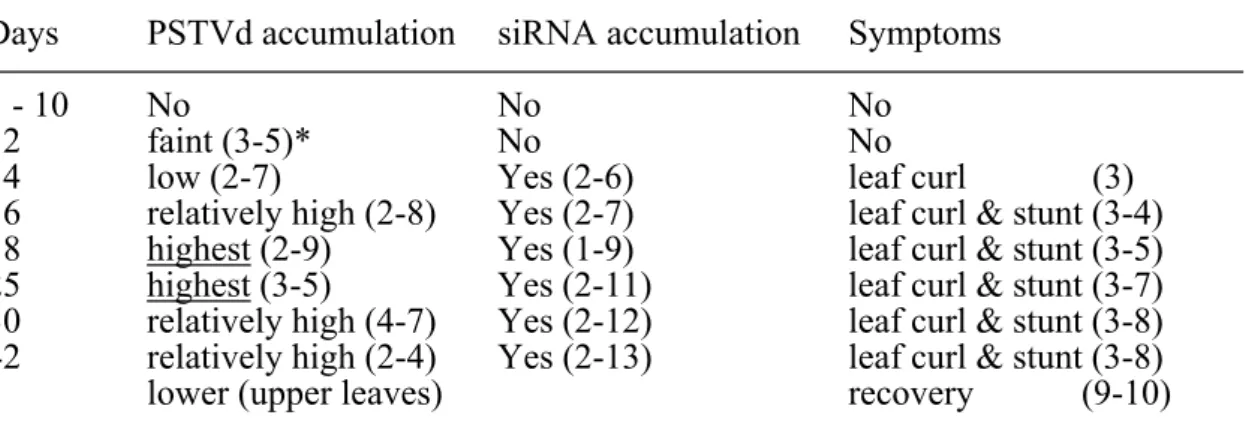
In the Chapter 2, we first performed three experiments on time-course analysis of viroid-specific siRNA accumulation in plants infected with PSTVd and HSVd, to have a clearer understanding of the relation between the timing of recovery and viroid-specific siRNA accumulation. The first experiment was a two-day interval analysis of PSTVd, PSTVd-specific siRNA and symptoms in tomato infected with PSTVd. The result was summarized in Table 1. In this experiment, when plants were infected with PSTVd in the cotyledon stage, the primary leaf curl appeared on the 3rd true leaf 14 days after inoculation.
PSTVd accumulation reached the detectable level in 3rd–5th leaves 12 days after inoculation, i.e., two days before the primary symptom expression. PSTVd-specific siRNA reached the detectable level in 2nd–7th leaves 14 days after inoculation, i.e., two days behind PSTVd detection, or the same day to primary symptom expression. Symptoms consisting of leaf curling with vein necrosis became severer in leaf number 4th-8th and plants stunted severely by 16, 18, 25 and 30 days after inoculation. PSTVd accumulation reached the maximum level in the leaf number 2nd-9th at 18 days after inoculation, and sub-maximum level in 3rd-5th at 25 days after inoculation. PSTVd accumulation decreased gradually to a certain level hereafter, although it still kept fairly good concentration. These results indicated that, in PSTVd-infected tomato plants, PSTVd accumulation reached the maximum level 18-25 days after inoculation in the leaf number 2nd-9th which showed severe leaf curling accompanied by vein necrosis, and then gradually decreased to a certain level in the upper leaves which started to recover from severe disease symptoms.
Table 1. Time-course analysis of PSTVd, PSTVd-specific siRNA and symptoms in tomato (cv. Rutgers) infected with PSTVd.
Days PSTVd accumulation siRNA accumulation Symptoms
1 - 10 No No No
12 faint (3-5)* No No
14 low (2-7) Yes (2-6) leaf curl (3)
16 relatively high (2-8) Yes (2-7) leaf curl & stunt (3-4) 18 highest (2-9) Yes (1-9) leaf curl & stunt (3-5) 25 highest (3-5) Yes (2-11) leaf curl & stunt (3-7) 30 relatively high (4-7) Yes (2-12) leaf curl & stunt (3-8) 42 relatively high (2-4) Yes (2-13) leaf curl & stunt (3-8) lower (upper leaves) recovery (9-10)
*: numbers in parenthesis indicate leaf number, i.e., (3-5) means leaf numer 3 to 5.
A defect in this experiment was insufficiency of LMW-RNA obtained from tiny primordial leaves. We reaped 5 plants every two days and combined them for extraction, however, the amounts of LMW-RNA from 5 tiny primordial leaves were less than 0.4 ug at the best, which was still below the detection limit of our experiment condition. At least 1 ug of LMW-RNA in a lane was essential to verify PSTVd-specific siRNA accumulation in our northern hybridization sensitivity. To improve the limitation, we have them performed the second experiment focusing on the day when PSTVd-specific siRNA first reached the detectable level or the day just after the induction of RNA silencing; i.e., 14 or 15 days after inoculation.
As described in Result section, we successfully extracted enough amount of LMW-RNA from 50 PSTVd-infected tomato plants of 15 days after inoculation. The analysis of PSTVd-specific siRNA accumulation in the tiny primordial leaves clearly indicated that PSTVd-specific siRNA was detectable in all the primordial leaves up to 9th at 15 days after inoculation, just the day when primary disease symptom was observed or the day just after the induction of RNA silencing targeting PSTVd replication. It should be noteworthy that the 9th primordial leaf has just begun to develop at the time when PSTVd-specific siRNA first reached the detectable level in all the leaves and primordial leaves except for the meristem, and recovery started later from the 9th and 10th leaves. It means that the recovery takes place according to the following process; i.e., (1) PSTVd-specific siRNA spread systemically in the plant immediately after the induction of RNA silencing targeting PSTVd replication, (2) primordial leaves being developed hereafter will be exposed to the RNA silencing from the beginning of their differentiation, and (3) suppression of PSTVd replication in these tissues later results in recovery from severe disease symptoms. If the recovery starts in such
primordial leaves after establishment of RNA silencing, we will meet the recovery phenomenon after the leaves fully expanded nearly a month later.
Accumulation of a short and a long siRNA unique to viroid-inducing RNA silencing In the second experiment, when PSTVd-inducing RNA silencing was established in tomato plants, two classes of PSTVd-specific siRNAs were detectable; i.e., short one of ca. 21 nucleotides and long one of ca. 25 nucleotides. It should be noteworthy that when we examined PSTVd-specific siRNA accumulation in all the expanded leaves and primordial leaves at 15 days after inoculation, just after the induction of RNA silencing, only a short siRNA was detectable in the upper primordial leaves being developed, on the other hand, both of the short and a long siRNAs were clearly detectable in the lower fully expanded leaves showing prominent disease symptoms. Similar analysis on 16, 18, and 25 days after inoculation revealed that only the short siRNA was detectable in the primordial leaves being developed, however, the long siRNA as well as the short one became detectable later as the leaves grew up to expand. It was also evident that accumulation of the long one behinds the short one by 2 to several days, or, in other words, the short one preceded the long one 2 to several days. Since the short one spread whole plants except for the meristem quickly after the induction, it looks more like an identified signaling molecule for systemic spreading of viroid-specific RNA silencing.
The similar analysis performed using cucumber plants infected with HSVd again indicated that only a short HSVd-specific siRNA of ca.21 nucleotides was evident in the primordial leaves being developed, however, both of the long and the short ones were clearly detectable in the lower expanded leaves.
The double viroid-specific siRNA accumulation pattern was also observed in hops infected with HSVd, AFCVd and HLVd in the later growing season. On the contrary, only a short CMV-specific siRNA of ca. 21 nucleotides was detectable in tomato plants infected with CMV, but no long CMV-specific siRNA was detectable in any of the leaves, even in fully expanded symptomatic leaves. Consequently, the pattern of siRNA accumulation in which a short one of ca.21 nucleotides first accumulated quickly after the induction of RNA silencing then a long one of ca.25 nucleotides followed slowly 2 – several days behind, was the one unique to viroid-inducing RNA silencing triggered in viroid-infected host plants.
IV.References
Diener TO (1971) Potato spindle tuber “virus”: a plant virus with properties of a free nucleic acid. III. Subcellular location of PSTV-RNA and the question of whether virions exist in extracts or in situ. Virology 43:75–89.
Diener TO (2001) The viroid: biological oddity or evolutionary fossil? Adv. Virus Res.
57:137–84.
Hadidi A, Flores R, Randles JW, Semancik JS, eds. (2003) Viroids. CSIRO Publ.:
Collingwood, Aust.
Itaya A, Folimonov A, Matsuda Y, Nelson RS, Ding B (2001) Potato spindle tuber viroid as inducer of RNA silencing in infected tomato. Mol. Plant-Microbe Interact.
14:1332–1334.
Papaefthimiou I, Hamilton AJ, Denti MA, Baulcombe DC, Tsagris M, Tabler M (2001) Replicating potato spindle tuber viroid RNA is accompanied by short RNA fragments that are characteristic of posttranscriptional gene silencing. Nucleic Acids Res.
29:2395–2400.
Mart´ınez de Alba AE, Flores R, Hern´andez C (2002) Two chloroplastic viroids induce the accumulation of the small RNAs associated with post-transcriptional gene silencing. J.
Virol. 76:13094–13096.
Markarian N, LiHW,DingSW,Semancik JS (2004) RNA silencing as related to viroid induced symptom expression. Arch. Virol. 149:397–406.
Woo Y-M, Itaya A, Owens RA, Tang L (1999) Characterization of nuclear import of potato spindle tuber viroid RNA in permeabilized protoplasts. Plant J. 17:627–635.
Ding B, Kwon M-O, Hammond R, Owens RA (1997) Cell-to-cell movement of potato spindle tuber viroid. Plant J. 12:931–936.
Flores R, Hern´andez C, Mart´ınez de Alba AE, Dar`os JA, Di Serio F (2005) Viroids and viroid-host interactions. Annu. Rev. Phytopathol. 2005. 43:4.1–4.23
Sano T, Matsuura Y (2004) Accumulation of short interfering RNAs characteristic of RNA silencing precedes recovery of tomato plants from severe symptoms of potato spindle tuber viroid infection. J. Gen. Plant Pathol. 70:50–53.