Fukushima Medical University
福島県立医科大学 学術機関リポジトリ
This document is downloaded at: 2021-11-08T00:21:17Z
Title Ratio of alpha2-macroglobulin levels in cerebrospinal fluid and serum: an expression of neuroinflammation in acute
disseminated encephalomyelitis( 本文 ) Author(s) 鈴木, 雄一
Citation
Issue Date 2019-09-30
URL http://ir.fmu.ac.jp/dspace/handle/123456789/1051
Rights
© 2019. This accepted manuscript version is made available under the CC-BY-NC-ND 4.0 license. Published version:
Pediatr Neurol. 2019 Sep;98:61-67. doi:
10.1016/j.pediatrneurol.2019.04.020 DOI
Text Version ETD
Ratio of alpha 2-macroglobulin levels in cerebrospinal fluid and serum: an expression of neuroinflammation in acute disseminated encephalomyelitis
Yuichi Suzuki1), Koichi Hashimoto1), Kyoka Hoshi2), Hiromi Ito2), Yoshinobu Kariya2), Kyohei Miyazaki1), Masatoki Sato1), Yukihiko Kawasaki1), Mari Yoshida3), Takashi Honda4), Yasuhiro Hashimoto2), Mitsuaki Hosoya1)
1) Department of Pediatrics, Fukushima Medical University School of Medicine, Fukushima, Japan
2) Department of biochemistry, Fukushima Medical University School of Medicine, Fukushima, Japan
3) Department of Neuropathology, Institute for Medical Science of Aging, Aichi Medical University, Aichi, Japan
4) Department of Human Life Sciences, Fukushima Medical University School of Nursing, Fukushima, Japan
Address:
Yuichi Suzuki [email protected]
Department of Pediatrics, Fukushima Medical University School of Medicine, 1 Hikariga-oka, Fukushima, Fukushima 960-1295, Japan
Word counts:
Abstract: 241 words Manuscript: 3358 words
Abstract
BACKGROUND: Acute encephalitis and encephalopathy (AEE) are life-threatening diseases in children. However, no laboratory examinations are performed for their early diagnosis and treatment. Alpha 2-macroglobulin (α2M) is a blood glycoprotein that increases at the early stages of inflammation. In this study, we investigated whether α2M was associated with AEE.
METHODS: We analyzed the association between AEE and α2M using cerebrospinal fluid (CSF) and serum samples from patients with acute disseminated encephalomyelitis (ADEM), infection-related acute encephalopathy (AE), febrile status epilepticus (FSE), and febrile seizure (FS). Samples were collected in the pediatric department of hospitals throughout Fukushima Prefecture between January 1, 1999, and May 31, 2012.
RESULTS: α2M levels in the CSF were quantified at 4.7 (3.8–8.4) μg/mL for ADEM, 2.1 (1.1–
2.3) μg/mL for AE, 1.1 (0.9–6.4) μg/mL for FSE, and 1.0 (0.8–1.1) μg/mL for FS. α2M levels in patients with ADEM were significantly higher than those in patients with AE and FS (p = 0.019 and p = 0.002, respectively). The ratio of the α2M level in the CSF to that in the serum in patients with ADEM was significantly higher than that in patients with FSE (p = 0.04). In patients with ADEM, α2M levels in the CSF decreased with treatment.
CONCLUSIONS: Our results suggest that the ratio of the α2M level in the CSF to that in the serum reflects the neuroinflammatory status of patients with ADEM and indicates the
expression of early neuroinflammation in ADEM.
Key words: Alpha 2-macroglobulin; neuroinflammation; encephalitis; encephalopathy; febrile seizure
Running title: Alpha 2-macroglobulin in ADEM
Introduction
Acute encephalitis and encephalopathy (AEE) can result in various sequelae, including death;
hence, early intervention is required. Although AEE occurs with a low frequency, it is common in infants in East Asia (1). It results from various causes, including viral infections and
connective tissue disease; moreover, pathologies related to immune conditions are different and have not yet been clarified. Therefore, AEE diagnosis is based on a comprehensive assessment of each patient’s clinical course, laboratory examinations, head imaging, and
electroencephalogram findings. However, in the acute phase, particularly when the patient is in a critical condition, diagnosis is often challenging because of the difficulty associated with conducting adequate examinations. In other words, these diseases are difficult to distinguish based on clinical presentations, despite the difference in pathologies.
Acute encephalitis is classified into viral and non-viral encephalitis. The most common viral encephalitis is herpes encephalitis, whereas the most common non-viral encephalitis types include acute disseminated encephalomyelitis (ADEM) and non-herpetic acute limbic encephalitis (ALE). In viral encephalitis, viral pathogen isolation or gene detection is directly linked to its diagnosis. The herpes simplex virus is the most common cause of encephalitis in western countries. Herpes simplex encephalitis occurs at all ages during infancy and childhood, with peak incidence during the first year of life (2). For non-viral encephalitis, diseases in which autoantibodies are involved in the pathology have recently been clarified, such as the
anti-N-methyl-D-aspartic acid receptor antibody in non-herpetic ALE. Nevertheless, the pathology of ADEM remains unclear.
Acute encephalopathy (AE) is classified as being either related or unrelated to infectious diseases. Infectious disease-related AE is a generic term for a wide range of cerebral
dysfunctions accompanying infectious diseases, although no finding has suggested pathogen invasion in the central nervous system (CNS). On the contrary, encephalopathy unrelated to infectious diseases is a brain dysfunction accompanying various non-infectious diseases, such as liver failure, renal failure, and hypertension. Febrile seizure (FS) is a generic term for the condition in which convulsions appear transiently during a fever. FS is a common disease in children and has a good prognosis without clinical sequelae. As the initial clinical symptoms of ADEM, AE, and FS are similar, these diseases are difficult to differentiate in the acute phase.
Many cytokines and biochemical proteins have been studied as biomarkers of AEE. Reportedly, levels of inflammatory cytokines in the cerebral spinal fluid (CSF) increase during the acute phase, and studies have reported the role of interferon-gamma in acute encephalitis; the role of interleukin-6 (IL-6) and IL-8 in bacterial meningitis; and the role of tumor necrosis factor (TNF)-alpha, soluble TNF receptor-1, and IL-6 in AE (3, 4). On the contrary, regarding
biochemical proteins in the CSF, S-100B, glial fibrillary acidic protein (GFAP), and tau protein
have been reported as brain injury biomarkers of AE (5–8). These biomarkers represent the repair process of damaged nerve cells and axons. Therefore, they are not suitable for expressing neuroinflammation in the acute phase.
Alpha 2-macroglobulin (α2M) can be an indicator of acute inflammation, which is a biological defense response, unlike the previously reported damaged substances resulting from cytotoxicity, such as S-100 B, GFAP, and tau protein. We focused on α2M, whose blood level increased in a short time at the onset of inflammation. An increase in α2M levels in the CSF in bacterial meningitis has been reported (3, 9, 10). Extensive studies have been conducted on the relationship between α2M levels in the CSF and CNS diseases in patients with bacterial meningitis, showing that α2M levels in the CSF reflected α2M produced in the serum during systemic inflammation due to the damage to the blood–brain barrier (BBB) (3, 10–12). However, there have been no reports on α2M levels in patients with ADEM.
In this study, we aimed to measure α2M levels in the CSF and serum samples of patients with ADEM, AE, febrile status epilepticus (FSE), or FS in the acute phase and to examine the association between neuroinflammation and α2M.
Materials and Methods Sample collection Participants
We analyzed preserved samples collected from 35 patients with ADEM, AE, FSE, and FS who were treated in the pediatric departments of hospitals throughout Fukushima Prefecture between January 1, 1999, and May 31, 2012. These samples were stored to assess cytokine levels and conduct viral polymerase chain reactions (PCRs). The first diagnosis was provided based on the findings on admission, and it was confirmed by the clinical course. A presumable diagnosis of ADEM was clinically made based on alterations to or a loss of consciousness lasting > 24 h, presenting with acute polysymptomatic neurological signs, and cranial/spinal imaging showing one or multiple lesions suggestive of demyelination. A presumable diagnosis of AE was
clinically established based on the acute onset of impaired consciousness accompanied by brain dysfunction, usually preceded by infection (1, 13). The FS group consisted of children who presented with fever and seizures; however, they were later found to be free from acute
neurological damage based on the course of clinical events, laboratory data, and brain imaging, where available. FSE was diagnosed in patients with FS who presented with seizures lasting for
> 30 min. CSF samples from patients with bacterial meningitis were used as positive controls, and patients with leukemia in remission with no neurological abnormality confirmed by regular examinations were used as negative controls. Patients with viral meningitis, myelitis, or vascular, metabolic, endocrine, or toxic disorders were excluded.
Preservation of CSF and serum samples
CSF and serum samples were all collected within 24 h after disease onset. These samples were immediately centrifuged after collection and stored at −80°C. Patients who provided samples in which macroscopic hemolysis was observed were excluded from the analysis.
Ethics
Preserved samples were used in this study. This study was conducted with the approval of the Ethics Committee of the Fukushima Medical University (approval no. 1684). Information related to this research was released on our university website, and patients were included in the study using an opt-out methodology.
Immunoblotting
The protein concentration of clinical samples was quantified using a BCA Protein Assay Kit (ThermoFisher Scientific, Waltham, MA, USA). CSF samples were diluted with a sample loading buffer and heated at 95°C for 5 min, and then electrophoresed at 200 mV, 20 mA/sheet, for 73 min using a 5%–20% gradient polyacrylamide gel (Wako, Osaka, Japan). Clinical
specimens with a total protein content of 0.4 μg and 2 ng of human plasma α2M (Sigma-Aldrich, St. Louis, MO, USA) as a standard were electrophoresed. The gel of <15 kDa was cut out from the main gel and subjected to silver staining (Wako) to detect transthyretin as a banding control.
It was then transferred to nitrocellulose membranes (Bio-Rad Laboratories, Hercules, CA, USA) at 300 V, 350 mA, for 45 min by wet western blotting. After blocking for 1 h at room
temperature under 3% milk phosphate-buffered saline containing Tween-20 (PBST), the membranes were reacted with 3 μg/mL of anti-α2M antibody (ICN/Cappel, Aurora, OH, USA) and diluted with 3% milk PBST for 2 h. After washing three times with PBST, the membranes were reacted with horseradish peroxidase (HRP)-labeled anti-goat IgG antibody (Jackson ImmunoResearch Laboratories, West Grove, PA, USA), diluted to 0.08 μg/mL with 3% milk PBST for 1 h, and then washed in the same way with PBST. Immunoreactive bands were detected using a SuperSignal® West Dura Extended Duration Substrate (ThermoFisher). The band intensity was calculated using an Image Saver 6 luminescence detector (ATTO, Tokyo, Japan).
Enzyme-linked immunosorbent assay (ELISA)
For ELISA, 100 μL of anti-human α2M antibody (2.0 μg/mL; ICN/Cappel Pharmaceuticals) diluted with 0.05 M carbonate–bicarbonate (pH 9.6) was added to each well of a 96-well C8 MaxiSorp™ immunomodule plate (Nunc, Roskilde, Denmark) and incubated at 4°C overnight.
After washing once with Tris-buffered saline containing Tween-20 (TBST), 300 μL of 10%
Block Ace (DS Pharma Biomedical, Osaka, Japan) was added to each well and incubated as a blocking step for 1 h. After washing five times with TBST, 0.75 μL of each CSF sample was applied to each well and incubated at 37°C for 1 h. After washing again five times with TBST, the plate was incubated with 100 μL of HRP-conjugated anti-human α2M antibody (2.0 μg/mL;
GeneTex, San Antonio, TX, USA) at room temperature for 1 h. After further washing five times with TBST, the plate was reacted with 3, 3', 5, 5'-tetramethylbenzidine peroxidase substrates (Kirkegaard and Perry Laboratories, Gaithersburg, MD, USA). The reaction was quenched using 1 M hydrochloric acid, and the absorbance at 450 nm was measured with a microplate reader (Bio-Rad Laboratories).
Immunohistochemistry
Formalin-fixed paraffin-embedded brain tissue infected with herpes encephalitis was cut into 5-μm-thick sections using a microtome and mounted on glass slides. Deparaffinized sections were incubated with or without anti-human α2M antibody (goat IgG, AF1938, R&D
SYSTEMS)followed by biotinylated rabbit anti-goat IgG (Nichirei, Tokyo, Japan). The antigens were visualized with avidin–biotin peroxidase complex and peroxidase DAB enzyme
histochemistry. Sections were then counterstained with cresyl violet and cover-slipped.
Statistical analysis
Statistical analysis was performed using IBM SPSS Statistics 21 (IBM Japan, Ltd., Tokyo, Japan). Continuous variables were expressed as medians (interquartile range). Comparisons between groups were made using the Kruskal–Wallis test. Thereafter, comparisons between two groups were made using the Mann–Whitney U test. A p-value < 0.05 was considered
statistically significant.
Results
Patient characteristics
Table 1 shows the characteristics of patients according to the diagnosis. In total, 35 patients (17 boys, 48.6%, and 18 girls, 51.4%) were enrolled. The age at disease onset ranged from infancy to school aged. No significant differences were found in their age at diagnosis.
General examination findings
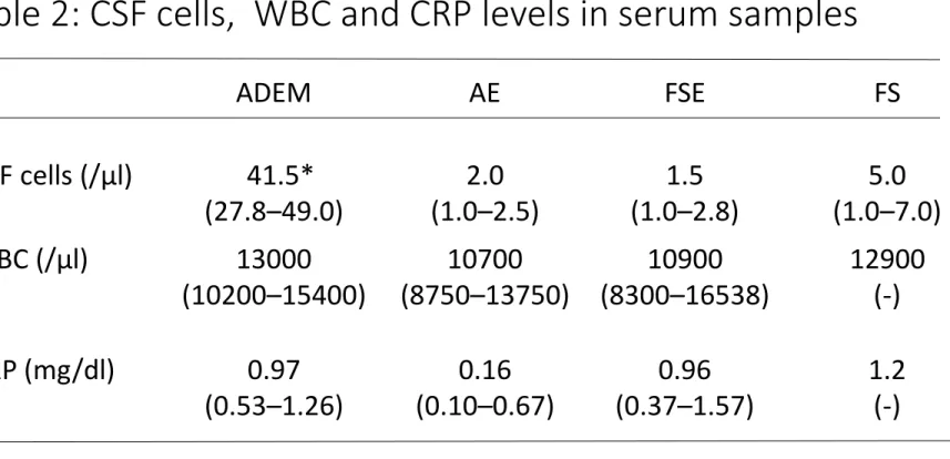
The number of cells in the CSF and the white blood cell count in blood, as well as the
C-reactive protein (CRP) levels in the blood samples, are shown in Table 2. The median number of cells in the CSF was 41.5 (27.8–49.0)/μL for patients with ADEM, 2.0 (1–2.5)/μL for those
with AE, 1.5 (1.0–2.8)/μL for those with FSE, and 5.0 (1.0–7.0)/μL for those with FS. The number of cells in the CSF samples from patients with ADEM was significantly higher than those from patients with AE, FSE, and FS (p = 0.02, 0.01, and 0.04, respectively). On the contrary, no significant differences were found in the number of cells in the white blood cell count or CRP levels among the blood samples.
Detection of α2M in CSF by western blotting
A 180-kDa α2M band (Fig. 1) was detected in all CSF samples from patients with AE, ADEM, FSE, and FS. Strong bands were detected in two samples (nos. 1 and 2) from patients with ADEM. Samples from patients with bacterial meningitis, in whom α2M levels in the CSF were reported to increase markedly, were used as positive controls (no. 11). A strong band was also detected in the other sample from patients with AE, but most other samples had weak bands. In addition, the bands of the control samples were weak (nos. 9 and 10).
Quantification of α2M in CSF and serum by ELISA
To confirm the results of the western blot analysis, we performed sandwich ELISA for α2M using the CSF and serum samples (Table 3). The median α2M level (interquartile range) in the CSF was 4.7 (3.8–8.4), 2.1 (1.1–2.3), 1.1 (0.9–6.4), and 1.0 (0.8–1.1) μg/mL in patients with ADEM, AE, FSE, and FS, respectively. The α2M level in patients with ADEM was significantly higher than those in patients with AE and FS (p = 0.019 and p = 0.002, respectively). For reference, the α2M level in the CSF from patients with bacterial meningitis (n = 4) and control patients (n = 18) was 24.0 (6.9–25.2) and 1.54 (1.3–2.0) μg/mL, respectively (data are not shown). On the contrary, the median α2M level in the serum of patients was 363.9 (352.3–
380.8) mg/dL in ADEM, 353.8 (328.7–429.7) mg/dL in AE, 356.4 (335.1–392.3) mg/dL in FSE, and 263.6 (174.7–284.9) mg/dL in FS. No significant differences were observed in α2M levels in the serum among the diseases. The ratio of the α2M level in the CSF to that in the serum was examined, and the median value of this ratio in patients with ADEM was significantly higher than those in patients with AE and FSE (p = 0.016 and p = 0.04, respectively).
Scatter diagram of α2M levels in CSF and serum
To investigate the correlation between α2M levels in the CSF and serum, we plotted α2M levels in 10 paired samples of the CSF and serum from five patients with ADEM and five patients with AE (Fig. 2). α2M levels in the CSF differed markedly among patients with ADEM, regardless of α2M in their serum. There was almost no correlation between α2M levels in the CSF samples and those in the serum samples (R = 0.264, p = 0.668) from patients with ADEM. On the contrary, in patients with AE, α2M levels in the CSF were low.
Receiver operating characteristics (ROC) curve
To distinguish ADEM from the other diseases (AE, FSE, and FS) based on the ratio of α2M levels in the CSF and in serum, ROC curve analysis was performed (Fig. 3). The α2M cutoff level, according to Youden’s index (sensitivity + specificity − 1), was 0.42 μg/mg. The
sensitivity and specificity were 1.00 and 0.79, respectively. Furthermore, to analyze α2M levels in the CSF, the cutoff level was set as 2.5 μg/mL. The sensitivity was 1.00 and specificity was 0.80, which were almost the same as those for the ratio of the α2M level in the CSF and that in the serum.
Changes in α2M band levels in the CSF of patients with ADEM after treatment
We examined the temporal changes in α2M band levels in the CSF (Fig. 4). Compared with CSF samples obtained at disease onset, α2M band levels decreased after steroid pulse therapy (no. 1 vs. no. 2 and no. 3 vs. no. 4). On the contrary, in samples obtained from a patient before being diagnosed with ADEM who did not receive therapeutic intervention, no significant decrease was found in the band levels even 10 days after disease onset (no. 5 vs. no 6).
α2M immunoreactive cells in herpes viral encephalitis
To examine α2M production in the CNS, the tissue sections of patients with herpes simplex encephalitis were subjected to immunohistochemistry. As shown in Fig. 5A, astrocyte-like cells (arrow) and microglia-like cells (circle) were immunoreactive against anti-α2M antibodies.
Vascular endothelial cells were also immunoreactive (asterisk). Many macrophage-like cells were also immunoreactive in the perivascular Virchow–Robin spaces of blood vessels (data not shown). No immunoreactive signal was observed without the first antibody (Fig. 5B).
Discussion
α2M is a glycoprotein with a molecular weight of 725.000, making it one of the most polymeric substances among the plasma proteins, and it exhibits a mass of approximately 180 kDa by electrophoresis (14). α2M is produced in the Kupffer cells of the liver during the early stage of inflammation, and it appears in the blood. Regarding its function in the blood in vivo, α2M is known to non-specifically capture proteases released from bacteria and act as a protease inhibitor, resulting in the inhibition of cellular immunity (15). α2M is characterized by its rapid response, which is more rapid than antigen–antibody reactions. After capturing proteases, α2M–
protease complexes are taken up by α2M receptors on the surfaces of hepatocytes, macrophages, fibroblasts, and mast cells. Therefore, the complexes disappear from the blood upon the
resolution of the inflammation (16, 17).
In this study, α2M levels in the CSF of patients with ADEM were higher than those in patients with AE, FSE, and FS(Fig. 1). α2M levels in the CSF and serum from patients with bacterial meningitis showed a significantly strong correlation (r = 0.74, p < 0.0001) (3). In other words, it appears that α2M in the case of bacterial meningitis is produced by the liver and that α2M levels in the CSF increase as a result of the concentration gradient of α2M in the serum. Kanoh et al.
concluded that α2M levels in the CSF of patients with bacterial meningitis increase as a result of the permeation of α2M from the serum to the CSF. α2M has a large molecular weight, so this permeation depends on the degree of inflammation-induced BBB damage (10). However, no studies on α2M levels in the CSF of patients with ADEM have been reported. A significant difference was found in α2M levels in the CSF between patients with ADEM and those with AE, whereas no significant differences were observed in serum α2M levels of patients with these diseases (Table 3).
ADEM is defined as the first episode of inflammatory demyelination, with polyfocal neurological deficits implicating the involvement of multiple sites of the CNS (18). There have been no reports on vascular permeability in patients with ADEM. Furthermore, the mechanism relating to the extent of BBB damage and the concentration gradient of serum α2M is yet to be elucidated. In patients with ADEM, α2M levels in the CSF are not correlated with those in the serum, suggesting that α2M is produced in the CNS (Fig. 2). Serum α2M plays an important role as a carrier protein for IL-6 and activates IL-6 produced at local inflammatory sites (19).
Our present results indicate that astrocyte-like cells may produce α2M in brain tissues (Fig. 5).
This observation is supported by neuroinflammation relating to a microglia–astrocyte–mast cell network (20). In addition to findings from patients with CNS infections, such as herpes simplex encephalitis, those from patients with neurodegenerative diseases, such as Alzheimer’s disease, multiple sclerosis, and Parkinson’s disease, also demonstrates neuroinflammation induced by glial cells of the brain (21). To date, there have been no reports on α2M production in the brain.
The results of our study suggest that α2M is, at least partly, produced in the CNS under inflammatory conditions.
In ADEM, it is unknown whether the increase in α2M is due to brain production or vascular permeability. The mechanism of neuroinflammation is related to microglia and mast cells, which act as macrophages in the brain and react with cytokines, chemokines, and
neurotransmitters. When lesions form in CNS, microglia are activated to produce inflammatory cytokines and mediate inflammatory responses to disease or injury. One of these responses may be α2M production.
We investigated whether α2M levels in the CSF could be used clinically. α2M levels in the CSF and the ratio of the α2M level in the CSF to that in the serum were significantly elevated in the acute phase of ADEM. On the contrary, a comparison of the α2M bands in the CSF samples
obtained at disease onset and after treatment showed that the bands were weaker after steroid pulse treatment. However, in a patient not receiving treatment, the α2M bands remained strong over time (Fig. 4). Changes in α2M levels in the CSF may have reflected the effect of
intervention. These data suggest that the ratio of the α2M level in the CSF to that in the serum reflects the neuroinflammatory status of patients with ADEM and indicates the expression of early neuroinflammation in ADEM.
Limitations
In this study, pathogens are not clearly known in any AEE cases. In addition, as our study uses only a small number of samples, further accumulation of data is warranted. Finally, the presence of α2M in the CSF does not indicate disease specificity; therefore, the results should be
interpreted quantitatively, considering the clinical course.
Acknowledgments
This research received financial support from the following: Japan Agency for Medical Research and Development [grant numbers 16hm0102042h0001, 17hm0102042h0002, and 18hm0102042h0003]; Japan Science and Technology Agency [grant numbers AS221Z00232F, AS231Z01053, 241FT0255, and 149]; Ministry of Education, Culture, Sports, Science, and Technology of Japan (a Grant-in-Aid for Scientific Research on Innovative Areas [grant number 23110002] and a Grant-in-Aid for Scientific Research [grant number 23590367]); the General Insurance Association of Japan [a medical grant research on traffic accidents]; the National Mutual Insurance Federation of Agricultural Cooperatives; and our department and the
Biochemistry Department of Fukushima Medical University School of Medicine [grant number 17K10120]. We would like to thank the pediatricians from Fukushima Prefecture for their support during sample collection, as well as the biochemical staff in our department for their technical support.
References
[1]. Mizuguchi M, Yamanouchi H, Ichiyama T, Shiomi M. Acute encephalopathy associated with influenza and other viral infections. Acta Neurol Scand Suppl. 2007;186:45-56
[2]. Roos KL. Encephalitis. Handb Clin Neurol. 2014;121:1377-81
[3]. Kanoh Y, Ohara T, Akahoshi T. Acute inflammatory biomarkers in cerebrospinal fluid as indicators of blood cerebrospinal fluid barrier damage in Japanese subjects with infectious meningitis. Clin Lab. 2011;57:37-46
[4]. Sato M, Hosoya M, Honzumi K, Watanabe M, Ninomiya N, Shigeta S, et al. Cytokine and cellular inflammatory sequence in enteroviral meningitis. Pediatrics. 2003;112:1103-7
[5]. Tsukahara H, Fujii Y, Matsubara K, Yamada M, Nagaoka Y, Saito Y, et al. Prognostic value of brain injury biomarkers in acute encephalitis/encephalopathy. Pediatr Int. 2013;55(4):461-4 [6]. Tanuna N, Miyata R, Kumada S, Kubota M, Takanashi J, Okumura A, et al. The axonal damage marker tau protein in the cerebrospinal fluid is increased in patients with acute encephalopathy with biphasic seizure and late reduced diffusion. Brain Dev. 2010;32:435-9 [7]. Shiihara T, Miyake T, Izumi S, Watanabe M, Kamayachi K, Kodama K, et al. Serum and cerebrospinal fluid S100B, neuron-specific enolase, and total tau protein in acute
encephalopathy with biphasic seizures and late reduced diffusion: a diagnostic validity. Pediatr Int. 2012;54:52-5
[8]. Vincent A, Bien CG, Irani SR, Waters P. Autoantibodies associated with diseases of the CNS: new developments and future challenges. Lancet Neurol. 2011;10:759-72
[9]. Kanoh Y, Ohara T, Kanoh M, Akahoshi T. Serum matrix metalloproteinase-2 levels indicate blood-CSF barrier damage in patients with infectious meningitis. Inflammation.
2008;31(2):99-104
[10]. Kanoh Y, Ohtani H. Levels of interleukin-6, CRP and alpha 2 macroglobulin in
cerebrospinal fluid(CSF) and serum as indicator of blood-CSF barrier damage. Biochem Mol Biol Int. 1997;43(2):269-78
[11]. Hiraoka A, Tominaga I, Hori K. Sodium dodecylsulfate capillary gel electrophoretic measurement of the concentration ratios of albumin and alpha2-macroglobulin in cerebrospinal fluid and serum of patients with neurological disorders. J Chromatogr A. 2000;895(1-2):339-44 [12]. Saso L, Leone MG, Mo MY, Grippa E, Cheng CY, Silvestrini B. Differential changes in alpha2-macroglobulin and hemopexin in brain and liver in response to acute inflammation.
Biochemistry (Mosc). 1999;64(7):839-44
[13]. Hoshino A, Saitoh M, Oka A, Okumura A, Kubota M, Saito Y, et al. Epidemiology of acute encephalopathy in Japan, with emphasis on the association of viruses and syndromes.
Brain Dev. 2012;34:337-43.
[14]. Borth W. α2-macroglobulin, a multifunctional binding protein with targeting
characteristics. FASEB J.1992;6(15):3345-53
[15]. Ohtani H, Saito M, Koshiba K. Alpha-2-macroglobulin deficiency in patients with advanced prostate cancer. Oncology.1985;42:341-4
[16]. Wolf BB, Lopes MB, VandenBerg SR, Gonias SL. Characterization and
immunohistochemical localization alpha 2-macroglobulin receptor (low-density lipoprotein receptor-related protein) in human brain. Am J Pathol. 1992;141(1):37-42
[17]. Hussaini IM, Srikumar K, Quesenberry PJ, Gonias SL. Colony-stimulating factor-1 modulates alpha 2-macroglobulin receptor expression in murine bone marrow macrophages. J Biol Chem. 1990;265(32):19441-6
[18]. Dale RC, Brilot F, Banwell B. Pediatric central nervous system inflammatory demyelination: acute disseminated encephalomyelitis, clinically isolated syndromes, neuromyelitis optica, and multiple sclerosis. Curr Opin Neurol. 2009;22(3):233-40.
[19]. Matsuda T, Hirano T, Nagasawa S, Kishimoto T. Identification of alpha 2-macroglobulin as a carrier protein for IL-6. J Immunol. 1989;142(1):148-52
[20]. Skaper SD, Facci L, Giusti P. Mast cells, glia and neuroinflammation: partners in crime?
Immunology. 2014;141:314-27
[21]. Capellano G, Carecchi M, Fleetwood T, Magistrelli L, Cantello R, Dianzani U, et al.
Immunity and inflammation in neurodegenerative diseases. Am J Neurodegener Dis.
2013;2(2):89-107
Figure legends
Fig. 1: Western blotting of CSF samples
(A) The same quantity of CSF protein (0.4 µg) was applied to each lane. Western blotting was performed using anti-sheep α2M antibody. Nos. 1 and 2 are for patients with ADEM, 3 and 4 are for patients with AE, 5 and 6 are for patients with FSE, and 7 and 8 for patients with FS. Migrating positions of molecular weight markers (150 and 250 kDa) are indicated by arrows (far left of the figure). The bands for leukemia in remission (nos. 9 and 10) and that for bacterial meningitis (no. 11) are shown on the far right side of the figure.
(B) Silver staining was performed and a gel < 15 kDa was used as a loading control.
α2M, alpha 2-macroglobulin; ADEM, acute disseminated encephalomyelitis; AE, acute encephalopathy; CSF, cerebrospinal fluid; FSE, febrile status epilepticus; FS, febrile seizure.
Fig. 2: Scatter diagram of α2M levels in the CSF and serum
α2M levels in the CSF and serum are plotted along the vertical and horizontal axes, respectively.
Closed circles represent α2M levels in patients with ADEM and open circles represent those in patients with AE. The boxed area in the figure is the reference range of α2M levels in the serum.
α2M, alpha 2-macroglobulin; ADEM, acute disseminated encephalomyelitis; AE, acute encephalopathy; CSF, cerebrospinal fluid.
Fig. 3: Receiver-operator characteristic curve for the ratio of the α2M level in the CSF to that in the serum. At a cutoff level of 0.42 μg/mg, the sensitivity and specificity for ADEM were 1.00 and 0.79, respectively
α2M, alpha 2-macroglobulin; ADEM, acute disseminated encephalomyelitis; CSF, cerebrospinal fluid.
Fig. 4: Changes in α2M band levels in the CSF after treatment for ADEM
Paired samples of the CSF (on disease onset and after treatment) were obtained from patients with ADEM. Western blotting was performed in the same manner as in Fig. 1. Nos. 1 and 3 show α2M bands at onset, and nos. 2 and 4 show their α2M bands after steroid pulse therapy.
Both α2M levels decreased after treatment. On the contrary, there was no change in α2M bands between nos. 5 and 6, obtained at a 10-day interval from a patient before being diagnosed with ADEM.
α2M, alpha 2-macroglobulin; ADEM, acute disseminated encephalomyelitis; CSF, cerebrospinal fluid.
Fig. 5: α2M immunoreactive cells in herpes simplex encephalitis
On encephalitis tissue, astrocyte-like cells (arrow) and microglia-like cells (circle) were immunoreactive against anti-α2M antibodies (A). Vascular endothelial cells were also immunoreactive (asterisk). No immunoreactive signal was observed without the first antibody and only signals of counter staining with cresyl violet were observed (B).
Table 1: Patient Characteristics
ADEM AE FSE FS
n 5 7 12 11
Age, y, median
(interquartile range)
4.4
(1.0–11.4)
2.3 (0.7–7.0)
1.1 (0.6–4.4)
2.6 (0.3–7.3)
Sex (B:G) 1:4 4:3 6:6 6:5
ADEM: acute disseminated encephalomyelitis; AE: infection-related acute
encephalopathy; FSE: febrile status epilepticus; FS: Febrile seizure simplex type B:boys, G:girls
Table 2: CSF cells, WBC and CRP levels in serum samples
ADEM AE FSE FS
CSF cells (/μl) 41.5*
(27.8–49.0)
2.0 (1.0–2.5)
1.5 (1.0–2.8)
5.0 (1.0–7.0)
WBC (/μl) 13000
(10200–15400)
10700 (8750–13750)
10900 (8300–16538)
12900 (-)
CRP (mg/dl) 0.97
(0.53–1.26)
0.16 (0.10–0.67)
0.96 (0.37–1.57)
1.2 (-) The number of CSF samples : ADEM (n=5), AE (n=7), FSE (n=12), and FS(n=11).
The number of serum samples : ADEM (n=3), AE (n=7), FSE (n=10), and FS (n=1).
CSF cells: Cerebral spinal fluid cells; WBC: white blood cell; CRP: c-reactive protein.
All concentrations are expressed as median level (interquartile range).
*P-values (<0.05) for CSF cells are detected vs AE, FSE, and FS.
Table 3:α2M levels in CSF, serum, and ratio in CSF to serum
ADEM AE FSE FS
CSF (μg/ml)
4.7 * (3.8–8.4)
2.1 (1.1–2.3)
1.1 (0.9–6.4)
1.0 (0.8–1.1) Serum
(mg/dl)
363.9 (352.3–380.8)
353.8 (328.7–429.7)
356.4 (335.1–392.3)
263.6 (174.7-284.9) CSF/Serum
(μg/mg)
1.46 **
(0.75–2.28)
0.28 (0.27–0.34)
0.3 (0.27–0.50)
0.17 (-)
The number of CSF samples : ADEM (n=5), AE (n=7), FSE (n=12), and FS(n=11).
The number of serum samples : ADEM (n=8), AE (n=6), FSE (n=13), and FS (n=3).
The ratio in CSF to serum(CSF/Serum) : ADEM (n=5), AE (n=5), FSE (n=8), and FS (n=1).
All concentrations and ratio are expressed as median level (interquartile range).
*P-values (<0.05) for CSF cells are detected vs AE and FS.
**P-values (<0.05) for CSF cells are detected vs AE and FSE.
250- 150-
Serum α2M
Fig.1
[kDa]
ADEM AE FSE FS Leukemia in
remission
Bacterial meningitis
1 2 3 4 5 6 7 8 9 10 11
(A) 15- (B)
Fig.2
α2M in CSF 〔μg/ml〕
Fig.3
5 25 50 100
血清α2M (ng)
α2M
1 2 3 4 5 6
Fig.4
(+) (+) (-) treatmentSteroid
Fig.5
*
A B
anti-human α2M antibody
(+) (ー)
1μl 0.25μl
CSF
Purified α2M
0.5ng
1μl 0.25μl 0.05ng
ICN(Cappel) R&D
- - - - - - - --
250
100 150
75 50 37
25 15 10
- - - - - - - --
the membranes were reacted with 3 μg/ml of anti-α2M antibody (cappel;ICN, Aurora, OH, USA) 100 μl of HRP-conjugated anti-human α2M antibody (2.0 μg/ml; GeneTex, San Antonio, TX, USA)
The specificity of anti-human alpha2M antibodies were verified by each producer. To further confirm the
specificity we performed westernblot analysis. On the blot R&D antibody used for immunohistochemistry reacts a single band, which comigrates with purified alpha2M.
ICN antibody used for westernblot analysis also reacts with a band comigrating with purified alpha2M. Faint signal around 60 kDa appears to be non-specific binding to albumin, which is contained at 1 microgram/mL.
Combination of Cappel capture antibody and Gene Tex detection antibody were used for ELISA. The ELISA
revealed that CSF contains 2 ng alpha2M/microliter, which is consistent with the signal on the blot, comparing
purified alpha2M. These results suggest that the
antibodies used are specific to alpha2M with minimum background signal.
Fig.R 1
y = 0.8623x + 0.0718 R² = 0.9016
0 0.5 1 1.5 2 2.5 3
0.0 0.5 1.0 1.5 2.0 2.5 3.0
Western blotting (μg/ml)
sandwich ELISA (µg/ml)
Correlation between Western blotting and sandwich ELISA(n=13)
α2M: Correlation between Western blotting and sandwich ELISA
Fig.R 2