Regulation of NCAPG by miR-99a-3p (passenger strand) inhibits cancer cell
aggressiveness and is involved in CRPC
(マイクロ RNA99a-3p(パッセンジャー鎖)は NCAPG を制御し、去勢抵抗性前 立腺癌の病態に関与する) 千葉大学大学院 医学薬学府 4 年博士課程 先端医学薬学専攻 (主任:市川 智彦 教授) 新井 隆之
Summary
Effective treatments for patients with castration-resistant prostate cancer (CRPC) have not yet been established. Novel approaches for identification of putative therapeutic targets for CRPC are needed. Analyses of RNA-sequencing of microRNA (miRNA) expression revealed that miR-99a-3p (passenger strand) is significantly downregulated in several types of cancers. Here, we aimed to identify novel miR-99a-3p regulatory networks and therapeutic targets for CRPC. Ectopic expression of miR-99a-3p significantly inhibited cancer cell proliferation, migration, and invasion in PCa cells. Non-SMC condensin I complex subunit G (NCAPG) was a direct target of miR-99a-3p in PCa cells. Overexpression of NCAPG was detected in CRPC clinical specimens and was significantly associated with shorter disease-free survival and advanced clinical stage. Knockdown of NCAPG inhibited cancer cell aggressiveness. The passenger strand miR-99a-3p acted as an antitumor miRNA in naïve PCa and CRPC. NCAPG was regulated by miR-99a-3p, and its overexpression was involved in CRPC pathogenesis. Involvement of passenger strand of miRNA in cancer pathogenesis is novel concept and identification of antitumor miRNA regulatory networks in CRPC might be provided novel prognostic markers and therapeutic targets for this disease.
Keywords: microRNA, miR-99a-5p, miR-99a-3p, non-SMC condensin I complex subunit G,
Introduction
In developed countries, prostate cancer (PCa) is one of the most commonly diagnosed cancers, identified by prostate-specific antigen (PSA) screening; PCa is also the thirdleading cause of cancer-related death among men (1). Most naïve PCa initially responds well to androgen-deprivation therapy (ADT). However, during ADT treatment, PCa cells acquire ADT treatment resistance and progress to a lethal pathology known as castration-resistant prostate cancer (CRPC) (2). Cancer cells that have reached CRPC can cause distant metastasis, and effective treatments for patients with CRPC have not yet been established (3). Identification of the molecular pathogenesis underlying acquisition of androgen-independent and metastatic signaling pathways based on advanced genomic approaches is essential for further
understanding of this disease.
MicroRNAs (miRNAs) are endogenous small RNA (molecules 18-23 bases in length) that act as central players regulating the expression control of protein-coding and protein-noncoding RNAs (4, 5). Interestingly, a single miRNA can directly regulate a vast number of RNAs in human cells (6). Therefore, aberrant expression of miRNAs can disrupt normal control of RNA expression in cancer cells. Furthermore, dysregulation of miRNAs is contributed to cancer cell malignancies, such as progression, metastasis, and treatment resistance (7-10).
Analysis of our original miRNA expression signature of cancers based on
RNA-sequencing revealed that several passenger strands of miRNAs, e.g., miR-145-3p, miR-150-3p,
miR-149-3p, miR-199a-3p, and miR-144-5p, are downregulated in several cancer tissues and act
as antitumor miRNAs in cancer cells (11-15). However, this is inconsistent with the paradigm that the guide strand of miRNA is loaded into the miRNA-induced silencing complex (RISC) and represses translation or degradation of target genes (16), whereas the passenger strand of miRNA is thought to be destroyed in the cytoplasm and to have no function (17-19).
We have sequentially identified the functional significance of passenger strands of miRNAs in cancer cells based on miRNA signatures (11-15). In this study, we focused on
miR-99a-5p (guide strand) whose expression was significantly downregulated in our miRNA
signature of metastatic CRPC (15), and investigated the functional roles including passenger strand miR-99a-3p in naïve PCa and CRPC cells. Previous studies have shown that the guide strand miR-99a-5p has antitumor roles in several cancers (20-23). In contrast, no studies have reported the role of the passenger strand miR-99a-3p in cancer cells. Novel strategies based on passenger strands of miRNAs will enhance our understanding of the molecular pathways underlying naïve PCa and CRPC pathogenesis.
Collection of clinical prostate specimens and cell lines
Clinical specimens were collected at Teikyo University Chiba Medical Center and Chiba University Hospital from 2013 to 2016. Patient characteristics and clinical features are
summarised in Table 1. The protocol of this study was approved by the Institutional Review Boards of Teikyo University and Chiba University. We have experimented with human PCa cell lines (PC3, DU145 and C4-2). The cells were maintained as previously reported (11, 15, 24, 25).
Quantitative real-time reverse transcription polymerase chain reaction (qRT-PCR)
The procedure of PCR quantification is described in our previous reports. (11, 15, 24-26). Expression levels of miR-99a-5p and miR-99a-3p normalized to expression of RNU48 were analyzed by TaqMan qRT-PCR. The expression levels of NCAPG and pri-miR-99a were assessed by being normalised with GAPDH or GUSB. Detailed product numbers of reagents used are shown in the Table S1.
Transfection with mature miRNA, small-interfering RNA (siRNA), or plasmid vectors
We used the mature miRNAs, siRNAs and plasmid vectors described below: Pre-miR miRNA precursor (hsa-miR-99a-5p; assay ID: PM10719 and hsa-miR-99a-3p; assay ID: PM12983; Applied Biosystems, Foster City, CA, USA), Stealth Select RNAi siRNAs;
si-NCAPG (cat. nos. HSS127430 and HSS184671; Invitrogen, Carlsbad, CA, USA), and negative
control miRNA/siRNA (P/N: AM17111; Applied Biosystems). RNAs were incubated with OPTI-MEM (Invitrogen) and Lipofectamine RNAiMax reagent (Invitrogen) at a concentration of 10 nM by reverse transfection. We used NCAPG plasmid vector designed by ORIGENE (cat. no. SC111395; Rockville, MD, USA). Transfection procedures were described as previous studies (11, 15, 24-26).
Cell proliferation, migration, and invasion assays
As functional analyses, cell proliferation, migration, and invasion assays were carried out based on our past reports (11, 15, 24-26). We confirmed all experiments in triplicate.
Confirmation of miRNAs incorporated into the RNA-induced silencing complex (RISC) by Ago2 immunoprecipitation
To investigate whether exogenous miR-99a-5p and miR-99a-3p were incorporated into the RISC, we carried out immunoprecipitation assays using a microRNA isolation kit for human Ago2 (Wako, Osaka, Japan). The procedure is described in our past reports. (11, 15).
Identification strategy of estimated target genes regulated by miR-99a-3p in PCa cells
To identify putative miR-99a-3p target genes, we used in silico database analyses and comprehensive gene expression analyses by microarray technologies, as described previously (11, 15, 24-26). The microarray data were deposited into the GEO database
(https://www.ncbi.nlm.nih.gov/geo/; accession number: GSE85614).
Western blotting
Immunoblotting was carried out with rabbit anti-NCAPG antibodies (1:750; ab56382; Abcam, Cambridge, UK). We used anti-glyceraldehyde 3-phosphate dehydrogenase (GAPDH) antibodies (1:10000, ab8245; Abcam) for an internal loading control. The experimental
procedures were performed as described in our past reports (11, 24-26).
Plasmid construction and dual-luciferase reporter assays
A partial wild type sequence of the NCAPG 3'-untranslated region (UTR) or a sequence having a deletion of the miR-99a-3p target site was inserted into the psiCHECK-2 vector
(C8021; Promega, Madison, Wis., USA). The procedures were reported previously (11, 24-26).
Immunohistochemistry
Tissue specimens were incubated overnight at 4°C with anti-NCAPG antibodies (1:150; ab56382; Abcam). The procedures were described previously (11, 15, 24-26).
The Cancer Genome Atlas (TCGA) database analyses of PCa
To identify the clinical significance of NCAPG, we applied to TCGA database. The gene expression and clinical data were analysed using cBioportal (http://www.cbioportal.org/) (27). The data were obtained on May 30, 2017.
Statistical analysis
The relationship between the 2 groups was analysed using the Mann-Whitney U test. The relationship of 3 variables or more was analysed using Bonferroni-adjusted Mann-Whitney U tests. The correlation between 2 groups was evaluated by Spearman's rank test. Survival
analyses by Kaplan-Meier method and log rank test was performed using JMP software (version 13; SAS Institute Inc., Cary, NC, USA). For all other analyses, Expert StatView (version 5, SAS Institute Inc.) was used.
Expression levels of miR-99a-5p and miR-99a-3p in PCa specimens and cell lines
In human genome, miR-99a is located on chromosome 21q21.1 and the mature sequences of miR-99a-5p and miR-99a-3p are AACCCGUAGAUCCGAUCUUGUG-3 'and
5'-CAAGCUCGCUUCUAUGGGUCUG-3', respectively (Fig. S1). We validated the expression levels of miR-99a-5p and miR-99a-3p in PCa tissues (hormone-sensitive prostate cancer [HSPC]: n = 16, CRPC: n = 4), normal tissues (n = 17), and PCa cell lines (PC3, DU145 and C4-2). Table 1 shows the patients’ characteristics. The expression levels of miR-99a-5p and
miR-99a-3p were markedly lower in PCa and CRPC tissues than in normal tissues (miR-99a-5p: P = 0.0001 and P < 0.0001, 99a-3p: P = 0.0047 and P = 0.0001; Figure 1A and 1B). miR-99a-5p and miR-99a-3p were expressed with positive correlation. (r = 0.771, P < 0.0001; Figure
1C). Furthermore, the expression level of pri-miR-99a, a precursor of miR-99a-5p/-3p, was also examined and the expression was downregulated in the PCa tissues (Fig. S2).
Both miR-99a-5p and miR-99a-3p bound to Ago2
To verify that both miR-99a-5p and miR-99a-3p functioned by incorporation into the RISC, we performed immunoprecipitation with antibodies targeting Ago2 which plays a key role of RISC (Fig. S3A). Quantification of miRNAs bound to Ago2 was detected by PCR methods. The amount of miR-99a-5p bound to Ago2 was remarkably higher than that in cells transfected with mock, miR-control, and miR-99a-3p (P < 0.0001; Fig. S3B). Similarly, the amount of miR-99a-3p bound to Ago2 was markedly higher than that in cells transfected with mock, miR-control, and miR-99a-5p (P < 0.0001; Fig. S3B).
Effects of restoring miR-99a-3p on cell proliferation, migration, and invasion activities in PCa cell lines
To confirm the tumor suppressive roles of miR-99a-5p and miR-99a-3p, we carried out ectopic expression assays by miRNA transfection into PC3, DU145 and C4-2 cells. According to the results of functional assays, cancer cell proliferation, migration activity, and invasion activity were all remarkably inhibited by transfection with miR-99a-3p compared with those of mock- or miR-control-transfected PC3, DU145 C4-2 cells (P < 0.0001, P < 0.0001, and P < 0.0001, respectively; Figure 1D-F, Fig. S4A and S4B). Cell proliferation assay was also
performed in LNCaP cells, and its ability was suppressed by transfection with miR-99a-3p (data not shown). In contrast, miR-99a-5p showed no significant antitumor effects (Figure 1D-F).
Search for putative oncogenes regulated by miR-99a-3p in PCa cells
We focused on miR-99a-3p, which showed marked antitumor effects. The selection strategy of miR-99a-3p target genes is shown in Figure 2A. Initially, we used the TargetScan
Human 7.1 database and found that 1,591 genes had theoretical target sites for miR-99a-3p in their 3’-UTRs. Next, we extracted genes whose expression levels were decreased by
transfection with miR-99a-3p by gene expression analysis (GEO accession number:
GSE85614). Genes that were markedly decreased by transfection into PC3 cells with
miR-99a-3p are shown in Table 2 (fold-change log2< -2.0). In this study, a total of 30 putative oncogenic targets of miR-99a-3p regulation were identified in PC cells. We investigated further whether it has related to the pathogenesis of PCa and these targets by using TCGA database. Among these targets, 17 genes (NCAPG, SGOL1, RRM2, ESCO2, ZNF695, CDK1, NEK2, FANCI, FAM64A,
ZWINT, PIGL, KIF11, MCM4, BRCA1, CDKN3, GRIA2 and MKI67) were involved in PCa
pathogenesis, high expression of these genes were significantly associated with disease-free survival rate (Figure 2B, 3A and 3B).
Finally, we focused on NCAPG, which showed the greatest reduction in expression following transfection with miR-99a-3p.
Clinical significance of NCAPG in PCa
According to TCGA database, NCAPG expression levels were closely related to prognosis and clinical stage in patients with PCa. High NCAPG expression group had remarkably shorter disease-free survival (DFS) than that of the low expression group in patients with PCa (P = 0.0009, Figure 2B). Moreover, the expression levels of NCAPG were markedly increased in cases with advanced T stage, advanced N stage, and high Gleason Score (Figure 2C). These results indicated that NCAPG may affect disease progression and malignancy in PCa.
NCAPG was directly regulated by miR-99a-3p in PCa cells
The expression of NCAPG mRNA was significantly decreased by miR-99a-3p
transfection compared to that of mock- or miR-control-transfected cells (Figure 4A). Consistent with this, NCAPG protein expression was reduced by miR-99a-3p transfection (Figure 4B).
To validate direct binding of miR-99a-3p in NCAPG mRNA, we performed luciferase reporter assays. The TargetScan database predicted that miR-99a-3p joined at position 462-468 in the 3’-UTR of NCAPG. The luminescence intensity was remarkably reduced by
co-transfection with miR-99a-3p and wild-type vector of 3’-UTR of NCAPG. In contrast, using the vector in which the target site of miR-99a-3p was deleted, the luminescence intensity did not change (Figure 4C).
Expression of NCAPG in PCa clinical specimens
We evaluated the expression levels of NCAPG in PCa tissues (HSPC: n = 16, CRPC: n = 4), normal tissues (n = 17), and PCa cell lines (PC3, DU145 and C4-2). NCAPG was markedly
upregulated in CRPC tissues compared with that in normal tissues and HSPC tissues (P = 0.0002, P = 0.0018, respectively; Figure 5A). Additionally, Spearman’s rank test indicated that
miR-99a-3p and NCAPG were expressed with negative correlation. (P = 0.0263, r = -0.370;
Figure 5B).
Furthermore, to analyse NCAPG protein expression, immunohistochemistry was performed with PCa clinical specimens (Table 1). In CRPC specimens, NCAPG protein was strongly expressed in metastatic tissues from patients with CRPC, compared with non-PCa or HSPC specimens (Figure 5C and 5D).
Effects of silencing NCAPG in PCa cell lines
We examined the effects of NCAPG knockdown in PC3, DU145 and C4-2 cells using 2 types of si-NCAPG (si-NCAPG-1 and si-NCAPG-2). Two siRNAs effectively downregulated
NCAPG mRNA and NCAPG protein expression in PC3, DU145 and C4-2 cells (Figure 6A and
6B). Additionally, functional assays indicated that cell proliferation, migration, and invasion were significantly inhibited by knockdown of NCAPG in comparison with mock- or si-control-transfected cells (Figure 6C-E, Fig. S5A and S5B). Even in LNCaP cells, cell proliferation assay was performed, and its ability was markedly suppressed by knockdown of NCAPG (data not shown).
Effects of cotransfection with NCAPG/miR-99a-3p in PC3 cells
We performed NCAPG rescue experiments by cotransfection with NCAPG and
miR-99a-3p into PC3 cells. Western blot analysis of NCAPG protein expression is shown in Figure 7A
and 7B. According to western blotting, NCAPG protein levels were recovered by cotransfection with NCAPG and miR-99a-3p in PC3 cells. Moreover, the proliferation, migration, and invasion capacities of PC3 cells were recovered by cotransfection with NCAPG and miR-99a-3p
compared with cells transfected with miR-99a-3p only (Figure 7C-E, Fig. S6A and S6B). These results indicated that NCAPG affected the aggressiveness of PC3 cells.
Discussion
One of the main challenges in the treatment of CRPC is the control of aggressive and lethal metastatic PCa cells. We believe that identifying genes and pathways involved in metastasis and the acquisition of treatment resistance will lead to the development of new therapeutic strategies. Based on this background, we have identified several antitumor miRNAs, e.g., miR-1, miR-133a, miR-26a, miR-26b, the miR-29 family, miR-205, miR-218, miR-221,
28-34). Among these oncogenic genes, the extracellular matrix-related genes laminin γ3 (LAMC3) and lysyl oxidase-like 2 (LOXL2) were found to be overexpressed in naïve PCa clinical
specimens and to enhance cancer cell migration and invasion in PCa cells (30, 31). Moreover, integrin α3 (ITGA3) and β1 (ITGB1), heterodimeric transmembrane receptors, were also overexpressed in naïve PCa clinical specimens, and integrin-mediated oncogenic signaling enhanced cancer cell aggressiveness (25). These molecules are putative therapeutic targets for patients with naïve PCa and CRPC.
In general miRNA biogenesis, guide strand of miRNA is incorporated into RISC (RNA-induced silencing complex) and acts as a fine-tuner of expression control. In contrast, passenger strand of miRNA is disassembled and has no function (17-19). In miRNA biology, miRNA strand selection process is still obscure that which strand become the guide strand or the passenger strand from a miRNA duplex. Recent studies suggested that the thermodynamic character of the duplex seems to play an important role (35). An important feature of the
miRNA guide strand is the U-bias at the 5 'end and excess purine, and the passenger strand has a C-bias at the 5' end and an excess of pyrimidine (35). The molecular dynamics of miRNA (guide strand and passenger strand) degradation and stabilization in normal and disease cells remain largely unknown.
Despite the previous theory that passenger strands of miRNA have no function, many studies have suggested that some passenger strands have actually functioning in the plant and human cells (36-38). Our recent studies showed that some passenger strands of miRNAs, e.g.,
miR-145-3p, miR-149-3p, miR-150-3p, miR-199a-3p and miR-144-5p, acted as anti-tumor
miRNAs in several types of cancers (11-15). miR-145-5p (guide strand) is known to act as an antitumor miRNA in a variety of cancers through targeting several oncogenes (39-41). We showed that both strands of pre-miR-145, i.e., miR-145-5p and miR-145-3p, were significantly downregulated in CRPC specimens compared with those in naïve PCa or non-PCa specimens (15). Our data demonstrated that miR-145-3p (passenger strand) had stronger antitumor effects than miR-145-5p (guide strand) in PCa cells (15). We also confirmed the antitumor effects of
145-3p in bladder and lung and cancers (42, 43). More recently, we showed that both miR-150-5p (guide strand) and miR-150-3p (passenger strand) acted as antitumor miRNAs through
targeting SPARC/osteonectin and cwcv and kazal like domains proteoglycan 1 (SPOCK1) in naïve PCa and CRPC cells (11). The involvement of passenger strand miRNAs in cellular processes regulation is a new conception in RNA research.
In this study, we focused on miR-99a-5p whose expression was significantly
downregulated in our miRNA signature of metastatic CRPC, and investigated the functional roles including passenger strand 99a-3p in PCa cells. As the results, we indicated that
miR-99a-5p and miR-99a-3p were obviously different in clinical specimens and cancer cell
lines. We do not see any clear answer as to why this kind of difference will arise. This challenge is an important issue for miRNA research. In addition, a more detailed study on the
concentration of miRNAs to be transfected into cancer cells and anti-tumor effects will be necessary.
The miR-99a-5p (guide strand) has been reported to have tumor-suppressive roles in various types of cancers, including PCa (20-23, 44). In non-small cell lung cancer, miR-99a-5p was reported to suppress cancer cell proliferation and metastasis by controling the AKT1 signaling pathway and insulin-like growth factor-1 receptor, which could also serve as a diagnostic biomarker (23, 44). Additionally, several recent reports demonstrated the antitumor effects of miR-99a-5p on mammalian target of rapamycin (mTOR) regulation (20-22). For example, miR-99a-5p directly regulates the mTOR pathway to induce G1-phase cell cycle arrest and suppress tumorigenicity in renal cell carcinoma (21). Additionally, in PCa, the miR-99 family, including miR-99a-5p, directly targets the chromatin-remodeling factors SMARCA5 and
SMARCD1 and the growth regulatory kinase mTOR, suppresses the expression of PSA, and
blocks PCa cell proliferation (45). Furthermore, inhibition of the miR-99a/let-7c/miR-125b-2 miRNA cluster promotes the induction of several androgen-induced genes and stimulates the initiation and progression of PCa (46).
In contrast, the passenger strand miR-99a-3p has been reported as a diagnostic marker of the chemotherapy response in patients with advanced colorectal cancer (47); however, there are no reports examining the functional significance of miR-99a-3p in cancer cells. Our previous studies of miRNA signatures showed that miR-99a-3p was significantly downregulated in bladder cancer, renal cell carcinoma and head and neck squamous cell carcinoma, suggesting
miR-99a-3p has anti-tumor roles in these cancers (48-50). Moreover, TCGA database revealed
that low expression of miR-99a-3p was significantly associated with poor prognosis in head and neck cancer and lung adenocarcinoma (Fig. S7). This is the first report demonstrating that
miR-99-3p may function as an antitumor miRNA in naïve PCa and CRPC cells.
Unique nature of miRNA, single miRNA controls vast number of genes in normal and cancer cells. We performed gene expression analyses and in silico database search to identify
miR-99a-3p regulated oncogenic genes in PCa cells. Interestingly, a large number of cohort
analyses by TCGA database showed several targets were deeply involved PCa pathogenesis. These genes might be important tools for elucidating the molecular pathogenesis of PCa and CRPC.
In this study, by focusing on miR-99a-3p, which had not been well studied in previous reports, we found that NCAPG was directly regulated by miR-99a-3p in PCa cells.
found to be essential for PCa pathogenesis, as demonstrated by analysis of TCGA database. Interestingly, our previous study indicated that NCAPG was regulated by miR-145-3p in PCa cells (15). Thus, NCAPG is a candidate gene controlled by multiple antitumor miRNAs in CRPC, and its function in the pathogenesis of PCa may be important. However, the cancer-promoting functions of this molecule are still not well known.
NCAPG is involved in mitotic chromosome condensation and is related to the cell cycle.
Mitotic chromosome condensation is an essential cellular property of all proliferating cells and results in reconstitution of chromosomes into rod-like mitotic chromosomes, ensuring
separation of sister chromatids during cell division. In vertebrates, there are two types of condensin complexes, type I and II complexes, both of which contain non-structural
maintenance of chromosomes (non-SMC) regulatory subunits. Defects in one of the subunits cause incomplete chromosome condensation (51, 52). NCAPG exists in the condensin I complex and is associated with proper segregation of sister chromatids in the condensation and fission of mitotic chromosomes (53). Previous studies showed that NCAPG was involved in the cell cycle and had cancer-promoting functions in several types of cancer (54, 55). A recent study showed that knockdown of NCAPG induced apoptosis, reduced cancer cell survival, and
suppressed the epithelial-mesenchymal transition (EMT) in cancer cells via upregulation of Bax, cleaved caspase-3, and E-cadherin and downregulation of cyclin A1, CDK2, Bcl-2, N-cadherin, and HOXB9 in hepatocellular carcinoma (55). Our present data showed that aberrant expression of NCAPG enhanced PCa cell aggressiveness. Thus, these data suggested that NCAPG had clinical significance in PCa pathogenesis and could have applications as a therapeutic target in CRPC.
In conclusion, both strands of pre-miR-99a, i.e., miR-99a-5p and miR-99a-3p, were significantly reduced in naïve PCa and CRPC clinical specimens. The passenger strand,
miR-99a-3p, had potent antitumor effects via targeting of the oncogene NCAPG in PCa cells. NCAPG was markedly elevated in CRPC and was involved in CRPC pathogenesis, suggesting
that NCAPG could have applications as a therapeutic target in CRPC. The involvement of passenger strand miRNAs in cancer cells is novel concept of naïve PCa and CRPC pathogenesis.
Acknowledgements
This study was supported by KAKENHI grants 17K16778(B), 17K16777(B), 16K20125(B), 17K11160(C), 16H05462(B), and 15K10801(C).
Conflict of interest
References
1. Siegel RL, Miller KD, Jemal A. Cancer Statistics, 2017. CA Cancer J Clin. 2017;67(1):7-30.
2. Crona DJ, Whang YE. Androgen Receptor-Dependent and -Independent Mechanisms Involved in Prostate Cancer Therapy Resistance. Cancers (Basel). 2017;9(6).
3. Crawford ED, Higano CS, Shore ND, Hussain M, Petrylak DP. Treating Patients with Metastatic Castration Resistant Prostate Cancer: A Comprehensive Review of Available Therapies. J Urol. 2015;194(6):1537-47.
4. Bartel DP. MicroRNAs: genomics, biogenesis, mechanism, and function. Cell. 2004;116(2):281-97.
5. Filipowicz W, Bhattacharyya SN, Sonenberg N. Mechanisms of post-transcriptional regulation by microRNAs: are the answers in sight? Nature reviews Genetics. 2008;9(2):102-14. 6. Friedman RC, Farh KK, Burge CB, Bartel DP. Most mammalian mRNAs are
conserved targets of microRNAs. Genome Res. 2009;19(1):92-105.
7. Nelson KM, Weiss GJ. MicroRNAs and cancer: past, present, and potential future. Mol Cancer Ther. 2008;7(12):3655-60.
8. Iorio MV, Croce CM. MicroRNAs in cancer: small molecules with a huge impact. Journal of clinical oncology : official journal of the American Society of Clinical Oncology. 2009;27(34):5848-56.
9. Esquela-Kerscher A, Slack FJ. Oncomirs - microRNAs with a role in cancer. Nat Rev Cancer. 2006;6(4):259-69.
10. Wiemer EA. The role of microRNAs in cancer: no small matter. European journal of cancer. 2007;43(10):1529-44.
11. Okato A, Arai T, Kojima S, Koshizuka K, Osako Y, Idichi T, et al. Dual strands of pre-miR150 (pre-miR1505p and pre-miR1503p) act as antitumor miRNAs targeting SPOCK1 in naive and castration-resistant prostate cancer. Int J Oncol. 2017;51(1):245-56.
12. Okato A, Arai T, Yamada Y, Sugawara S, Koshizuka K, Fujimura L, et al. Dual Strands of Pre-miR-149 Inhibit Cancer Cell Migration and Invasion through Targeting FOXM1 in Renal Cell Carcinoma. Int J Mol Sci. 2017;18(9).
13. Koshizuka K, Hanazawa T, Kikkawa N, Arai T, Okato A, Kurozumi A, et al. Regulation of ITGA3 by the anti-tumor miR-199 family inhibits cancer cell migration and invasion in head and neck cancer. Cancer Sci. 2017.
14. Matsushita R, Seki N, Chiyomaru T, Inoguchi S, Ishihara T, Goto Y, et al. Tumour-suppressive microRNA-144-5p directly targets CCNE1/2 as potential prognostic markers in bladder cancer. Br J Cancer. 2015;113(2):282-9.
miR-145-3p regulatory networks on survival in patients with castration-resistant prostate cancer. Br J Cancer. 2017;117(3):409-20.
16. Gregory RI, Chendrimada TP, Cooch N, Shiekhattar R. Human RISC couples microRNA biogenesis and posttranscriptional gene silencing. Cell. 2005;123(4):631-40. 17. Chendrimada TP, Gregory RI, Kumaraswamy E, Norman J, Cooch N, Nishikura K, et al. TRBP recruits the Dicer complex to Ago2 for microRNA processing and gene silencing. Nature. 2005;436(7051):740-4.
18. Hutvagner G, Zamore PD. A microRNA in a multiple-turnover RNAi enzyme complex. Science. 2002;297(5589):2056-60.
19. Matranga C, Tomari Y, Shin C, Bartel DP, Zamore PD. Passenger-strand cleavage facilitates assembly of siRNA into Ago2-containing RNAi enzyme complexes. Cell. 2005;123(4):607-20.
20. Hu Y, Zhu Q, Tang L. MiR-99a antitumor activity in human breast cancer cells through targeting of mTOR expression. PLoS One. 2014;9(3):e92099.
21. Cui L, Zhou H, Zhao H, Zhou Y, Xu R, Xu X, et al. MicroRNA-99a induces G1-phase cell cycle arrest and suppresses tumorigenicity in renal cell carcinoma. BMC Cancer.
2012;12:546.
22. Zhao J, Chen F, Zhou Q, Pan W, Wang X, Xu J, et al. Aberrant expression of microRNA-99a and its target gene mTOR associated with malignant progression and poor prognosis in patients with osteosarcoma. Onco Targets Ther. 2016;9:1589-97.
23. Chen C, Zhao Z, Liu Y, Mu D. microRNA-99a is downregulated and promotes proliferation, migration and invasion in non-small cell lung cancer A549 and H1299 cells. Oncology letters. 2015;9(3):1128-34.
24. Goto Y, Kojima S, Kurozumi A, Kato M, Okato A, Matsushita R, et al. Regulation of E3 ubiquitin ligase-1 (WWP1) by microRNA-452 inhibits cancer cell migration and invasion in prostate cancer. Br J Cancer. 2016;114(10):1135-44.
25. Kurozumi A, Goto Y, Matsushita R, Fukumoto I, Kato M, Nishikawa R, et al. Tumor-suppressive microRNA-223 inhibits cancer cell migration and invasion by targeting
ITGA3/ITGB1 signaling in prostate cancer. Cancer Sci. 2016;107(1):84-94.
26. Arai T, Okato A, Kojima S, Idichi T, Koshizuka K, Kurozumi A, et al. Regulation of spindle and kinetochore-associated protein 1 by antitumor miR-10a-5p in renal cell carcinoma. Cancer Sci. 2017;108(10):2088-101.
27. Gao J, Aksoy BA, Dogrusoz U, Dresdner G, Gross B, Sumer SO, et al. Integrative analysis of complex cancer genomics and clinical profiles using the cBioPortal. Science signaling. 2013;6(269):pl1.
Tumour suppressors miR-1 and miR-133a target the oncogenic function of purine nucleoside phosphorylase (PNP) in prostate cancer. Br J Cancer. 2012;106(2):405-13.
29. Kato M, Goto Y, Matsushita R, Kurozumi A, Fukumoto I, Nishikawa R, et al. MicroRNA-26a/b directly regulate La-related protein 1 and inhibit cancer cell invasion in prostate cancer. Int J Oncol. 2015;47(2):710-8.
30. Kato M, Kurozumi A, Goto Y, Matsushita R, Okato A, Nishikawa R, et al. Regulation of metastasis-promoting LOXL2 gene expression by antitumor microRNAs in prostate cancer. J Hum Genet. 2017;62(1):123-32.
31. Nishikawa R, Goto Y, Kojima S, Enokida H, Chiyomaru T, Kinoshita T, et al. Tumor-suppressive microRNA-29s inhibit cancer cell migration and invasion via targeting LAMC1 in prostate cancer. Int J Oncol. 2014;45(1):401-10.
32. Nishikawa R, Goto Y, Sakamoto S, Chiyomaru T, Enokida H, Kojima S, et al. Tumor-suppressive microRNA-218 inhibits cancer cell migration and invasion via targeting of LASP1 in prostate cancer. Cancer Sci. 2014;105(7):802-11.
33. Nishikawa R, Goto Y, Kurozumi A, Matsushita R, Enokida H, Kojima S, et al. MicroRNA-205 inhibits cancer cell migration and invasion via modulation of centromere protein F regulating pathways in prostate cancer. Int J Urol. 2015;22(9):867-77.
34. Goto Y, Kojima S, Nishikawa R, Kurozumi A, Kato M, Enokida H, et al. MicroRNA expression signature of castration-resistant prostate cancer: the microRNA-221/222 cluster functions as a tumour suppressor and disease progression marker. Br J Cancer.
2015;113(7):1055-65.
35. Frank F, Sonenberg N, Nagar B. Structural basis for 5'-nucleotide base-specific recognition of guide RNA by human AGO2. Nature. 2010;465(7299):818-22.
36. Liu WW, Meng J, Cui J, Luan YS. Characterization and Function of MicroRNA( *)s in Plants. Frontiers in plant science. 2017;8:2200.
37. McCall MN, Kim MS, Adil M, Patil AH, Lu Y, Mitchell CJ, et al. Toward the human cellular microRNAome. Genome Res. 2017;27(10):1769-81.
38. Marzi MJ, Ghini F, Cerruti B, de Pretis S, Bonetti P, Giacomelli C, et al. Degradation dynamics of microRNAs revealed by a novel pulse-chase approach. Genome Res.
2016;26(4):554-65.
39. Feng Y, Zhu J, Ou C, Deng Z, Chen M, Huang W, et al. MicroRNA-145 inhibits tumour growth and metastasis in colorectal cancer by targeting fascin-1. Br J Cancer. 2014;110(9):2300-9.
40. Avgeris M, Stravodimos K, Fragoulis EG, Scorilas A. The loss of the
tumour-suppressor miR-145 results in the shorter disease-free survival of prostate cancer patients. Br J Cancer. 2013;108(12):2573-81.
41. Cui XB, Li S, Li TT, Peng H, Jin TT, Zhang SM, et al. Targeting oncogenic PLCE1 by miR-145 impairs tumor proliferation and metastasis of esophageal squamous cell carcinoma. Oncotarget. 2016;7(2):1777-95.
42. Matsushita R, Yoshino H, Enokida H, Goto Y, Miyamoto K, Yonemori M, et al. Regulation of UHRF1 by dual-strand tumor-suppressor microRNA-145 (145-5p and miR-145-3p): Inhibition of bladder cancer cell aggressiveness. Oncotarget. 2016.
43. Mataki H, Seki N, Mizuno K, Nohata N, Kamikawaji K, Kumamoto T, et al. Dual-strand tumor-suppressor microRNA-145 (miR-145-5p and miR-145-3p) coordinately targeted MTDH in lung squamous cell carcinoma. Oncotarget. 2016;7(44):72084-98.
44. Yu SH, Zhang CL, Dong FS, Zhang YM. miR-99a suppresses the metastasis of human non-small cell lung cancer cells by targeting AKT1 signaling pathway. J Cell Biochem.
2015;116(2):268-76.
45. Sun D, Lee YS, Malhotra A, Kim HK, Matecic M, Evans C, et al. miR-99 family of MicroRNAs suppresses the expression of prostate-specific antigen and prostate cancer cell proliferation. Cancer Res. 2011;71(4):1313-24.
46. Sun D, Layer R, Mueller AC, Cichewicz MA, Negishi M, Paschal BM, et al. Regulation of several androgen-induced genes through the repression of the miR-99a/let-7c/miR-125b-2 miRNA cluster in prostate cancer cells. Oncogene. 2014;33(11):1448-57. 47. Molina-Pinelo S, Carnero A, Rivera F, Estevez-Garcia P, Bozada JM, Limon ML, et al. MiR-107 and miR-99a-3p predict chemotherapy response in patients with advanced colorectal cancer. BMC Cancer. 2014;14:656.
48. Itesako T, Seki N, Yoshino H, Chiyomaru T, Yamasaki T, Hidaka H, et al. The microRNA expression signature of bladder cancer by deep sequencing: the functional significance of the miR-195/497 cluster. PLoS One. 2014;9(2):e84311.
49. Goto Y, Kurozumi A, Nohata N, Kojima S, Matsushita R, Yoshino H, et al. The microRNA signature of patients with sunitinib failure: regulation of UHRF1 pathways by microRNA-101 in renal cell carcinoma. Oncotarget. 2016;7(37):59070-86.
50. Koshizuka K, Nohata N, Hanazawa T, Kikkawa N, Arai T, Okato A, et al. Deep sequencing-based microRNA expression signatures in head and neck squamous cell carcinoma: dual strands of pre-miR-150 as antitumor miRNAs. Oncotarget. 2017;8(18):30288-304.
51. Hirano T, Kobayashi R, Hirano M. Condensins, chromosome condensation protein complexes containing XCAP-C, XCAP-E and a Xenopus homolog of the Drosophila Barren protein. Cell. 1997;89(4):511-21.
52. Thadani R, Uhlmann F, Heeger S. Condensin, chromatin crossbarring and chromosome condensation. Curr Biol. 2012;22(23):R1012-21.
Functional dissection of the Drosophila melanogaster condensin subunit Cap-G reveals its exclusive association with condensin I. PLoS Genet. 2013;9(4):e1003463.
54. Liang ML, Hsieh TH, Ng KH, Tsai YN, Tsai CF, Chao ME, et al. Downregulation of miR-137 and miR-6500-3p promotes cell proliferation in pediatric high-grade gliomas. Oncotarget. 2016;7(15):19723-37.
55. Liu W, Liang B, Liu H, Huang Y, Yin X, Zhou F, et al. Overexpression of nonSMC condensin I complex subunit G serves as a promising prognostic marker and therapeutic target for hepatocellular carcinoma. Int J Mol Med. 2017;40(3):731-8.
Figure legends
Figure 1. Expression of miR-99a-5p/3p in clinical prostate specimens and functional analysis of miR-99a-5p/3p in PCa cell lines.
(A) Expression levels of miR-99a-5p in PCa clinical specimens and cell lines determined using qRT-PCR. RNU48 was used as an internal control. (B) Expression levels of miR-99a-5p in PCa clinical specimens and cell lines. (C) Correlations among the relative expression levels of
miR-99a-5p and miR-99a-3p. (D-F) Cell proliferation, migration, and invasion assays in cells
transfected with miR-99a-5p/3p. *P < 0.0001 and **P < 0.001.
Figure 2. Identification of miR-99a-3p target genes and relationship between NCAPG and clinicopathological factors.
(A) Flow chart of the strategy for identification of miR-99a-3p target genes. (B) Kaplan-Meier patient survival curves for disease-free survival rates based on NCAPG expression in patients with PCa from TCGA database. (C) According to TCGA database, the expression levels of
NCAPG were significantly increased in cases of advanced T stage, advanced N stage, and high
Gleason score. *P < 0.01, **P < 0.001, ***P < 0.0001.
Figure 3. Kaplan-Meier survival curves based on expression of 16 genes, excluding
NCAPG, in patients with PCa.
Kaplan-Meier patient survival curves for disease-free survival rates based on expression of 16 genes, excluding NCAPG, in patients with PCa, according to TCGA database.
Figure 4. Direct regulation of NCAPG by miR-99a-3p in PCa cells.
(A) NCAPG mRNA expression was evaluated using qRT-PCR in PC3, DU145 and C4-2 cells 48 h after transfection with miR-99a-3p. GAPDH was used as an internal control. *P < 0.0001. (B) NCAPG protein expression was evaluated by western blotting in PC3, DU145 and C4-2 cells 72 h after transfection with miR-99a-3p. (C) miR-99a-3p binding sites in the 3’-UTR of NCAPG mRNA. Dual luciferase reporter assays in PC3 using vectors encoding a putative miR-99a-3p target site in the NCAPG 3’-UTR (positions 462-468). Data were normalised by expression ratios of Renilla/firefly luciferase activities. *P < 0.0001.
Figure 5. Expression of NCAPG in clinical PCa specimens.
(A) Expression levels of NCAPG in PCa clinical specimens and cell lines. GUSB was used as an internal control. (B) The negative correlation between miR-99a-3p and NCAPG. (C)
NCAPG in mCRPC specimens.
Figure 6. Effects of NCAPG silencing in PCa cell lines.
(A) NCAPG mRNA expression was evaluated using qRT-PCR analysis of PC3, DU145 and C4-2 cells 48 h after transfection with si-NCAPG-1 or si-NCAPG-C4-2. GAPDH was used as an internal control. *P < 0.0001. (B) NCAPG protein expression was evaluated by western blot analysis of PC3, DU145 and C4-2 cells 72 h after transfection with 1 or si-NCAPG-2. GAPDH was used as a loading control. (C-E) Cell proliferation, migration, and invasion assays following transfection with si-NCAPG-1 and si-NCAPG-2. *P < 0.0001.
Figure 7. Effects of cotransfection with NCAPG/miR-99a-3p in PCa cell lines.
(A) NCAPG protein expression was evaluated by western blot analysis of PC3 cells 48 h after forward transfection with the NCAPG vector. GAPDH was used as a loading control. (B) NCAPG protein expression was evaluated by western blot analysis of PC3 cells 72 h after reverse transfection with miR-99a-3p and 48 h after forward transfection with the NCAPG vector. (C) Cell proliferation was determined using XTT assays 72 h after reverse transfection with miR-99a-3p and 48 h after forward transfection with the NCAPG vector. *P < 0.0001. (D) Cell migration activity was assessed by wound-healing assays 48 h after reverse transfection with miR-99a-3p and 24 h after forward transfection with the NCAPG vector. **P < 0.001. (E) Cell invasion activity was characterized by invasion assays 48 h after reverse transfection with
miR-99a-3p and 24 h after forward transfection with the NCAPG vector. **P < 0.001.
Table 1: Patient characteristics
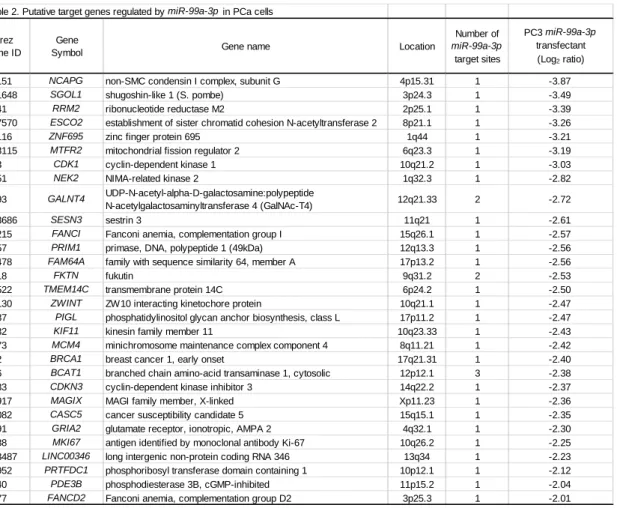
Table 2: Putative target genes regulated by miR-99a-3p in PCa cells
Fig. S1: Schematic representation of the chromosomal location of human miR-99a.
miR-99a is located on human chromosome 21q21.1. The mature miRNAs miR-99a-5p (guide
strand) and miR-99a-3p (passenger strand) are derived from pre-miR-99a.
Fig. S2: Expression levels of pri-miR-99a in PCa clinical specimens and cell lines.
Expression of pri-miR-99a was downregulated in PCa tissues than normal tissues. GUSB was used as an internal control.
Fig. S3: Both strands of miR-99a-5p and miR-99a-3p incorporated into the RISC
incorporated miRNAs were prepared with high purity using immunoprecipitation with anti-human Ago2 monoclonal antibodies. Expression of the miRNAs was quantified with qRT-PCR.
miR-31-5p was used as an internal control. *P < 0.0001. (B) Expression of miR-99a-5p was
significantly higher than that in cells transfected with mock, miR control, and miR-99a-3p. Likewise, expression of miR-99a-3p was significantly higher than that in cells transfected with mock, miR control, and miR-99a-5p. *P < 0.0001.
Fig. S4: Phase micrographs of wound healing and invasion assays following transfection with miR-99a-5p/3p in PCa cell lines.
(A) Phase micrographs of PC3, DU145 and C4-2 cells 24 h after monolayer wound healing. (B) Phase micrographs of invading PC3, DU145 and C4-2 cells.
Fig. S5: Phase micrographs of wound healing and invasion assays following transfection with si-NCAPG in PCa cell lines.
(A) Phase micrographs of PC3, DU145 and C4-2 cells 24 h after monolayer wound healing. (B) Phase micrographs of invading PC3, DU145 and C4-2 cells.
Fig. S6: Phase micrographs of wound healing and invasion assays following cotransfection with NCAPG/miR-99a-3p in PC3 cells.
(A) Phase micrographs of PC3 cells 24 h after monolayer wound healing. (B) Phase micrographs of invading PC3 cells.
Fig. S7: Kaplan-Meier survival curves based on miR-99a-3p expression in patients with Head and Neck squamous cell carcinoma and Lung adenocarcinoma.
Kaplan-Meier curves for overall survival rates (OS) showed that the OS of high miR-99a-3p expression group was significantly longer than that of low expression group. Data was extracted from OncoLnc website (http://www.oncolnc.org/).
Table 1. Patient characteristics Age PSA (years) (ng/mL) 1 Biopsy non-PCa 57 5.71 - - - RT-PCR 2 Biopsy non-PCa 74 9.45 - - - RT-PCR 3 Biopsy non-PCa 70 8.58 - - - RT-PCR 4 Biopsy non-PCa 73 4.8 - - - RT-PCR 5 Biopsy non-PCa 67 6.91 - - - RT-PCR 6 Biopsy non-PCa 50 7.05 - - - RT-PCR 7 Biopsy non-PCa 74 9.91 - - - RT-PCR 8 Biopsy non-PCa 76 20.9 - - - RT-PCR 9 Biopsy non-PCa 59 4.5 - - - RT-PCR 10 Biopsy non-PCa 75 1.1 - - - RT-PCR 11 Biopsy non-PCa 60 7.29 - - - RT-PCR 12 Biopsy non-PCa 73 38.7 - - - RT-PCR 13 Biopsy non-PCa 69 11.9 - - - RT-PCR 14 Biopsy non-PCa 77 23.3 - - - RT-PCR 15 Biopsy non-PCa 61 4.57 - - - RT-PCR 16 Biopsy non-PCa 59 7.37 - - - RT-PCR 17 Biopsy non-PCa 65 5.06 - - - RT-PCR 18 Biopsy HSPC 70 75.7 4+5 4 1 1 IV RT-PCR 19 Biopsy HSPC 78 1800 4+5 4 1 1 IV RT-PCR 20 Biopsy HSPC 75 68.4 5+4 4 1 0 IV RT-PCR 21 Biopsy HSPC 62 38.7 4+5 2b 1 0 IV RT-PCR 22 Biopsy HSPC 70 25.5 4+5 3b 0 0 III RT-PCR 23 Biopsy HSPC 88 888 4+5 3b 1 1 IV RT-PCR 24 Biopsy HSPC 69 33.9 4+5 4 0 1 IV RT-PCR 25 Biopsy HSPC 62 62.3 4+5 3b 1 0 IV RT-PCR 26 Biopsy HSPC 78 5 4+5 2c 0 1b IV RT-PCR 27 Biopsy HSPC 64 449 4+5 3b 1 1 IV RT-PCR 28 Biopsy HSPC 81 365 4+5 4 1 1 IV RT-PCR 29 Biopsy HSPC 76 715 5+4 4 1 1 IV RT-PCR 30 Biopsy HSPC 79 555 4+5 3 1 1 IV RT-PCR 31 Biopsy HSPC 63 1120 4+5 2c 0 1b IV RT-PCR 32 Biopsy HSPC 67 4.95 4+5 4 1 1b IV RT-PCR 33 Biopsy HSPC 70 19.5 5+5 4 1 1c IV RT-PCR 34 Biopsy CRPC 69 15.8 5+4 3b 1 1 IV RT-PCR 35 Biopsy CRPC 72 212 5+4 4 1 1 IV RT-PCR 36 Biopsy CRPC 71 4.4 4+5 4 1 1 IV RT-PCR 37 Biopsy CRPC 68 7.54 4+5 4 1 1b IV RT-PCR 38 Prostatectomy HSPC 65 5.3 4+5 2a 0 0 II IHC
39 Prostatectomy HSPC 61 21.48 4+4 3a 0 0 III IHC
40 Autopsy CRPC 64 4100 4+5 4 1 1c IV IHC
41 Autopsy CRPC 75 4690 4+5 4 1 1c IV IHC
Remarks Patient
No. Procedure Diagnosis T N M Stage
Gleason score
Table 2. Putative target genes regulated by miR-99a-3p in PCa cells
Entrez Gene ID
Gene
Symbol Gene name Location
Number of miR-99a-3p target sites PC3 miR-99a-3p transfectant (Log2 ratio)
64151 NCAPG non-SMC condensin I complex, subunit G 4p15.31 1 -3.87
151648 SGOL1 shugoshin-like 1 (S. pombe) 3p24.3 1 -3.49
6241 RRM2 ribonucleotide reductase M2 2p25.1 1 -3.39
157570 ESCO2 establishment of sister chromatid cohesion N-acetyltransferase 2 8p21.1 1 -3.26
57116 ZNF695 zinc finger protein 695 1q44 1 -3.21
113115 MTFR2 mitochondrial fission regulator 2 6q23.3 1 -3.19
983 CDK1 cyclin-dependent kinase 1 10q21.2 1 -3.03
4751 NEK2 NIMA-related kinase 2 1q32.3 1 -2.82
8693 GALNT4 UDP-N-acetyl-alpha-D-galactosamine:polypeptide
N-acetylgalactosaminyltransferase 4 (GalNAc-T4) 12q21.33 2 -2.72
143686 SESN3 sestrin 3 11q21 1 -2.61
55215 FANCI Fanconi anemia, complementation group I 15q26.1 1 -2.57
5557 PRIM1 primase, DNA, polypeptide 1 (49kDa) 12q13.3 1 -2.56
54478 FAM64A family with sequence similarity 64, member A 17p13.2 1 -2.56
2218 FKTN fukutin 9q31.2 2 -2.53
51522 TMEM14C transmembrane protein 14C 6p24.2 1 -2.50
11130 ZWINT ZW10 interacting kinetochore protein 10q21.1 1 -2.47
9487 PIGL phosphatidylinositol glycan anchor biosynthesis, class L 17p11.2 1 -2.47
3832 KIF11 kinesin family member 11 10q23.33 1 -2.43
4173 MCM4 minichromosome maintenance complex component 4 8q11.21 1 -2.42
672 BRCA1 breast cancer 1, early onset 17q21.31 1 -2.40
586 BCAT1 branched chain amino-acid transaminase 1, cytosolic 12p12.1 3 -2.38
1033 CDKN3 cyclin-dependent kinase inhibitor 3 14q22.2 1 -2.37
79917 MAGIX MAGI family member, X-linked Xp11.23 1 -2.36
57082 CASC5 cancer susceptibility candidate 5 15q15.1 1 -2.35
2891 GRIA2 glutamate receptor, ionotropic, AMPA 2 4q32.1 1 -2.30
4288 MKI67 antigen identified by monoclonal antibody Ki-67 10q26.2 1 -2.25
283487 LINC00346 long intergenic non-protein coding RNA 346 13q34 1 -2.23
56952 PRTFDC1 phosphoribosyl transferase domain containing 1 10p12.1 1 -2.12
5140 PDE3B phosphodiesterase 3B, cGMP-inhibited 11p15.2 1 -2.04
Table S1. Product numbers of reagents
Reagant Type Agency Assay ID Catalog nomber
hsa-miR-99a-5p TaqMan MicroRNA Assay Applied Biosystems 000435 4427975
hsa-miR-99a-3p TaqMan MicroRNA Assay Applied Biosystems 002141 4427975
RNU48 TaqMan MicroRNA Assay Applied Biosystems 001006 4427975
has-miR-99a TaqMan Pri-miRNA Assay Applied Biosystems Hs03302729_pri 4427012
NCAPG TaqMan probes and primers Applied Biosystems Hs00254617_m1 4331182
GAPDH TaqMan probes and primers Applied Biosystems Hs02786624_g1 4351370
Cancer Medicine. 2018 May; 7(5):1988-2002. doi: 10.1002/cam4.1455.