A
telectasis is one of the common complications occurring in the perioperative period [1,2], in intensive care [3,4], and among trauma patients [5-9].In some cases, atelectasis may cause desaturation or respiratory failure, which may influence the patient’s outcome [3,10,11]. Atelectasis can be seen on com- puted tomography (CT) images in the dependent lung as it progresses to acute respiratory distress syndrome (ARDS) [12]. We can divide atelectasis into 6 types based on the differing underlying mechanisms [13,14]:
resorption (absorption), adhesive, passive, compres- sive, cicatrization, and gravity-dependent atelectasis.
Among these types, high-inspired oxygen is known as one of the etiologies of resorption (absorption) atelecta- sis [1], and it occurs mainly in dependent lung regions [15].
A relationship between the oxygen concentration and atelectasis formation has been described in several investigations. In animal studies, the high-inspired oxygen concentration causes absorption atelectasis in the setting of mechanical ventilation [16] or early ARDS [17]. A clinical study reported that hyperoxia and hypergravity are independent risk factors of reversible atelectasis formation [18]. In a clinical setting, it was reported that (1) atelectasis developed soon after the
CopyrightⒸ 2020 by Okayama University Medical School.
http ://escholarship.lib.okayama-u.ac.jp/amo/
Original Article
Relationship between a High-inspired Oxygen Concentration and Dorsal Atelectasis in High-energy Trauma Patients
Kenzo Ishiia,b*, Hiroshi Morimatsub, Kazumi Onoa,c and Koji Miyashod
Departments of aAnesthesiology and Oncological Pain Medicine, and dCritical Care and Emergency Medicine, Fukuyama City Hospital, Fukuyama, Hiroshima 721-8511, Japan, bDepartment of Anesthesiology and Resuscitology, Okayama University Graduate School of Medicine, Dentistry and Pharmaceutical Sciences, Okayama 700-8558, Japan,
cDepartment of Anesthesiology & Intensive Care Medicine, Kawasaki Medical School, Kurashiki, Okayama 701-0192, Japan We performed a retrospective cohort study of 911 high-energy trauma patients who underwent chest CT scans at least twice after admission. We hypothesized that in high-energy trauma patients, a high-inspired oxygen concentration delivered after admission results in dorsal atelectasis. The study’s primary outcome was dorsal atelectasis formation diagnosed based on CT images. We defined dorsal atelectasis as the presence of atelectasis at ≥10 mm thick on CT images. We defined high-inspired oxygen concentration as >60% oxygen delivered between two CT scans. Four hundred sixty-five patients (51.0%) developed atelectasis according to the second CT scan, and 338 (37.1%) received a high-inspired oxygen concentration. A univariate analysis showed that the rate of the high-inspired oxygen concentration in the atelectasis group was significantly higher than that in the non-atelectasis group (43.4% vs. 30.1%, p<0.001). However, a logistic regression analysis showed that there was no significant relationship between the oxygen concentration and the formation of dorsal atelectasis (OR: 1.197, 95%CI: 0.852-1.683, p=0.30). Age, the Injury Severity Score, BMI, and smoking were found to be risk factors of dorsal atelectasis formation in high-energy trauma patients. There was no relationship between the oxygen concentration and atelectasis formation in our series of high-energy trauma patients.
Key words: trauma patient, dorsal atelectasis, oxygen concentration
Received December 26, 2018 ; accepted August 19, 2019.
*Corresponding author. Phone : +81-84-941-5151; Fax : +81-84-941-5159
E-mail : [email protected] (K. Ishii) Conflict of Interest Disclosures: No potential conflict of interest relevant to this article was reported.
induction of general anesthesia, and (2) 100% oxygen was a significant cause of atelectasis during the induc- tion of anesthesia [19,20]. The use of 30-40% oxygen or the addition of positive end-expiratory pressure (PEEP) to 100% oxygen at the general anesthesia is thus recommended [21]. In ARDS patients, the use of 100%
oxygen (vs. 60%) worsened oxygenation [22].
Conservative oxygen therapy (COT) (i.e., a target SpO2 value of 90-92%) might be associated with decreased radiological evidence of atelectasis [23,24]
and with reduced intensive care unit (ICU) [25], in-hospital, and 30-day mortality [26]. In light of these reports, high concentration oxygen appears to be one of the leading causes or risk factors for atelectasis forma- tion, and there may be clinical evidence of the efficacy of COT, which is oxygen restriction.
Even among trauma patients, pulmonary complica- tions are significant problems that might cause worse outcomes [27] and respiratory failure (e.g., injury/
ARDS) [28]. Atelectasis had been described as a fre- quent cause of early post-traumatic lung dysfunction [6]. In terms of the inspired oxygen concentration, 100% oxygen with a high flow rate is usually recom- mended for these patients [29]. However, no studies have reported an association between the delivery of a high-inspired concentration of oxygen and atelectasis formation in such patients. We conducted the present study to analyze the relationship between a high-in- spired oxygen concentration and dorsal atelectasis for- mation in high-energy trauma patients. We hypothe- sized that a high-inspired oxygen concentration delivered after admission results in dorsal atelectasis in high-energy trauma patients.
Patients and Methods
This was a single-center retrospective cohort study conducted to investigate patients treated at Fukuyama City Hospital during the period from April 2008 to March 2010. The Institutional Review Board of Fukuyama City Hospital approved this study (approval no. 187; April 21, 2014). Patient informed consent was waived because of the study’s retrospective design.
We provided an opportunity for patients to opt out of the study. The trial registration number UMIN000025255 was registered retrospectively on December 13, 2016.
We included high-energy trauma patients who were
transported and admitted to our emergency depart- ment and had undergone at least 2 chest computed tomography (CT) scans. The definition of ‘high energy trauma’ was adapted from the criteria of the Fire and Disaster Management Agency of Japan’s Ministry of Internal Affairs and Communications [29]. At our institution at that time, almost all high-energy trauma patients underwent two CT scans: at their arrival to the emergency department, and approx. 24 h after admis- sion.
We assessed the incidence of dorsal atelectasis in the patients by comparing these two CT scans of each patient. We measured the thickness of atelectasis from the dorsal chest wall in each CT image and compared the atelectasis thickness between the high-oxygen group and the low-oxygen group. In this study, we defined
‘dorsal atelectasis’ as the presence of atelectasis with a thickness of ≥10 mm, taking the measurement results into account. We excluded patients who did not undergo a second CT scan, those who died within 24 h from their injury, those who were not categorized as high-energy trauma, and those who were re-trans- ported to our hospital after having been accommodated in another hospital.
We evaluated the following parameters as baseline characteristics: sex, age, Injury Severity Score (ISS), smoking history, history of respiratory disease, and body mass index (BMI). We investigated severely injured patients whose Abbreviated Injury Score (AIS) of any part of the body was assessed as ≥3, the patients with a thoracic AIS score of ≥3, and the patients with an abdominal AIS score of ≥3. We also examined the relationship between the surgical procedure under gen- eral anesthesia (GA), mechanical ventilation (MV) in the ICU, and the order of complete bed rest.
We determined whether a high-inspired oxygen concentration was delivered between each patient’s first and second CT scan. In this study, we defined ‘high- inspired oxygen concentration’ as >60% oxygen deliv- ered between the two CT scans. If the patient was mechanically ventilated (invasive positive pressure ven- tilation [IPPV] or non-invasive positive pressure venti- lation [NPPV]), a high-inspired oxygen concentration was defined as 60-100% of the fraction of inspired oxy- gen.The absence of a need for mechanical ventilation (i.e., ‘without MV’) was defined as the delivery of oxy- gen ≥6 L/min with the use of a non-rebreathing mask.
Following the ‘pre-hospital oxygen administration pro- tocol’ of our medical control institution, the emergency medical service administered 100% oxygen (>10 L/min using a non-rebreathing mask) to all of the high-energy trauma patients, and all patients enrolled in this study continued to receive the same flow rate of oxygen until the first CT scan.
If a patient required ventilation by endotracheal intubation before the first CT examination, he or she was ventilated with 100% oxygen until admission to the emergency ICU. During the study period, we usually adjusted the oxygen flow at the time of the patient’s admission to the ICU or high-care unit after the first CT examination. However, because there was no policy of adjusting the oxygen flow after admission, the oxygen flow adjustment was decided by the emergency physi- cian in charge. We often administered high oxygen to high-energy trauma patients after admission. When we evaluated the incidence of atelectasis revealed by CT, we were blinded to the high-/low-oxygen status of the patients.
The primary outcome of the study was dorsal atelec- tasis formation in the high-energy trauma patients, diagnosed based on the CT scan results. The study’s secondary outcome was dorsal atelectasis formation in the subgroup of patients who did not exhibit atelectasis in the first CT scan (excluding initial atelectasis). We also performed a subgroup analysis regarding the rela- tionship between the high-inspired concentration of oxygen and atelectasis formation in the patients divided according to age, the patient severity (ISS), BMI, and with/without mechanical ventilation. We defined the age, ISS, and BMI subgroups as <65 years versus ≥65 years, ISS <9 versus ≥9, and BMI <25 versus ≥25, respectively.
We did not calculate the sample size at the start of the study because it was not available. We conducted the study for a period of 3 years. The data are expressed as the mean and standard deviation (SD) or the number of patients and percentage (%). Comparisons between the atelectasis group and non-atelectasis group were conducted using the Chi-squared test, Student’s t-test, or a repeated measure analysis of variance (ANOVA).
We performed a multivariate logistic regression analysis to determine the incidence of dorsal atelectasis as an independent variable with the following factors:
patient sex, age, ISS, smoking, history of respiratory disease, BMI, surgical procedure under general anes-
thesia between CT scans, mechanical ventilation between CT scans, ordered complete bed rest, and duration of oxygen delivery (the time between CT scans). In this analysis, we selected the ISS as the severity of the trauma. We did not select AIS ≥3 (any part), Thoracic AIS ≥3, or Abdominal AIS ≥3, because the ISS was calculated from the AIS. We used SPSS software for the statistical analyses.
Results
We enrolled 1,416 trauma patients during the study period, and we excluded 457 patients, including those without high-energy trauma (n=234), burn patients (n=4), those who died within 24 h from injury (n=48), and those who were re-transported to our hospital after having been accommodated in another hospital (n=169).
We also excluded 48 patients with missing data, includ- ing patients who did not undergo a second CT. We thus studied a final total of 911 high-energy trauma patients who were admitted to the emergency department of our hospital and who underwent CT at least twice (Fig.1).
The mean age of the patients was 48 (SD 23.3) years.
The number of men was 599 (65.8%), the number of smokers was 425 (46.7%), and the mean BMI of the
1,416 trauma patients
959 high-energy trauma patients
We analyzed 911 high-energy trauma patients We excluded 457 patients
• Without high-energy trauma(234)
• Burn injury (4)
• Death within 24 hours from injury (48)
• Re-transportation to our hospital (169)
• Others (2)
We excluded 48 patients
• With missing data
• Without undergoing a second CT
Fig. 1 The cases of 911 high-energy trauma patients admitted to our hospitalʼs emergency department and underwent at least two CT scans were analyzed. We enrolled 1,416 trauma patients; 505 patients were excluded for various reasons, leaving a final total of 911 patients.
patients was 22.5 (SD 4.1). The mean ISS was 13.9 (SD 11.7) points. The number of patients who had an AIS of
≥3 of any part of the body was 542 (59.5%). The num- ber of patients who had a thoracic AIS of ≥3 was 266 (29.1%), and the number of patients who had an abdominal AIS of ≥3 was 82 (9%). The number of patients who underwent a surgical procedure under general anesthesia between CT scans was 288 (31.6%), and 195 (21.4%) patients received mechanical ventila- tion between scans. The mean duration of oxygen delivery (i.e., the time from the first CT to the second CT [time between CT scans]) was 17.3 h (SD 5.5 h) (Table 1).
The number of patients with a high-inspired oxygen concentration delivered between CT scans was 338 (37.1%) (high-oxygen group), and 573 (62.9%) patients received a low oxygen concentration (low-oxygen group). The results of the measurement of atelectasis thickness in the high- and low-oxygen groups are illus- trated in Fig.2. The mean atelectasis thickness values of the high-oxygen group at the first and second CT exams
were 6.13 (SD 8.82) and 12.57 (12.36), and those of the low-oxygen group at the first and second CT exams were 3.57 (6.82), and 8.84 (11.13), respectively. There were significant differences between the values at the first and second CT scans in both groups according to the repeated measure ANOVA (both p<0.001).
Among the 911 high-energy trauma patients, 465 (51.0%) patients exhibited dorsal atelectasis in the sec- ond CT scan (the atelectasis group), and 446 (49.0%) patients had no dorsal atelectasis (the non-atelectasis group). In the atelectasis group, 252 cases (27.7%) already had atelectasis at the first CT scan; this corre- sponds to 54.4% of the atelectasis group. In the non-at- electasis group, there was no case in which atelectasis was recognized in the first CT. Compared to the non-atelectasis group, the atelectasis patients were older [mean 58.5 (SD 18.0) years vs. 37.8 (23.6) years], had higher ISS values [17.6 (12.3) vs. 10.1 (9.7) points], and had higher BMI values [23.7 (4.0) vs. 21.8 (3.88) kg/m2].
Between CT scans, the atelectasis patients were
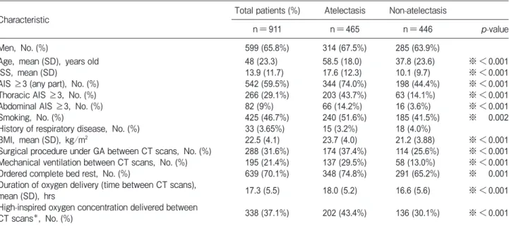
Table 1 Patient characteristics, primary outcomes according to the univariate analysis, and comparison of the atelectasis and non- atelectasis groups
Characteristic Total patients (%) Atelectasis Non-atelectasis
n=911 n=465 n=446 p-value
Men, No. (%) 599 (65.8%) 314 (67.5%) 285 (63.9%)
Age, mean (SD), years old 48 (23.3) 58.5 (18.0) 37.8 (23.6) ※<0.001
ISS, mean (SD) 13.9 (11.7) 17.6 (12.3) 10.1 (9.7) ※<0.001
AIS ≥3 (any part), No. (%) 542 (59.5%) 344 (74.0%) 198 (44.4%) ※<0.001
Thoracic AIS ≥3, No. (%) 266 (29.1%) 203 (43.7%) 63 (14.1%) ※<0.001
Abdominal AIS ≥3, No. (%) 82 (9%) 66 (14.2%) 16 (3.6%) ※<0.001
Smoking, No. (%) 425 (46.7%) 240 (51.6%) 185 (41.5%) ※ 0.002
History of respiratory disease, No. (%) 33 (3.65%) 15 (3.2%) 18 (4.0%)
BMI, mean (SD), kg/m2 22.5 (4.1) 23.7 (4.0) 21.2 (3.88) ※<0.001
Surgical procedure under GA between CT scans, No. (%) 288 (31.6%) 174 (37.4%) 114 (25.6%) ※<0.001 Mechanical ventilation between CT scans, No. (%) 195 (21.4%) 137 (29.5%) 58 (13.0%) ※<0.001
Ordered complete bed rest, No. (%) 639 (70.1%) 348 (74.8%) 291 (65.2%) ※ 0.001
Duration of oxygen delivery (time between CT scans),
mean (SD), hrs 17.3 (5.5) 18.0 (5.2) 16.6 (5.6) ※<0.001
High-inspired oxygen concentration delivered between
CT scans*, No. (%) 338 (37.1%) 202 (43.4%) 136 (30.1%) ※<0.001
*High-inspired oxygen concentration: mechanical ventilation (IPPV or NPPV) with high fractions of inspired oxygen (FIO2) ranging from 60% to 100%, or spontaneous breathing with 6liters/minute or more oxygen delivery using a non-rebreathing mask.
※p<0.05. There was a significant difference in the inspired oxygen concentration between the atelectasis and non-atelectasis groups according to the univariate analysis, and almost all of the other factors were significantly different between the atelectasis and non-atelec- tasis groups except for gender and history of respiratory disease. AIS, Abbreviated Injury Score; BMI, body mass index; CT, computed tomography; GA, general anesthesia; IPPV, invasive positive pressure ventilation; ISS, Injury Severity Score; NPPV, non-invasive posi- tive pressure ventilation.
more likely to have undergone a surgical procedure under general anesthesia [174 (37.4%) vs. 114 (25.6%)]
and mechanical ventilation [137 (29.5%) vs. 58 (13%)]
and to be ordered complete bed rest [348 (74.8%) vs.
291 (65.2%)]. The duration of oxygen delivery (i.e., the time between CT scans) was significantly longer in the atelectasis group compared to the non-atelectasis group: mean 18.0 (SD 5.2) h versus 16.6 (5.6) h, respec- tively (Table 1).
Primary outcome. When we compared the inci- dence of high-inspired oxygen concentration adminis- tration between the groups in a univariate analysis (Table 1), we observed that the patients in the atelecta- sis group received a significantly higher concentration of oxygen between CT scans compared to the non-atel- ectasis group: 202 (43.4%) vs. 136 (30.1%), respec- tively. In the logistic regression analysis, we selected the ISS as a severity index instead of AIS ≥3 (any part), Thoracic AIS ≥3, or Abdominal AIS ≥3, because the ISS was calculated from the AIS and these factors were highly related to each other. We thus performed the multivariate logistic regression analysis using sex, age, ISS, smoking, history of respiratory disease, BMI, sur- gical procedure under general anesthesia between CT
scans, mechanical ventilation between CT scans, ordered complete bed rest, and duration of oxygen delivery (time between CT scans) to obtain the odds ratio (OR) of atelectasis formation.
In that analysis (Table 2), age (OR: 1.045, 95%CI:
1.037-1.053, p<0.001), ISS (OR: 1.064, 95%CI:
1.045-1.084, p<0.001), smoking (OR: 1.875, 95%CI:
1.305-2.694, p<0.001), BMI (OR: 1.183, 95%CI:
1.132-1.237, p<0.001), and duration of oxygen deliv- ery (time between CT scans) (OR: 1.057, 95%CI:
1.027-1.089, p<0.001) showed a significant difference between the atelectasis group and the non-atelectasis group. By contrast, the high-inspired oxygen concen- tration showed no significant difference in the OR of atelectasis formation in the multivariate logistic regres- sion analysis (OR: 0.754, 95%CI: 0.549-1.035, p=0.081) (Table 2).
Secondary outcome. We performed a subgroup analysis of the patients who did not show atelectasis in their first CT scan (excluding initial atelectasis). The results are shown in Fig.2 and Table 3. In this sub- group, the mean atelectasis thicknesses of the high-ox- ygen group (n=214) at the first and second CT exams were 0.73 (SD 2.24) and 6.83 (9.02), and those of the
(n=573) (n=338)
0.00 4.00 8.00 12.00
First CT Second CT
Total patients; n=911
High oxygen Low oxygen mm (mean)
.00 4.00 8.00 12.00
Second CT First CT
Excluding the patients with initial atelectasis; n=659
High oxygen Low oxygen mm (mean)
(n=445) (n=214)
Fig. 2 The thickness of atelectasis at the first and second CT examinations in the high-oxygen and low-oxygen groups. Among the 911 patients, the thicknesses of atelectasis at the first and second CT scans [mean (SD)] were 6.13 (8.82) and 12.57 (12.56) in the high-oxy- gen (n=338) group and 3.57 (6.82) and 8.84 (11.13) in the low-oxygen (n=573) group. Those values when the initial-atelectasis patients were excluded were 0.73 (2.44) and 6.83 (9.02) in the high-oxygen group (n=214) and 0.46 (2.06) and 5.39 (8.90) in the low-oxygen group (n=445), respectively. According to a repeated measure ANOVA, all groups had a significant increase in the thickness of atelectasis at the second CT scan (vs. the first CT). There was a significant difference between the high- and low-oxygen groups in the analysis of total patients (p<0.001) and among the patients who did not exhibit atelectasis at the first CT scan (i.e., excluding the patients with initial atelectasis) (p<0.001).
low-oxygen group (n=445) at the first and second CT exams were 0.46 (2.06) and 5.39 (8.90), respectively.
There was a significant difference between the 2 groups according to the repeated measure ANOVA (p<0.001) (Fig.2).
We performed a multivariate logistic regression analysis to determine the relationship between atelecta- sis formation and oxygen concentration in the patients who did not have atelectasis in the first CT scan (excluding initial atelectasis) (Table 3). Age (OR: 1.045, 95%CI: 1.035-1.055, p<0.001), ISS (OR: 1.051, 95%CI: 1.028-1.075, p<0.001), smoking (OR: 1.752, 95%CI: 1.128-2.721, p<0.001), BMI (OR: 1.167, 95%CI: 1.106-1.232, p<0.001), and the
duration of oxygen delivery (time between CT scans) (OR: 1.075, 95%CI: 1.036-1.114, p<0.001) showed a significant difference between the atelectasis group and the non-atelectasis group. There was also no significant relationship between atelectasis formation and the oxy- gen concentration in the patients who did not have atelectasis in the first CT scan (OR: 0.886, 95%CI:
0.581-1.351, p=0.574).
Subgroup analyses based on the patients’ age, ISS, BMI and mechanical ventilation were also performed in the multivariate logistic regression analysis for atelecta- sis formation and oxygen concentration. None of the subgroups showed a higher risk of dorsal atelectasis formation in high oxygen concentration; age (<65 years
Table 2 The results of the multivariate logistic regression to determine the independent variables of the incidence of dorsal atelectasis
OR 95%CI p-value
High concentration of oxygen delivered between CT scans 1.197 0.852-1.683 0.300
Male 0.867 0.591-1.272 0.465
Age 1.045 1.037-1.053 ※<0.001
ISS 1.064 1.045-1.084 ※<0.001
Smoking 1.875 1.305-2.694 ※ 0.001
History of respiratory disease 0.848 0.342-2.106 0.723
BMI 1.183 1.132-1.237 ※<0.001
Surgical procedure under GA between CT scans 1.094 0.758-1.578 0.632
Mechanical ventilation between CT scans 1.231 0.751-2.018 0.409
Ordered complete bed rest 1.398 0.971-2.013 0.072
Duration of oxygen delivery (time between CT scans) 1.057 1.027-1.089 ※<0.001
※p<0.05. Age, ISS, smoking, BMI, and duration of oxygen delivery (time between CT scans) were each an independent risk factor for atelectasis formation in the high-energy trauma patients. There was no significant relationship between the inspired oxygen concentration and atelectasis formation. BMI, body mass index; GA, general anesthesia; ISS, Injury Severity Score.
Table 3 The results of multivariate logistic regression to determine the independent variables of incidence of dorsal atelectasis in the patients excluding initial atelectasis
OR 95%CI p-value
High concentration of oxygen delivered between CT scans 0.886 0.581-1.351 0.574
Male 0.816 0.518-1.286 0.381
Age 1.045 1.035-1.055 ※<0.001
ISS 1.051 1.028-1.075 ※<0.001
Smoking 1.752 1.128-2.721 ※ 0.013
History of respiratory disease 1.005 0.347-2.913 0.992
BMI 1.167 1.106-1.232 ※<0.001
Surgical procedure under GA between CT scans 1.511 0.977-2.336 0.063
Mechanical ventilation between CT scans 1.071 0.573-2.001 0.831
Ordered complete bed rest 1.237 0.801-1.910 0.337
Duration of oxygen delivery (time between CT scans) 1.075 1.036-1.114 ※<0.001
※p<0.05. Age, ISS, smoking, BMI, and duration of oxygen delivery (time between CT scans) were each an independent risk factor for atelectasis formation the patients who had not exhibited atelectasis at the first CT scan. There was no significant relationship between the inspired oxygen concentration and atelectasis formation in this subgroup.
old, OR: 1.302, 95%CI: 0.877-1.930, p=0.19; ≥65 years old, OR: 1.374, 95%CI: 0.768-2.459) p=0. 84;
ISS (<9, OR: 0.885, 95%CI: 0.497-1.578, p=0.68;
≥9, OR: 1.417, 95%CI: 0.926-2.167), BMI (<25, OR: 1.263, 95%CI: 0.861-1.852, p=0.23; ≥25, OR: 1.091, 95%CI: 0.524-2.273, p=0.82), and mechanical ventilation (MV, OR: 1.229, 95%CI:
0.540-2.798, p=0.62; without MV: OR: 1.224, 95%CI: 0.832-1.802, p=0.31) (Fig.3).
Discussion
We conducted a retrospective cohort study of high-energy trauma patients to evaluate the relation- ship between the inspired oxygen concentration and dorsal atelectasis. Our analyses revealed that the atelec- tasis group received a higher inspired oxygen concen- tration compared to the non-atelectasis group.
However, this relationship was not an independent risk
factor in the multivariate logistic regression analysis including the subgroup analyses based on age, ISS, BMI, and with/without MV.
We studied only high-energy trauma patients.
Several studies have shown that the incidence of atelec- tasis formation after severe trauma ranges from 7.4% to 60% [5-9]. Fifty percent of our patients showed atelec- tasis formation after high-energy trauma; thus, our study population would be relevant to previous reports.
It has also been shown that the risk factors of the com- plications [30] and atelectasis formation [31] of trauma patients are age, the severity of the trauma [30], chest trauma, abdominal injury, high APACHE 2 score, and the duration of mechanical ventilation [31]. We ana- lyzed similar factors in the present study, and our find- ings demonstrated that age, ISS, BMI, and smoking were independent risk factors of atelectasis formation in high-energy trauma patients. To the best of our knowl- edge, no previous studies of trauma patients have
Subgroup
All patients (n=911)
Risk Lower for Atelectasis Formation Risk Higher for Atelectasis Formation
OR (95%CI), p-value 1.197 (0.852-1.683), p=0.30
1.302 (0.877-1.930), p=0.19 1.374 (0.768-2.459), p=0.84
0.885 (0.497-1.578), p=0.68 1,417 (0.926-2.167), p=0.11
1.263 (0.861-1.852), p=0.23 1.091 (0.524-2.273), p=0.82
1.229 (0.540-2.798), p=0.62 1.224 (0.832-1.802), p=0.31
0 0.5 1 1.5 2 2.5 BMI
BMI <25 (n=703) BMI ≥25 (n=206) ISS
ISS <9 (n=337) ISS ≥9 (n=574)
Oxygen delivery MV (n=195) Without MV
(n=716)
Age
<65 year (n=622)
≥65 year (n=289)
: OR: Range of 95%CI
Fig. 3 Subgroup analyses: The odds ratio (OR) of atelectasis formation with high-inspired oxygen administration according to the multi- variate logistic regression analysis adjusted for confounders* in all patients and each subgroup. We performed subgroup analyses based on patient age, ISS, BMI, and with/without mechanical ventilation (MV). The adjusted confounders* in the multivariate logistic regression analysis were sex, age, ISS, smoking, history of respiratory disease, BMI, the surgical procedure under general anesthesia between CT scans, mechanical ventilation between CT scans, ordered complete bed rest, and the duration of oxygen delivery (time between CT scans). Each subgroup analysis was calculated without its own item. There was no significant relationship between atelectasis formation and the oxygen concentration in each subgroup analysis.
investigated the relationship between oxygen concen- trations and atelectasis formation.
Several investigations of the perioperative oxygen concentration and atelectasis formation have been reported. Rothen et al. reported that after anesthesia induction, there was a higher amount of atelectasis (mean 4.2 [SD 5.6] cm2) in the 100% oxygen group compared to the 30% oxygen group (mean 0.2 [SD 0.4]
cm2) [19]. Edmark et al. also reported that during and after pre-oxygenation and the induction of anesthesia from the awake stage to 14 min after the start of pre-ox- ygenation, atelectasis increased progressively in all patients, especially when 100% oxygen was used [20].
According to these 2 studies, a high oxygen concentra- tion is the major cause of atelectasis formation after the induction of anesthesia.
However, several research groups have stated that they found no relationship between the use of a high concentration of oxygen during anesthesia and postop- erative atelectasis formation. Edmark et al. reported that a lower oxygen concentration in the recovery phase did not reduce postoperative atelectasis compared to a fraction of inspired oxygen at 100% before the removal of the laryngeal mask airway (LMA) [32]. Meyhoff et al.
stated that the administration of 80% oxygen during anesthesia did not increase postoperative atelectasis formation compared to 30% oxygen [33]. Finally, according to a systematic review of the intraoperative high-inspired oxygen fraction and postoperative pul- monary function, the risk of atelectasis was not increased with a higher fraction of inspired oxygen [34].
When we considered the effect of oxygen on atelec- tasis formation, the timing and duration of oxygen exposure as well as the inspired oxygen concentration were strongly influenced [31]. In our present study, the relationship between atelectasis formation and high-in- spired oxygen concentration showed no significant dif- ference between the atelectasis group and the non-atel- ectasis group in the multivariate logistic analysis, although there was a significant difference between both groups in the univariate analysis. Therefore, the oxygen concentration was not a major cause of atelectasis for- mation in the present high-energy trauma patients after admission. We speculate that the relationship between the inspired oxygen concentration and dorsal atelectasis formation is not independent factors each other in the timing of after admission.
Dorsal atelectasis was shown by the first CT scan (performed at hospital arrival) in 27.7% of our 911 patients. All of the patients in this study received a high concentration of oxygen before their first CT exam. The results of our subgroup analyses excluding the patients who had already had atelectasis at the first CT were almost the same as those obtained for the total patient series. We therefore speculate that the patients those who had atelectasis at the first CT were also signifi- cantly influenced by several factors of atelectasis ana- lyzed in the primary outcome. The relationship between the inspired oxygen concentration in a pre-hospital setting and dorsal atelectasis remains unclear.
Patients were not distinguished by the use or non- use of mechanical ventilation in this study.
Guntaragorn Hongrattanae et al. reported that mechan- ical ventilation was one of the major risk factors of atel- ectasis formation [31]. The incidence of dorsal atelecta- sis in the present study’s mechanical ventilation group (n=195) was 72.3%, and that in the non-mechanical ventilation group (n=716) was 45.5%. Moreover, there was no significant relationship between the use/non-use of mechanical ventilation and atelectasis formation in the multivariate logistic analysis or in the subgroup analysis. Although the incidence of dorsal atelectasis was high in our mechanical ventilation group, dorsal atelectasis also occurred in almost half of the patients in the non-mechanical ventilation group. We thus suspect that the combination of mechanical ventilation and a high concentration of oxygen is not a cause of dorsal atelectasis formation.
Our logistic regression analysis revealed that the duration of oxygen delivery (the time between CT scans) showed a significant difference in the risk of dor- sal atelectasis formation. We compared high- and low-oxygen delivery groups during this period.
Although the duration of oxygen delivery could be more important as a risk of atelectasis formation com- pared to the oxygen concentration, it might merely represent the difference in the duration of bed rest, or only the time difference from the patient’s injury. We therefore could not conclude that the duration of oxy- gen delivery (time between CT scans) was an indepen- dent risk factor for atelectasis in this study.
Our multivariate analysis results demonstrated that age, ISS, BMI, and smoking were independent vari- ables of the incidence of dorsal atelectasis. Although an
AIS ≥3 (any part), a Thoracic AIS ≥3, and an Abdominal AIS ≥3 might be risk factors for atelectasis formation, those factors and the ISS are closely related to one another in part because the ISS is calculated from the AIS. We therefore selected the ISS as the parameter representing the severity of trauma. If we had analyzed the injury parts and their severity one by one, some might have been found to be a risk factor for atelectasis formation.
The limitations of this study are as follows. This was a retrospective cohort study performed at a single cen- ter. Because of the observational nature of the study, we were not able to confirm a cause-and-effect relation- ship. The data quality might not be sufficient or opti- mally controlled, because of the study’s retrospective design. However, the number of patients was close to 1,000, and all of the patients included in the study underwent at least two CT scans.
Second, there are 2 factors in the formation of dorsal atelectasis: absorption atelectasis and gravity-depen- dent atelectasis [15]. Many of the patients included in this study seemed to have both factors, but we did not individually address absorption or gravity-dependent atelectasis. Moreover, because of the study’s retrospec- tive nature, it was challenging to make a detailed evalu- ation of the stored CT scan images. We thus could not distinguish between absorption atelectasis and gravi- ty-dependent compression atelectasis. We did not nec- essarily need to divide the cases of atelectasis into these 2 factors; we simply intended to investigate the effect of oxygen concentration on dorsal atelectasis formation.
Third, our definition of dorsal atelectasis was atelec- tasis with a thickness of ≥10 mm from the dorsal chest wall in CT images. Because this was a retrospective investigation and it was difficult to perform a quantita- tive analysis using CT images obtained from the patients’ electronic medical records, there was not enough data for a quantitative analysis. We therefore analyzed the incidence of atelectasis formation using our definitions.
Finally, we should mention the definition of oxygen concentration. In this study, we focused on the fact that the oxygen supply between the first and the second CT scans was determined regardless of the severity of trauma. We considered 6 L/min of oxygen using a non-rebreathing mask as representing 60% inspired oxygen. However, the exact oxygen concentrations of the non-rebreathing mask are not known. Although we
classified the patients as above or below the oxygen concentration of 60%, the “higher” oxygen concentra- tion might result in a stronger effect.
In conclusion, we performed a retrospective obser- vational study to analyze the relationship between a high oxygen concentration and atelectasis formation in high-energy trauma patients. The results of a multivar- iate logistic regression analysis demonstrated that there was no significant relationship between the oxygen con- centration and the formation of dorsal atelectasis. We observed that patient age, severity (ISS), BMI, and smoking are factors related to dorsal atelectasis forma- tion in high-energy trauma patients.
Acknowledgments. We thank Rie Fujita and Rieko Sasaki for the data management.
References
1. Rigg JR: Pulmonary atelectasis after anaesthesia: pathophysiol- ogy and management. Can Anaesth Soc J (1981) 28:305-313.
2. JANES RM: Postoperative pulmonary complications. Am J Surg (1955) 89: 297-303.
3. Serejoa LG, da Silva-Júniora FP, Bastosa JP, Bruina GS, Motab RM and Bruina PF: Risk factors for pulmonary complications after emergency abdominal surgery. Respir Med (2007) 101:808-813.
4. Golfieri R, Giampalma E, Labate A, dʼArienzo PA, Jovine E, Grazi G, Mazziotti A, Maffei M, Muzzi C, Tancioni S, Sama C, Cavallari A and Gavelli G: Pulmonary complications of liver trans- plantation: radiological appearance and statistical evaluation of risk factors in 300 cases. Eur Radiol (2000) 10: 1169-1183.
5. Shorr RM, Crittenden M, Indeck M, Hartunian SL and Rodriguez A:
Blunt Thoracic Trauma, analysis of 515 patients. Ann Surg (1987) 206: 200-205.
6. Reske AW, Reske AP, Heine T, Spieth PM, Rau A, Seiwerts M, Busse H, Gottschaldt U, Schreiter D, Born S, Gama de Abreu M, Josten C, Wrigge H and Amato MB: Computed tomographic assessment of lung weights in trauma patients with early posttrau- matic lung dysfunction. Crit Care (2011) 15: R71.
7. Marts B, Durham R, Shapiro M, Mazuski JE, Zuckerman D, Sundaram M and Luchtefeld WB: Computed Tomography in the Diagnosis of Blunt Thoracic Injury. Am J Surg (1994) 168: 688- 8. Groeneveld AB: Increased permeability-oedema and atelectasis in 692.
pulmonary dysfunction after trauma and surgery: a prospective cohort study. BMC Anesthesiol (2007) 7: 7.
9. Karaaslan T, Meuli R, Androux R, Duvoisin B, Hessler C and Schnyder P: Traumatic Chest Lesions in Patients with Severe Head Trauma: A Comparative Study with Computed Tomography and Conventional Chest Roentgenograms. J Trauma (1995) 39:
1081-1086.
10. Muders T and Wrigge H: New insights into experimental evidence on atelectasis and causes of lung injury. Best Practice & Research Clinical Anaesthesiology (2010) 24: 171-182.
11. McAlister FA, Bertsch K, Man J, Bradley J and Jacka M: Incidence of and Risk Factors for Pulmonary Complications after Nonthoracic
Surgery. Am J Respir Crit Care Med (2005) 171:514-517.
12. Piantadosi CA and Schwartz DA: The Acute Respiratory Distress Syndrome. Ann Intern Med. (2004) 141:460-470.
13. Woodring JH and Reed JC: Types and Mechanisms of Pulmonary Atelectasis. Journal of Thoracic Imaging (1996) 11: 92-108.
14. Schindler MB: Treatment of atelectasis: where is the evidence?
Critical Care (2005) 9: 341-342.
15. Hedenstierna G and Edmark L: Mechanisms of atelectasis in the perioperative period. Best Pract Res Clin Anaesthesiol (2010) 24:
157-169.
16. Duggan M, McNamara PJ, Engelberts D, Pace-Asciak C, Babyn P, Post M and Kavanagh BP: Oxygen Attenuates Atelectasis-induced Injury in the In Vivo Rat Lung. Anesthesiology (2005) 103: 522- 17. Derosa S, Borges JB, Segelsjö M, Tannoia A, Pellegrini M, 531.
Larsson A, Perchiazzi G and Hedenstierna G: Reabsorption atel- ectasis in a porcine model of ARDS: regional and temporal effects of airway closure, oxygen, and distending pressure. J Appl Physiol (2013) 115: 1464-1473.
18. Dussault C, Gontier E, Verret C, Soret M, Boussuges A, Hedenstierna G and Montmerle-Borgdorff XS: Hyperoxia and hypergravity are independent risk factors of atelectasis in healthy sitting humans: a pulmonary ultrasound and SPECT/CT study. J Appl Physiol (2016) 121: 66-77.
19. Rothen HU, Sporre B, Engberg G, Wegenius G, Reber A, and Hedenstierna G: Prevention of atelectasis during general anaes- thesia. Lancet (1995) 345: 1387-1391.
20. Edmark L, Auner U, Enlund M, Ostberg E and Hedenstierna G:
Oxygen concentration and characteristics of progressive atelecta- sis formation during anaesthesia. Acta Anaesthesiol Scand (2011) 55: 75-81.
21. Hedenstierna G: Oxygen and anesthesia: what lung do we deliver to the post-operative ward? Acta Anaesthesiol Scand (2012) 56:
675-685.
22. Aboab J, Jonson B, Kouatchet A, Taille S, Niklason L and Brochard L: Effect of inspired oxygen fraction on alveolar dere- cruitment in acute respiratory distress syndrome. Intensive Care Med (2006) 32: 1979-1986.
23. Damiania E, Donatia A and Girardisb M: Oxygen in the critically ill: friend or foe? Curr Opin Anesthesiol (2018) 31:129-135.
24. Suzuki S, Eastwood GM, Goodwin MD, FRANZCR, Geertje D.
Noë, Smith PE, Glassford N, Schneider AG and Bellomo R:
Atelectasis andmechanical ventilationmode during conservative oxygen therapy: A before-and-after study. Journal of Critical Care (2015) 30:1232-1237.
25. Girardis M, Busani S, Damiani E, Donati A, Rinaldi L, Marudi A, Morelli A, Antonelli M and Singer M: Effect of Conservative vs
Conventional Oxygen Therapy on Mortality Among Patients in an Intensive Care Unit, The Oxygen-ICU Randomized Clinical Trial.
JAMA (2016) 316: 1583-1589.
26. Chu DK, Kim LH, Young PJ, Zamiri N, Almenawer SA, Jaeschke R, Szczeklik W, Schünemann HJ, Neary JD and Alhazzani W:
Mortality and morbidity in acutely ill adults treated with liberal ver- sus conservative oxygen therapy (IOTA): a systematic review and meta-analysis. Lancet 2018 (391): 1693-1705.
27. Holland MC, Mackersie RC, Morabito D, Campbell AR, Kivett VA, Patel R, Erickson VR and Pittet JF: The Development of Acute Lung Injury Is Associated with Worse Neurologic Outcome in Patients with Severe Traumatic Brain Injury. J Trauma (2003) 55:
106-111.
28. Engström J, Reinius H, Ström J, Bergström MF, Larsson IM, Larsson A and Borg T: Lung complications are common in inten- sive care treated patients with pelvis fractures: a retrospective cohort study. Scandinavian Journal of Trauma, Resuscitation and Emergency Medicine (2016) 24:52.
29. JPTEC council: JPTEC Guidebook, 2nd Ed, Hashime Sato, Tokyo (2016) pp17.
30. de Aguiar Júnior W, Saleh CM and Whitaker IY: Risk Factors for Complications of Traumatic Injuries. J Trauma Nurs (2016) 23:
275-283.
31. Hongrattana G, Reungjui P, Tumsatan P and Ubolsakka-Jones C:
Incidence and risk factors of pulmonary atelectasis in mechanically ventilated trauma patients in ICU: a prospective study. Int J Evid Based Healthc (2019) 17: 44-52.
32. Edmark L, Auner U, Lindbäck J, Enlund M and Hedenstierna G:
Post-operative atelectasis-a randomised trial investigating a ven- tilatory strategy and low oxygen fraction during recovery. Acta Anaesthesiol Scand (2014) 58: 681-688.
33. Meyhoff CS, Wetterslev J, Jorgensen LN, Henneberg SW, Høgdall C, Lundvall L, Svendsen PE, Mollerup H, Lunn TH, Simonsen I, Martinsen KR, Pulawska T, Bundgaard L, Bugge L, Hansen EG, Riber C, Gocht-Jensen P, Walker LR, Bendtsen A, Johansson G, Skovgaard N, Heltø K, Poukinski A, Korshin A, Walli A, Bulut M, Carlsson PS, Rodt SA, Lundbech LB, Rask H, Buch N, Perdawid SK, Reza J, Jensen KV, Carlsen CG, Jensen FS and Rasmussen LS; PROXI Trial Group: Effect of High Perioperative Oxygen Fraction on Surgical Site Infection and Pulmonary Complications After Abdominal Surgery. JAMA (2009) 302: 1543-1550.
34. Hovaguimian F, Lysakowski C, Elia N and Tramèr MR: Effect of Intraoperative High Inspired Oxygen Fraction on Surgical Site Infection, Postoperative Nausea and Vomiting, and Pulmonary Function-Systematic Review and Meta-analysis of Randomized Controlled Trials. Anesthesiology (2013) 119: 303-316.