• 検索結果がありません。

Expansion of Stent and Lumen Diameters on Follow-up after Carotid Artery Stenting in Patients with Carotid Artery Stenosis

N/A
N/A
Protected

Academic year: 2021

シェア "Expansion of Stent and Lumen Diameters on Follow-up after Carotid Artery Stenting in Patients with Carotid Artery Stenosis"

Copied!
11
0
0

読み込み中.... (全文を見る)

全文

(1)

DOI: 10.5797/jnet.oa.2016-0038

Expansion of Stent and Lumen Diameters on Follow-up after Carotid Artery Stenting in Patients with Carotid Artery Stenosis

Kenta Fujimoto,

1

Hiroyuki Hashimoto,

1

Mitsuhisa Nishiguchi,

1

Toshitaka Inui,

1

Naoki Tani,

1

Kaoru Horiuchi,

1

Megumi Chatani,

1

Ryuta Matsuoka,

1

Takahide Yaegaki,

1

Ichiro Nakagawa,

2

and Hiroyuki Nakase

2

Purpose: We examined postoperative stent and lumen expansions after carotid artery stenting (CAS) in patients with carotid artery stenosis. Furthermore, we investigated factors influencing the stent and lumen expansions in a follow-up period.

Subjects: 134 cases (128 patients) who underwent CAS and performed follow-up cerebral angiography 12 months after CAS were enrolled into this study. The stenosis rate based on the stent and lumen diameters on follow-up angiography as a percentage of that immediately after CAS was evaluated.

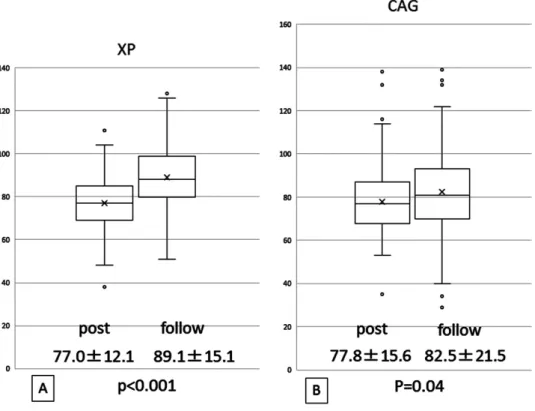
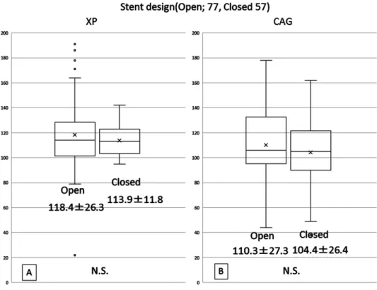
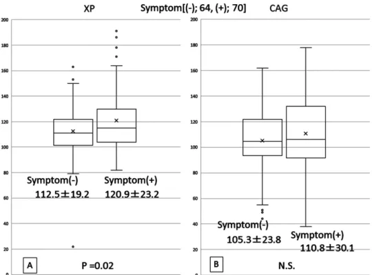
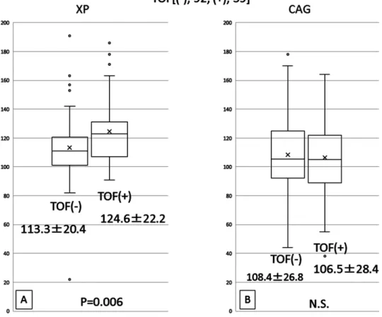
Results: Both the stent and lumen diameters were significantly dilated 12 months after CAS (p <0.001). There were no significant stent-type-related differences in the stent expansion rate. In the symptomatic stenosis group, this expansion rate was significantly higher than in the asymptomatic stenosis group (p = 0.02). With respect to the presence or absence of a high signal intensity on time of flight (TOF) magnetic resonance (MR) images, the stent expansion rate was significantly higher in the high signal intensity group (p = 0.006). In patients with a plaque/sternocleidomastoid muscle signal intensity ratio of ≥1.50 on plaque images, it was significantly higher than in those with a value of <1.50 (p = 0.006).

However, there were no significant differences in the lumen expansion rate among the groups.

Conclusion: Both the stent and vascular lumen were dilated 12 months after CAS. Plaque fragility influenced the stent expansion rate; however, there were no significant factor-related differences in the vascular lumen expansion rate.

Keywords▶  carotid artery stenting, self-expansion, plaque

Introduction

Since carotid artery stenting (CAS) for stenosis of the carotid artery became covered by health insurance in April 2008, it has commonly been performed in Japan. Initially, this procedure was primarily performed for patients at high risk for carotid endarterectomy (CEA) based on the results of the SAPPHIRE study.

1)

However, in 2010, the CREST study

2)

showed that CAS was as effective as CEA,

promoting its widespread application. On the other hand, the CREST

2)

and ICSS

3)

studies indicated that the inci- dence of perioperative stroke after CAS was higher than after CEA. Therefore, a reduction in the incidence is a key to an improvement in the results of CAS. Various studies have reported plaque assessment, medications, embolic protection devices, different cell-design stents, and proce- dures. Concerning procedures, studies using transcranial Doppler monitoring reported that distal embolism occurred most commonly on post-dilation after stenting.

4,5)

Several studies suggested that the incidence of embolic complica- tions can be decreased by omitting or restricting post- dilation although an insufficient stent dilatation also can occur even using self-expanding stents.

6,7)

There was one report about postoperative stent expansion 3 months after CAS; however, the long-term degree of postoperative stent expansion after CAS has not been elucidated.

8)

Our CAS strategy is targeting a residual stenosis rate of

≤30% using self-expanding stent considering both risk of embolic complications related to marked post-dilation and

1

Department of Neurosurgery, Osaka General Medical Center, Osaka, Osaka, Japan

2

Department of Neurosurgery, Nara Medical University, Kashihara, Nara, Japan

Received: March 18, 2016; Accepted: September 28, 2016 Corresponding author: Fujimoto Kenta. Department of Neuro- surgery, Osaka General Medical Center, 3-1-56 Bandai-higashi, Sumiyoshi-ku, Osaka, Osaka 558-8558, Japan

Email: [email protected]

©2017 The Editorial Committee of Journal of Neuroendovascular Therapy. All rights reserved.

Journal of Neuroendovascular Therapy 2017; 11: 65–75

Online November 22, 2016

(2)

that of early complications related to insufficient dilation.

In this study, we examined whether a stent itself expands or whether the vascular lumen is dilated in follow-up cerebral angiography based on the hypothesis that self-expansion may be serially achieved especially in patients with soft plaques. Furthermore, we investigated factors influencing postoperative self-expansion including the plaque fragility assessed by MRI and the stent design.

Subjects and Methods

Subjects

In our hospital, CAS for stenosis of the internal carotid artery has been performed for patients with ≥50% symp- tomatic stenosis or ≥75% asymptomatic stenosis (symp- tomatic: ≤50%, asymptomatic: ≤25% in accordance with the measurement method adopted in this study, as described below) according to the indication criteria adopted in the North American Symptomatic Carotid Endarterectomy Trial (NASCET)

9)

after obtaining approval from the Ethics Review Board and informed consent between January 2006 and April 2008, when CAS had not been covered by health insurance. After CAS became covered by health insurance, it was indicated for patients with ≥50% symp- tomatic stenosis or ≥80% asymptomatic stenosis, as deter- mined in the NASCET, at high risk for CEA, according to indication criteria in Japan. After the CREST study, CAS was selected as a first-choice procedure. CAS had been performed for 253 cases from January 2006, when CAS was introduced in our hospital, until December 2014. Of these, 134 cases (128 patients; 116 males, 18 females, age:

52–86 years, mean age: 72 years) in whom follow-up cere- bral angiography could be conducted, excluding treatment for restenosis, were enrolled into this study.

Assessment of the degree of stenosis

To measure the degree of stenosis, the vascular diameter at the stenotic site was compared with the distal internal carotid artery diameter in accordance with the NASCET

9)

using lateral views on cerebral angiography. However, after dilation, the lumen at the stenotic site sometimes dilates to a diameter larger than the distal carotid artery diameter; therefore, the formula of stenotic site/distal diameter × 100 was adopted. When the lumen diameter at the stenotic site became larger than the distal carotid artery diameter, it was recorded as a value of ≥100. Further- more, the stenosis rate on follow-up cerebral angiography after 1 year as a percentage of that immediately after CAS

was compared. To evaluate the degree of stenosis on plain X-ray, the stent diameter (D2) at the stenotic site was calculated as a percentage of the distal normal internal carotid artery diameter (D1), (D2/D1 × 100) in accordance with cerebral angiography-based assessment. On plain X-ray after 1 year, it was also calculated, assuming the same position as observed on plain X-ray immediately after CAS. The stenosis rate after 1 year as a percentage of that immediately after CAS was investigated. To compare changes between two groups, Student’s t-test was used.

A p value of 0.05 was regarded as significant.

Plaque assessment

Before CAS, carotid artery ultrasonography was performed for all patients. After January 2010, plaque assessment by the Black-blood method was conducted using MRI preop- eratively, and the plaque/sternocleidomastoid muscle signal intensity ratio (SIR) was calculated. On T1-weighted mag- netic resonance (MR) images (Black-blood method), the pixel values of the plaque were measured at six points. Sim- ilarly, those of the sternocleidomastoid muscle were measured at six points, and the ratio was calculated.

10)

In this study, the patients were divided into two groups: a group with an SIR of ≥1.5, at which plaque fragility may increase, and a group with an SIR of <1.5.

10,11)

Furthermore, some studies reported that plaques detected as a high signal intensity on time of flight (TOF) images were fragile

12,13)

and we also confirmed findings on TOF images, and retro- spectively examined the presence or absence of a high sig- nal intensity in the plaque area in previous patients.

Furthermore, it was indicated that plaques in patients with symptomatic lesions were more fragile than in those with asymptomatic lesions; therefore, the results were examined with respect to the presence or absence of symptoms.

14)

Techniques

In our hospital, CAS had been performed using a PRECISE

stent (Cordis, Miami Lakes, FL, USA) under distal balloon

protection with a Guardwire (Medtronic, Eden Prairie,

MN, USA) between January 2006 and April 2008, when

CAS had not been covered by health insurance. Pre-

dilation was conducted, and when ≥70% dilation (<30% in

accordance with NASCET measurement) was achieved

after stenting, post-dilation was omitted. From April 2008

until June 2010, a PRECISE stent was used under distal

filter protection with an Angioguard (Cordis). Pre-dilation

was conducted, and when ≥70% dilation was achieved

after stenting, post-dilation was omitted. From June 2010,

(3)

Self-expansion of Carotid Ar tery Stents after Carotid Ar tery Stenting

rate immediately after CAS × 100 [%]) was compared between the stent and lumen diameters. Assuming that self-expansion may be more marked in patients with softer plaques or when adopting a stent with a stronger radial force, we examined stent expansion with respect to the plaque/sternocleidomastoid muscle SIR on MR images, obtained using the Black-blood method, presence or absence of a high signal intensity on TOF images, presence or absence of symptoms, and stent design.

Results

The subjects were 134 cases (128 patients), with a mean age of 72 years (range: 52–86 years). The male-to-female ratio was 116:18. Follow-up cerebral angiography was performed 6 to 24 months after CAS and 134 of 253 cases (53.0%) were adopted this criteria in this study. The aver- age stenosis rate, measured using the above measurement method, on preoperative cerebral angiography was 22.9%

± 11.7% (range: 50 to 0%), whereas that on plain X-ray immediately after CAS was 77.0% ± 12.1%, but that on follow-up was 89.1% ± 15.1%, showing a significant increase (p <0.001) (Fig. 1A). The mean stenosis rate on cerebral angiography immediately after CAS was the device for distal filter protection was switched to a

Filterwire EZ (Boston Scientific, Natick, MA, USA). After November 2010, distal balloon protection was selected when a collateral pathway was present and distal filter pro- tection when it was absent. For tortuous lesions, a PRECISE stent was used, but a Carotid Wallstent (Boston Scientific) was used in patients in whom its insertion was considered possible. Pre-dilation was conducted using a balloon mea- suring 3.5 to 4.0 mm in diameter, and post-dilation using a balloon 0 to 0.5 mm smaller than the distal internal carotid artery diameter.

Follow-up

For medication, two of three antiplatelet drugs (clopidogrel at 75 mg, aspirin at 100 mg, and cilostazol at 200 mg) were administered for more than 1 week before CAS. The regi- men was switched to monotherapy 3 months after CAS.

Follow-up ultrasonography was performed once within 1 week after CAS, and, subsequently, every 3 months.

When restenosis or stent plaques were suspected, cerebral angiography was conducted. When there was no problem, cerebral angiography was performed after 12 months.

The stenosis rate on follow-up as a percentage of that immediately after CAS (stenosis rate on follow-up/stenosis

Fig. 1 Stent (A) and lumen (B) expansion 12 months after CAS, average ± standard deviation (%). CAG:

carotid angiography; CAS: carotid artery stenting; XP: X-ray photograph

(4)

Fig. 2 Stent (A) and lumen (B) expansion at 12 months after CAS according to stent type, average ± standard deviation (%). CAG: carotid angiography; CAS: carotid artery stenting; XP: X-ray photograph; NS: not significant

77.8% ± 15.6%, but that on follow-up was 82.5% ± 21.5%, showing a significant increase (p = 0.04) (Fig. 1B). We examined the stenosis rates immediately after CAS and on follow-up with respect to the plaque properties, stent design, and presence or absence of symptoms. Concerning the stent design, an open-cell stent was selected for 77 cases (Group O) and a closed-cell stent for 57 (Group C). The mean stent expansion rates were 118.4% ± 26.3% in Group O and 113.9% ± 11.8% in Group C. There was no signifi- cant difference in the expansion rate between the two groups (Fig. 2A). The mean lumen expansion rates on cerebral angiography were 110.3% ± 27.3% and 104.4% ± 26.4%, respectively, showing no significant difference (Fig. 2B).

Concerning symptoms, symptomatic stenosis was pres- ent in 70 cases and asymptomatic stenosis in 64 cases.

The mean stent expansion rates in the former and latter were 120.9% ± 23.2% and 112.5% ± 19.2%, respectively (p = 0.02), showing a significant difference (Fig. 3A).

However, the mean lumen expansion rates were 110.8% ± 30.1% and 105.3% ± 23.8%, respectively, showing no sig- nificant difference (Fig. 3B).

MR images of plaques were obtained in 90 cases. The plaque/sternocleidomastoid muscle SIR was ≥1.50, at

which the incidences of plaque hemorrhage and lipid-rich necrotic core may increase, in 28 cases, and it was <1.50 in 62.

13)

In cases with an SIR of ≥1.50, the mean stent expansion rate was 125.4% ± 21.1%. In those with an SIR of <1.50, it was 112.5% ± 18.8%; the value was signifi- cantly higher in the former (p = 0.006) (Fig. 4A). The mean lumen expansion rates in the former and latter were 102.4% ± 27.4% and 110.6% ± 25.3%, respectively, show- ing no significant difference (Fig. 4B).

TOF images were obtained in 131 cases. A high signal intensity was detected in 39 cases ((+) group), but not in 92 ((-) group). The mean stent expansion rates in the (+) and (-) groups were 124.6% ± 22.2% and 113.3% ± 20.4%, respectively, showing a significant difference (p = 0.006) (Fig. 5A). However, the mean lumen expansion rates were 106.5% ± 28.4% and 108.4% ± 26.8%, respectively, showing no significant difference (Fig. 5B).

The high-signal-intensity appearance rate on diffusion- weighted MR images after CAS in the subjects was 15.3%

(20/131). There were no neurological complications during

CAS or immediately after CAS, but minor stroke was

observed in three cases within 30 days after CAS, and

hyperperfusion syndrome in one case. When adopting our

measurement method, ≤50% (NASCET: ≥50%) restenosis

(5)

Self-expansion of Carotid Ar tery Stents after Carotid Ar tery Stenting

Fig. 3 Stent (A) and lumen (B) expansion at 12 months after CAS according to symptom, average ± stan- dard deviation (%). CAG: carotid angiography; CAS: carotid artery stenting; XP: X-ray photograph;

NS: not significant

Fig. 4 Stent (A) and lumen (B) expansion at 12 months after CAS according to plaque/muscle SIR on MR

plaque imaging, average ± standard deviation (%). CAG: carotid angiography; CAS: carotid artery stent-

ing; MR: magnetic resonance; XP: X-ray photograph; NS: not significant; SIR: signal intensity ratio

(6)

in accordance with the measurement method adopted in this study (Fig. 6A). Under Angioguard protection, pre-dilation was conducted using an Amia (Cordis) measuring 4.0 × 30 mm, and a PRECISE stent measuring 9 × 40 mm was inserted. As dilation was considered sufficient, post-dilation was not performed. The stenosis rate immediately after CAS was 61% (Fig. 6B). Follow-up ultrasonography did not show any abnormalities, and cerebral angiography was conducted after 18 months. The stent had expanded from 59% to 130% (Figs. 6D and 6E), and the lumen had also expanded from 61% to 132% (Fig. 6C).

Case 2

A 63-year-old male presented with amaurosis fugax of the right eye was referred to our hospital for evaluation.

Carotid artery ultrasonography revealed a low-echoic plaque at the bifurcation of the right carotid artery. MR images of the plaque showed a high signal intensity, with an SIR of 1.65 (Fig. 7A). On TOF MRA, a high signal intensity was detected (Fig. 7B). On cerebral angiography, the stenosis rate was 5% (NASCET: 95%) in accordance was noted in six cases (4.5%). Of these, it was symptom-

atic in one case (0.8%), and additional treatment was performed for five cases (3.8%). The incidence of ≥50%

restenosis was 2.4% (6/253), including cases in whom follow-up digital subtraction angiography (DSA) was not done. For all cases who underwent cerebral angiography within 6 months for some reason, additional cerebral angi- ography was performed during the study period. No patient received additional treatment within 6 months.

Representative Case

Case 1

A 70-year-old man was referred from the Department of Ophthalmology with amaurosis fugax of the left eye.

Carotid artery ultrasonography revealed a low-echoic plaque at the bifurcation of the left carotid artery. The peak systolic velocity (PSV) was 338.9 cm/s, confirming accel- eration. MR images of the plaque were not evaluated, and there was no high signal intensity on TOF MRA. The steno- sis rate on cerebral angiography was 11% (NASCET: 89%)

Fig. 5 Stent (A) and lumen (B) expansion at 12 months after CAS according to TOF MRA, average ± standard deviation (%). CAG: carotid angiography; CAS: carotid artery stenting; MRA: magnetic resonance angiography; NS: not significant; TOF: time of flight; XP: X-ray photograph; (+): high signal intensity;

(-): no high signal intensity

(7)

Self-expansion of Carotid Ar tery Stents after Carotid Ar tery Stenting

There were no changes in the stenosis rate or shape. The stent seemed to sink into the plaque (Fig. 8C). The lesion was asymptomatic, with a stenosis rate of 58% (NASCET:

42%), and additional treatment was considered unneces- sary. Follow-up is being continued.

Discussion

Recently, CAS has commonly been performed, and several studies, including the CREST study, reported that its effi- cacy was similar to that of CEA.

1,2)

The most important issue of CAS to be overcome is the prevention of thrombo- embolic complications due to distal embolism. For this purpose, efforts to select high-risk patients and modify medications/procedures/devices to reduce the incidence of distal embolism have been made in the literature.

with the measurement method adopted in this study (Fig. 8A). Under Guardwire protection, pre-dilation was conducted using an Amia measuring 4.0 × 30 mm, leading to transient cardiac arrest. The chest was hit, and the heart beat promptly resumed. After the intravenous injection of atropine sulfate at 5 mg, a PRECISE stent measuring 9 × 40 mm was inserted. Post-dilation was not conducted, and the procedure was completed. There were no neurolog- ical symptoms, and there were no clinical problems.

However, the stenosis rate immediately after CAS was 53% (Fig. 8B). During the follow-up, plain X-ray showed stent expansion, but ultrasonography revealed intra-stent thrombus formation; therefore, cerebral angiography was performed after 12 months. The stent had expanded from 59% to 130% (Figs. 8D and 8E), but lumen expansion was slight: from 53% (immediately after CAS) to 58%.

Fig. 6 CAG and plain cervical XP of Case 1. (A) Initial CAG, (B) CAG immediately after CAS, (C) CAG

18 months after CAS, (D) XP immediately after CAS, and (E) XP 18 months after CAS. CAG: carotid

angiography; CAS: carotid artery stenting; XP: X-ray photograph

(8)

Fig. 7 MRI and MRA of the Case 2. (A) Black-blood plaque imaging of the plaque showed high intensity (arrow) and its plaque/muscle signal intensity ratio was 1.65. (B) TOF MRA showed a high- signal-intensity plaque (arrow). TOF: time of flight

For the selection of high-risk patients, it is important to evaluate plaque properties based on preoperative diagnostic imaging findings. Some studies indicated the importance of plaque assessment using MRI, and, recently, it has been essential as preoperative assessment.

10,11,15)

Furthermore, others reported that TOF MRI is commonly used for preoperative plaque assessment because plaques detected as a high signal intensity on TOF images were fragile.

12,13)

Concerning procedures, several studies reported that post-dilation after stenting in the process of CAS was involved in distal embolism.

4,5,16)

Therefore, in our hospital, initially, CAS was performed using a PRE- CISE stent alone, and post-dilation was omitted when

≤30% (NASCET) residual stenosis was present. Since the introduction of a Carotid Wallstent, post-dilation has been conducted using a balloon smaller than the distal internal carotid artery diameter. As a result, residual stenosis has been observed in many patients immediately after CAS, and multimodalities including ultrasonography, CT angi- ography, cerebral angiography, and plain X-ray have been combined for follow-up evaluation.

The most accurate diagnostic modality to evaluate the stent and the lumen diameter is cerebral angiography;

however, it is invasive, used contrast medium, and requir- ing admission; hence, it is difficult to do this procedure for all patients in practice. Instead of cerebral angiography, ultrasonography for the lumen diameter and plain X-ray for stent diameter were used. However, it is unclear whether the degree of subsequent lumen expansion is reflected by the stent expansion. Therefore, we examined changes immediately after CAS and on follow-up in 134 cases who could be followed up using cerebral angiogra- phy. Concerning the timing of follow-up, a study indicated that marked restenosis frequently occurred within 6 months after CAS, and that the incidence of restenosis decreased 12 months or more after CAS;

17)

based on these results, we decided to perform follow-up cerebral angiography at 12 months after CAS, considering that the incidence of restenosis should be less frequently.

Self-expanding stents are used for CAS; they may also

expand after treatment. However, it is difficult to predict the

degree of expansion. Previous studies reported that the

perioperative clinical results were favorable despite post-

dilatation procedure.

6,7,18,19)

However, few studies have

examined the degree of stent expansion on follow-up.

8)

In this study, we investigated stent expansion and changes

(9)

Self-expansion of Carotid Ar tery Stents after Carotid Ar tery Stenting

et al.

8)

indicated that stent expansion was more marked in patients in whom a closed-cell stent with a weak radial force was used. They selected the closed-cell stent for soft plaques, and there may have been a selection bias. In addition, the results suggest that plaque properties more markedly influence expansion compared with the stent design. Of our series, a PRECISE stent was selected for all patients in the initial phase, and, after 2011, a Carotid Wallstent was used regardless of plaque properties if possible; therefore, there might have been less stent-option-related selection bias.

Plaque properties

When a plaque is soft, a stent may expand more markedly.

In this study, stent expansion was significantly more marked in a group with a plaque/sternocleidomastoid mus- cle SIR of ≥1.5 on assessment using the Black-blood method, in which plaques may have been soft, and in a group with a high signal intensity on TOF images. Further- more, a study reported that plaques in symptomatic stenotic in the vascular lumen on cerebral angiography 1 year after

CAS in patients who underwent CAS in our hospital, and in whom cerebral angiography findings could be evaluated.

The stent stenosis rates immediately after CAS and on follow-up cerebral angiography were 77.0% ± 12.1% and 89.1% ± 15.1%, respectively, showing expansion (p <0.001).

Furthermore, the lumen stenosis rates on cerebral angi- ography immediately after CAS and on follow-up were 77.8% ± 15.6% and 82.5% ± 21.5%, respectively, showing a significant enlargement (p = 0.04). During the follow-up period, both the stent and lumen diameters on cerebral angi- ography had increased (Fig. 1). The stent design and plaque properties may be closely involved in expansion.

Stent design

As an open-cell stent shows a stronger radial force compared with a closed-cell stent,

20)

its expansion may be more marked.

However, in this study, there was no significant stent-type- related difference in the expansion rate. In contrast, Tanno

Fig. 8 CAG and plain cervical XP of Case 2. (A) Initial CAG, (B) CAG immediately after CAS, (C) CAG

12 months after CAS, (D) XP immediately after CAS, and (E) XP 12 months after CAS. CAG: carotid

angiography; CAS: carotid artery stenting; XP: X-ray photograph

(10)

Conclusion

After CAS, both the stent and lumen diameters were expanded. The stent expansion rates in patients with a high signal intensity on TOF images, in whom plaques were considered fragile, those with a high signal intensity on MRI (Black-blood method), and those with symptomatic stenosis were significantly higher, but there was no significant stent- type-related difference. The lumen diameter also expanded with an increase in the stent diameter. However, the lumen did not readily expand under conditions facilitating stent expansion. Although postoperative stent/lumen expansion may be achieved on follow-up, the degree of the expansion is unpredictable in practice. Therefore, ≥30% (NASCET) acceptable expansion should be obtained during CAS.

Disclosure Statement

The authors completed the self-reporting of conflict of interest (COI) to the Japan Neurosurgical Society. There is no conflict of interest to be disclosed regarding this study.

References

1) Yadav JS, Wholey MH, Kuntz RE, et al: Protected carotid- artery stenting versus endarterectomy in high-risk patients.

N Engl J Med 2004; 351: 1493–1501.

2) Brott TG, Hobson RW, Howard G, et al: Stenting versus endarterectomy for treatment of carotid-artery stenosis.

N Engl J Med 2010; 363: 11–23.

3) International Carotid Stenting Study investigators, Ederle J, Dobson J, et al: Carotid artery stenting compared with end- arterectomy in patients with symptomatic carotid stenosis (International Carotid Stenting Study): an interim analysis of a randomised controlled trial. Lancet 2010; 375: 985–997.

4) Ackerstaff RG, Suttorp MJ, van den Berg JC, et al: Pre- diction of early cerebral outcome by transcranial Doppler monitoring in carotid bifurcation angioplasty and stenting.

J Vasc Surg 2005; 41: 618–624.

5) Rubartelli P, Brusa G, Arrigo A, et al: Transcranial Doppler monitoring during stenting of the carotid bifurcation: eval- uation of two different distal protection devices in prevent- ing embolization. J Endovasc Ther 2006; 13: 436–442.

6) Jin SC, Kwon OK, Oh CW, et al: A technical strategy for carotid artery stenting: suboptimal prestent balloon angio- plasty without poststenting balloon dilatation. Neurosur- gery 2010; 67: 1438–1442; discussion 1442–1443.

7) Spacek M, Zimolova P, Veselka J: Carotid artery stenting without post-dilation. J Interv Cardiol 2012; 25: 190–196.

lesions were more fragile than in asymptomatic lesions.

14)

In this study, stent expansion was more marked in the symptomatic stenosis group. However, there was no signif- icant difference in lumen expansion.

As the reason why lumen expansion did not always accompany stent expansion, neointima formation in the lumen may have induced hyperplasia, thrombus attach- ment, or plaque protrusion into the stent.

Several studies using a self-expanding stent for the coronary artery and involving follow-up reported that stent expansion led to neointimal ingrowth, causing narrowing of the lumen.

21–23)

Hong et al.

23)

inserted a self-expanding nitinol stent into the coronary artery in an animal experi- ment using pigs, and examined its pathological sections.

They confirmed an increase in the stent diameter 6 months after stenting, but the neointimal reaction of the vascular lumen reached a peak 4 to 8 weeks after stenting, and, after 6 months, the stent strut had reached the outer membrane in most pigs. In this study, in Case 2, the stent expanded, and sank into the plaque; the stent strut may have reached an area adjacent to the outer membrane.

Overall, lumen expansion during follow-up was con- firmed, as shown in Fig. 1B. Thus, even when chronic- phase self-expansion is insufficient, self-expansion may be achieved through continuous follow-up rather than early additional treatment. However, there are some patients in whom expansion is not achieved. And of our series, the stent sank into the plaque in some patients, as demonstrated in Case 2. In our hospital, 30% stenosis (NASCET) is tar- geted, and the procedure should be completed after obtain- ing an acceptable degree of expansion.

Limitations

Of our series, 119 cases in whom follow-up cerebral angi-

ography was not done (47%) were excluded. Cerebral

angiography has been performed if follow-up ultrasonog-

raphy suggested abnormalities even in the presence of

renal dysfunction; however, this may have affected the

results. In our hospital, several angio-suits have been used,

and there may be an error on measurement of absolute val-

ues, depending on the distance between the panel and

lesion. Therefore, we have assessed the degree of stenosis,

as a relative value, but not the absolute diameter at the

most stenotic site; absolute-value assessment has not been

conducted. Furthermore, stent options and techniques were

modified between the early and late phases of the present

study period, and this may have contributed to the results.

(11)

Self-expansion of Carotid Ar tery Stents after Carotid Ar tery Stenting

identifying lipid-rich necrotic cores and intraplaque hemor- rhage in advanced human carotid plaques. Circulation 2001; 104: 2051–2056.

16) Vitek JJ, Roubin GS, Al-Mubarek N, et al: Carotid artery stenting: technical considerations. AJNR Am J Neuroradiol 2000; 21: 1736–1743.

17) Harrer JU, Morschel R, Mull M, et al: High rate of resteno- sis after carotid artery stenting in patients with high-grade internal carotid artery stenosis. Medium-term follow-up.

J Neurol 2008; 255: 1309–1314.

18) Baldi S, Zander T, Rabellino M, et al: Carotid artery stent- ing without angioplasty and cerebral protection: a single- center experience with up to 7 years’ follow-up. AJNR Am J Neuroradiol 2011; 32: 759–763.

19) Bussière M, Pelz DM, Kalapos P, et al: Results using a self-expanding stent alone in the treatment of severe symp- tomatic carotid bifurcation stenosis. J Neurosurg 2008;

109: 454–460.

20) Wissgott C, Schmidt W, Behrens P, et al: Experimental investigation of modern and established carotid stents. Rofo 2014; 186: 157–165.

21) Roguin A, Grenadier E, Linn S, et al: Continued expansion of the nitinol self-expanding coronary stent: angiographic analysis and 1-year clinical follow-up. Am Heart J 1999;

138: 326–333.

22) von Birgelen C, Airiian SG, de Feyter PJ, et al: Coronary wallstents show significant late, postprocedural expansion despite implantation with adjunct high-pressure balloon inflations. Am J Cardiol 1998; 82: 129–134.

23) Hong MK, Beyar R, Kornowski R, et al: Acute and chronic effects of self-expanding nitinol stents in porcine coronary arteries. Coron Artery Dis 1997; 8: 45–48.

8) Tanno Y, Mori T, Iwata T, et al: [Spontaneous dilatation of carotid artery stents three months after the procedure, with- out the need for Post-CAS balloon dilatation]. No Shinkei Geka 2015; 43: 1019–1025. (in Japanese)

9) North American Symptomatic Carotid Endarterectomy Trial Collaborators: Beneficial effect of carotid endarterectomy in symptomatic patients with high-grade carotid stenosis.

N Engl J Med 1991; 325: 445–453.

10) Yoshida K, Narumi O, Chin M, et al: Characterization of carotid atherosclerosis and detection of soft plaque with use of black-blood MR imaging. AJNR Am J Neuroradiol 2008;

29: 868–874.

11) Akutsu N, Hosoda K, Fujita A, et al: A preliminary prediction model with MR plaque imaging to estimate risk for new ischemic brain lesions on diffusion-weighted imaging after endarterectomy or stenting in patients with carotid stenosis. AJNR Am J Neuroradiol 2012; 33:

1557–1564.

12) Yamada K, Song Y, Hippe DS, et al: Quantitative eval- uation of high intensity signal on MIP images of carotid atherosclerotic plaques from routine TOF-MRA reveals elevated volumes of intraplaque hemorrhage and lipid rich necrotic core. J Cardiovasc Magn Reson 2012; 14: 81.

13) Yoshimura S, Yamada K, Kawasaki M, et al: High-intensity signal on time-of-flight magnetic resonance angiography indicates carotid plaques at high risk for cerebral embolism during stenting. Stroke 2011; 42: 3132–3137.

14) Bosiers M, de Donato G, Deloose K, et al: Does free cell area influence the outcome in carotid artery stenting? Eur J Vasc Endovasc Surg 2007; 33: 135–141; discussion 142–143.

15) Yuan C, Mitsumori LM, Ferguson MS, et al: In vivo

accuracy of multispectral magnetic resonance imaging for

Fig. 1   Stent  (A) and lumen (B) expansion 12 months after CAS, average ± standard deviation (%)
Fig. 2   Stent  (A) and lumen (B) expansion at 12 months after CAS according to stent type, average ± standard  deviation (%)
Fig. 3   Stent  (A) and lumen (B) expansion at 12 months after CAS according to symptom, average ± stan- stan-dard deviation (%)
Fig. 5   Stent  (A) and lumen (B) expansion at 12 months after CAS according to TOF MRA, average ± standard  deviation  (%)
+4

参照

関連したドキュメント

A previous study has demonstrated successful retrieval of unexpanded stent in left main coronary artery with an initial goose-neck snare 23 ; however, no effective device

It is shown that plasma endothelial lipase (EL) activity inversely correlated with HDL-C levels, and EL activity in CAD patients was significantly higher than in non CAD

The present results also provide in situ hybridization evidence that at least chief cells and possibly both chief cells and sustentacular cells of the carotid body express NGF mRNA..

Methods: IgG and IgM anti-cardiolipin antibodies (aCL), IgG anti-cardiolipin-β 2 glycoprotein I complex antibody (aCL/β 2 GPI), and IgG anti-phosphatidylserine-prothrombin complex

It is a new contribution to the Mathematical Theory of Contact Mechanics, MTCM, which has seen considerable progress, especially since the beginning of this century, in

Thus, in Section 5, we show in Theorem 5.1 that, in case of even dimension d &gt; 2 of a quadric the bundle of endomorphisms of each indecomposable component of the Swan bundle

Ntouyas, Boundary value problems for nonlinear fractional differential equations and inclusions with nonlocal and fractional integral boundary conditions, Opuscula Math, 33, No..

We will study the spreading of a charged microdroplet using the lubrication approximation which assumes that the fluid spreads over a solid surface and that the droplet is thin so