Posted at the Institutional Resources for Unique Collection and Academic Archives at Tokyo Dental College, Available from http://ir.tdc.ac.jp/
Title
Which Factors Affect the Long-Term Survival of Patients With Oral Squamous Cell Carcinoma With Distant Metastasis?
Author(s) Alternative
Sekikawa, S; Kawachi, H; Ogane, S; Saito, H; Takano, M; Nomura, T; Katakura, A; Takano, N; Shibahara, T
Journal Journal of oral and maxillofacial surgery, 78(3):
469-478
URL http://hdl.handle.net/10130/5404
Right
1 Full Length Article
Which factors affect the long-term survival of patients with oral squamous cell carcinoma with
distant metastasis?
Shoichi Sekikawa DDS, PhD, Clinical Fellow1,2, Homare Kawachi DDS, PhD, Assistant Professor3,
Satoru Ogane DDS, PhD, Lecturer3, Hirokazu Saito DDS, PhD, Assistant Professor4, Masayuki
Takano DDS, PhD, Associate professor1,3, Takeshi Nomura DDS, PhD, Professor and Chairman3,4,
Akira Katakura DDS, PhD, Professor and Chairman3,5, Nobuo Takano DDS, PhD, Director3,
Takahiko Shibahara DDS, PhD, Professor and Chairman1,3
1Department of Oral and Maxillofacial Surgery, Tokyo Dental College
2Department of Dentistry and Oral Surgery, Tokyo Metropolitan Tama Medical Center
3Oral Cancer Center, Tokyo Dental College
4Department of Oral Medicine, Oral and Maxillofacial surgery, Tokyo Dental College
5Department of Oral Pathobiological Science and Surgery, Tokyo Dental College
Corresponding author: Shoichi Sekikawa
Department of Oral and Maxillofacial Surgery, Tokyo Dental College
2-9-18 Kanda-Misakicho, Chiyoda-ku, Tokyo 101-0061, Japan
e-mail: [email protected]
Conflict of interest statement
None declared.
2
This study did not receive any specific grant from funding agencies in the public, commercial, or
1 Abstract
Purpose: The development of distant metastases (DM) in patients with oral squamous cell carcinoma
(OSCC) leads to dismal prospects for survival. This study aimed to identify risk factors for DM
development and long-term survival.
Patients and Methods: This study was a retrospective cohort study of OSCC patients at a single
institution. The predictor variables were age, sex, TN classification, histological grade, neck
dissection, infiltrative growth pattern (INF), vascular/lymphatic invasion, perineural invasion (PI),
extranodal extension, local recurrence, nodal metastasis, DM, the time to diagnosis of DM, surgery
for DM. The primary outcome variables were five-year overall survival (5y-OS) and median survival
time (MST), which were estimated using the Kaplan-Meier method. Cox hazard models were used to
identify risk factors for DM development.
Results: The cohort involved 526 patients, 402 of whom were available for analysis, with 37 of these
402 patients developing DM. On multivariate analysis, cN1-2 (HR: 3.36), moderate/poor
differentiation (HR: 2.51), INF c (HR: 3.27), vascular/lymphatic invasion (HR: 2.95), and PI (HR:
2.17) were independent predictors for DM development. The 5y-OS was 84.6% for non-DM patients
and 9.7% for DM patients, with a MST of 16.9 months. In DM patients with cN0, the 5y-OS was
18.2%, and the MST was 37.2 months, while in DM patients with cN1-2, the 5y-OS was 4.7%, and
the MST was 12.9 months. In patients with time to DM diagnosis ≥10.0 months, the 5y-OS of was
20.0%, and the MST was 38.6 months, while in patients with time to DM diagnosis <10.0 months,
the MST of was 11.7 months. The 5y-OS of patients who underwent a pulmonary metastasectomy
was 60.0%; the MST of the non-surgery group was 16.0 months.
Conclusion: In DM patients, cN0 and late time to DM diagnosis were associated with long-term
survival. Pulmonary metastasectomy may be worth considering to improve survival.
2
Distant metastasis; oral cancer; prognosis; risk factors; squamous cell carcinoma
Introduction
Oral cancer accounts for 117 384 deaths and 354 864 new diagnoses annually, and lip and
oral cancer is the 16th leading cause of cancer deaths worldwide [1]. Oral squamous cell carcinoma
(OSCC) is the most common form of oral cancer [2]. The overall and five-year survival rates for
OSCC are both approximately 60% but vary from 10% to 82% depending on the clinical stage, age,
race, comorbidity, and primary site [3]. Of several prognostic factors, it has been shown that distant
metastasis (DM) is one of the strongest for predicting poor survival [4]. DM is found in 8.3-12.1% of
primary cases without DM at the initial diagnosis after definitive surgery [4-8]. Although the overall
survival rate in oral cancer has improved in the last 20 years thanks to improvements in diagnostic
modalities and treatment methods [3, 9], patients with DM development have a poor prognosis, and
generally only palliative treatments may be offered [4]. N classification [10], extranodal extension
(ENE) [10, 11], and poor differentiation [12] are reportedly independent risk factors for DM
development although they are controversial [11, 13]. Therefore, patients with node-negative (N0)
disease are considered to have a low risk of DM development; however, some of these patients go on
to have DM and a consequently poor prognosis [14].
The aim of the present study was to evaluate the outcomes, including DM development and
long-term survival, in patients with OSCC after definitive surgery. The authors hypothesized that
node-negative disease would influence long-term survival of OSCC patients with DM. The specific
aim of this study was to identify the clinicopathological factors associated with the risk of DM
development and the prognosis of patients with DM, including those with clinical N0 (cN0) disease.
Patients and methods
3
This study was a retrospective cohort study enrolling patients with OSCC who underwent
surgery at Tokyo Dental College Oral Cancer Center between 2007 and 2016. The patients were
enrolled according to the reporting recommendations for tumor marker prognostic studies
(REMARK) guidelines [15]. To be included in this study sample, patients had to meet the following
inclusion criteria: 1) histologically confirmed OSCC; 2) primary definitive surgery at Tokyo Dental
College Oral Cancer Center between 2007 and 2016; and 3) availability of clinicopathological
information, including age, sex, T classification, stage, primary site, cancer recurrence, nodal
metastasis, various pathological features, survival status, survival duration, and cause of death.
Patients who did not undergo primary definitive surgery or the lacked information above were
excluded. The tumor stages were defined according to the TNM Classification of Malignant Tumors,
7th Edition (UICC) [16]. According to the National Comprehensive Cancer Network guidelines [17],
elective neck dissection (END) was considered when the depth of invasion was 4 mm or more, and
postoperative adjuvant therapy (tri-weekly cisplatin 100 mg/m2 + radiotherapy) was performed when
adverse risk features were found, including the involvement of two or more nodes, positive margins,
and ENE in our hospital. The diagnosis of DM was based on computed tomography (CT) or positron
emission tomography-CT (PET-CT) findings or histologic evidence. As a rule, at our institution, we
perform a monthly follow-up and head and neck CT scan for one year postoperatively; moreover, in
advanced cases, a CT scan of the lung area is also performed. At two years postoperatively, CT is
performed once every three to four months. A PET-CT is performed if needed, and in advanced cases
it is performed at least once every six months. The presence of multiple lung nodules of different
sizes on images were considered to indicate pulmonary metastases. A fine needle aspiration biopsy
was done if feasible for isolated lung nodules to distinguish it from primary lung cancer. Patients
were excluded if their tumor could not be distinguished as a metastasis or primary lung cancer. For
patients in our hospital in whom DM developed, depending on their performance status (PS) [18],
4
and systemic therapy, including chemotherapy and targeted molecular therapy or best supportive care,
was considered for inoperable cases. According to the criteria of Thomford et al. [19], distant
metastatic lesions, such as an isolated lung metastasis in patients with good PS, were resected if
feasible. This study was approved by the Research Ethics Committee of Tokyo Dental College (I
16-07). Informed consent was provided in compliance with the Helsinki Declaration.
Study variables
The primary predictor variables were demographic characteristics (age, sex), T classification
(T1-2/T3-4), cN classification (cN0/cN1-2), histological grade (Well/Moderate, Poor), neck
dissection (absent/present), infiltrative growth pattern (INF) (INFa, b/INFc) [20], vascular/lymphatic
invasion (absent/present), perineural invasion (PI) (absent/present), ENE (absent/present), local
cancer recurrence (absent/present), nodal metastasis (absent/present), DM (absent/present), the time
to diagnosis of DM after definitive surgery (< 10.0 months/≥ 10.0 months), surgical treatment for
DM (absent/present) and mortality (absent/present).
The primary outcome variables were five-year overall survival (5y-OS) and the median
survival time (MST). A receiver operating characteristics (ROC) curve was generated to determine
the cut-off value of time to diagnosis of DM to predict a poor prognosis in patients with DM.
Additional outcome variables were the development of distant metastasis, the five-year distant
metastasis-free survival rate (DMFS) and the DM failure rate.
Statistical analysis
The correlation between the clinicopathological features and DM was examined using the
univariate Cox proportional hazards regression model. All the outcome variables were estimated
using the Kaplan-Meier method and compared by the log-rank test. Multivariate Cox proportional
5
to the model being rejected, to identify the risk factors for DM development. The parameters initially
included in the Cox regression analysis were T classification, cN classification, histological grade,
neck dissection, INF, vascular/lymphatic invasion, PI, and local cancer recurrence. The proportional
hazards assumption was evaluated and satisfied for all variables used. All the tests were two-sided,
and all p-values < 0.05 were considered significant. All statistical analyses were performed with EZR
ver. 1.37 (Saitama Medical Center, Jichi Medical University, Saitama, Japan) [21], a graphical user
interface for R (The R Foundation for Statistical Computing, Vienna, Austria). More precisely, it is a
modified version of R commander designed to add statistical functions frequently used in
biostatistics.
Results
Patient characteristics, clinicopathological features, and primary site related to DM development
This retrospective cohort enrolled 526 patients with OSCC, 402 of whom were available for
analysis. Of these, 304 patients had cN0 disease. The mean age of the patients was 67 years (range:
23-95 years). The median duration of follow-up in all 402 cases was 40.3 months (range: 2.6-133.0
months). The median duration of follow-up in cN0 304 cases was 39.9 months (range: 2.6-133.0
months).
Thirty-seven (9.2%) of the 402 patients had DM. Among the 304 patients with cN0 disease,
14 (4.6%) had DM. The median time within which DM was diagnosed after definitive surgery was
8.3 months (1.1 to 36.2 months) in all the patients and 15.8 months (2.2 to 36.2 months) in patients
with cN0 disease, and there was a significant difference between the two groups (p = 0.00892,
Mann-Whitney U test). Twenty-nine cases were detected by CT, and eight cases were detected by
PET-CT. Most of the lung lesions were found by CT, and lesions in other organs were found by
PET-CT. The clinicopathological features of the 402 patients are shown in Table 1. Local recurrence
6
are shown in Fig. 1. The most frequent primary location was the tongue in 192 patients (47.8%),
followed by the lower gingiva in 93 patients (23.1%), upper gingiva in 55 patients (13.7%), floor of
the mouth in 28 patients (7.0%), buccal mucosa in 22 patients (5.5%), and other region in 12 patients
(2.9%). The incidence of DM was highest in patients with tongue SCC (16 (8.3%) of 192 patients),
followed by the lower gingiva (12 (12.9%) of 93 patients). There was no significant difference in the
incidence of DM by primary site (p = 0.541, Fisher’s exact test).
Site of DM
The most frequent metastatic site was the lung, which was found in 33 (89.2%) of 37 cases,
followed by bone (18.9%), liver (10.8%), and kidney (8.1%), skin (5.4%), brain (2.7%), heart (2.7%),
mediastinum (2.7%), muscle (2.7%), parotid gland (2.7%). In patients with lung metastases, multiple
metastases (24 of 33 cases) occurred more frequently than isolated metastases (9 of 33 cases) and
tended to be less frequent in patients with cN0 disease. Of all the patients, multiple DM sites were
found in 13 patients (35.1%). In patients with cN0 disease, multiple DM sites were found in four
patients (28.6%). In patients with cN1-2 disease, multiple DM sites were found in nine patients
(39.1%).
Survival analysis
The 5y-OS was 84.6% in patients with no DM and 9.7% in patients with DM. The MST was
16.9 months in patients with DM, who had a significantly worse prognosis than patients with no DM
(p < 0.00001, Fig. 2A). The 5y-OS was 82.1% in patients with cN0 disease and 61.2% in patients
with cN1-2 disease (p = 0.00011, Fig. 2B), and the DMFS was 81.1% in patients with cN0 and
60.6% in patients with cN1-2 (p = 0.00003, Fig. 2C). In addition, in patients who developed DM, the
5y-OS of patients with cN0 disease at the initial diagnosis was 18.2%, and their MST was 37.2
7
(p = 0.02760, Fig. 2D). The cut-off value of the time to diagnosis of DM after definitive surgery was
10.0 months based on the ROC curve analysis. The 5y-OS of patients with time to diagnosis ≥ 10.0
months was 20.0%, and their MST was 38.6 months while the MST of patients with time to
diagnosis < 10.0 months was 11.7 months (p < 0.00001, Fig. 2E). Surgical treatment for distant
metastatic lesions was performed in five patients with an isolated lung metastasis; the 5y-OS of the
surgery group was 60.0%, and the MST of the non-surgery group was 16.0 months (p = 0.00682, Fig.
2F). The surgical group had significantly longer survival.
Analysis of the DM failure rate
The DM failure rates in patients with cN0 disease and those with cN1-2 disease were
compared using Kaplan-Meier analysis and the log-rank test. The DM failure rate in patients with
cN0 disease was 5.8% while that in patients with cN1-2 disease was 24.5% (p < 0.00001, Fig. 3A).
Next, in patients with cN0 disease, the DM failure rate was 13.0% in patients who underwent END
and 3.4% in patients who did not undergo END (p = 0.00444, Fig. 3B). END did not reduce the
incidence of DM. In addition, in patients with pathological node-positive disease (pN+), the DM
failure rate of patients with ENE was 50.6% while in patients without ENE it was 19.8% (p =
0.00081, Fig. 3C).
Risk factors for developing DM
Based on risk factors identified on univariate analysis, a multivariate stepwise Cox
regression analysis was performed. It was found that cN1-2 disease (HR 3.36; 95%CI 1.66-6.80; p =
0.00077), moderate or poor differentiation (HR 2.51; 95%CI 1.18-5.35; p = 0.01669), INF c (HR
3.27; 95%CI 1.49-7.17; p = 0.00314), presence of vascular/lymphatic invasion (HR 2.95; 95%CI
1.36-6.40; p = 0.00634), and presence of perineural invasion (HR 2.17; 95%CI 1.05-6.50; p =
8 Discussion
In general, patients with DM development have dismal survival outcomes. Generally, only
palliative treatments may be offered to patients with DM, and there is no consensus on the
management of these patients. Most patients with head and neck cancer, including oral cancer, who
have DM development, will die within about one to 12 months after the diagnosis of DM [13, 22-24].
The MST of patients with DM was reported to be 12 months by Leon et al. [10] In the present study,
the 5y-OS of patients with DM was 9.7%, and their MST was 16.9 months, which also underscored
the very poor prognosis of patients with DM. However, there were groups with relatively long
survival times among patients with DM. Patients with cN0 disease at the initial diagnosis and
patients with the diagnosis of DM ≥10 months after surgery had relatively long survival times, and
their MST was about three times longer than that of patients with cN1-2 disease or patients with a
diagnosis of DM <10 months after surgery.
Furthermore, the 5y-OS of patients who received surgical treatment for a lung metastasis
was good, at 60%. The N classification (presence of nodal metastasis) is a strong independent
indicator of poor prognosis in patients with OSCC [3, 25]. Therefore, the pathological staging of the
nodal status is the gold standard by which risk may be stratified and treatment strategy designed. In
the present study as well, patients with pN-positive disease, especially those positive for ENE, were
at high risk of DM development. However, pN staging depends on surgical procedures, such as neck
dissection or sentinel lymph node biopsy, including elective dissection. Thus, the cN classification,
which does not depend on surgical treatment, is useful, and the cN classification is also reportedly a
significant predictor of poor prognosis although it is not a pathological classification [14, 26]. The
Kaplan-Meier and multivariate analyses in the present study also showed that cN classification is a
significant predictor of developing DM. Although positive ENE, moderate or poor differentiation,
9
development, the cN classification is considered highly useful since it does not depend on surgery.
On the other hand, although controlling occult metastases [27] was thought possibly to lead to less
DM, END failed to reduce DM. However, since END is usually performed in accordance with the
depth of tumor invasion, a selection bias cannot be excluded. Nonetheless, it is possible that END
may be found to be significant if an analysis adjusted for confounders, such as a propensity score
analysis, were performed; additional investigation is necessary [28]. If DM develops in patients with
OSCC, the time to DM detection is usually relatively short. For example, it was reported that the
median time to detection was 13 months and 11 months by Lim et al. [12] and Takahashi et al. [29],
respectively. In the present study, it was 8.3 months, somewhat short but similar to the findings of
previous reports. In addition, cases with a time to diagnosis of DM < 10 months showed a
significantly worse prognosis. Since 16 (76.2%) of 21 patients with a time to diagnosis <10 months
were cN+, it was suggested that cN+ cases developed DM earlier and had a poor prognosis. In
contrast, four (80.0%) of five patients who underwent surgical treatment for DM had a time to
diagnosis of DM ≥10 months.
There are some reports that patients who underwent a pulmonary metastasectomy had
relatively long-term survival, and Mazer et al. [30] and Wedman et al. [31] reported that the 5y-OS in
these patients was 43% and 59%, respectively. In addition, Petrella et al. [32] reported that surgery is
a valid curative alternative if the general principles of lung metastasectomy are respected because
pulmonary metastatic cancers respond poorly to chemotherapy. Although previous reports have the
limitations inherent in small-scale studies, surgical treatment for an isolated lung metastasis may be
worth considering to improve survival outcomes. However, while Shiono et al. [33] suggested the
efficacy of surgical treatment for pulmonary metastatic lesions, they simultaneously noted that
appropriate patient selection is needed because there are cases which respond poorly to surgical
treatment. Identifying appropriate criteria for the surgical treatment of patients with DM is necessary
10
The most frequently involved organ in OSCC metastases is the lung, followed by bone and
liver [10, 13, 34]. In this study, lung metastases were seen in 89.2% of the total patients, followed by
bone metastases (18.9%) and liver metastases (10.8%). Therefore, as mentioned above, surgery for
pulmonary metastasis is considered to be a valid curative alternative [35]. Furthermore, patients with
relatively late DM development may obtain relief, and because the operability and salvage treatment
are improved by early detection, postoperative routine surveillance is recommended [36, 37].
However, the incidence of DM in head and neck SCC (HNSCC) cases, including OSCC, is relatively
low compared to other tumor types [38]. The guidelines set forth by the NCCN and ASCO suggest
that there is no benefit to routine surveillance imaging for all HNSCC cases [17, 39]. On the other
hand, it was reported that lung cancer mortality in high-risk patients was reduced by low-dose chest
CT screening conducted in the National Lung Screening Trial [40]. Since it is undisputable that DM
development has a serious impact on the prognosis of patients with OSCC, follow-up strategies are
controversial. Identifying patients with a high risk of DM development and selecting candidates for
routine surveillance through additional tests are necessary. Routine surveillance may be required for
patients with positive N staging and/or risk factors, such as moderate or poor differentiation, INF c,
vascular/lymphatic invasion or perineural invasion on postoperative pathologic examination, because DM occurs in approximately 2.6-9.7% of cases, including patients with cN0 disease [26, 41]. Given
the behavior of these pathological factors, it is possible that the ability of cancer cells to migrate to,
and invade, the mesenchyma is related to DM development. It may be worthwhile to consider novel
candidate markers related to the epithelial-mesenchymal transition, such as tumor budding [42] and
the worst pattern of invasion [43], in DM development.
The chief strength of this study was that it included a relatively large number of patients
specifically with OSCC alone who underwent definitive surgery. Many previous studies included
patients with pharyngeal and laryngeal cancers to increase the case number [5, 6, 10, 24], but these
11
study also has several limitations. It was conducted at a single institution, and the number of events,
including the incidence of DM, was insufficient. The small number of events may have affected the
power of the multivariate analysis. In addition, the tumor stages in this study were defined according
to the 7th edition the UICC criteria. Because the 8th edition of the UICC staging criteria includes the
depth of invasion [44], different predictors may have been obtained if the 8th edition criteria had
been used. Additional research is needed with a larger cohort involving a greater number of events in
the future.
In conclusion, although patients with DM development had a poor prognosis, some groups
of patients with DM were found to have relatively long-term survival, such as those with cN0 disease
at initial diagnosis and/or a long time to diagnosis of DM. Positive N staging, moderate or poor
differentiation, INF c, ENE, presence of vascular/lymphatic invasion, and perineural invasion were
12
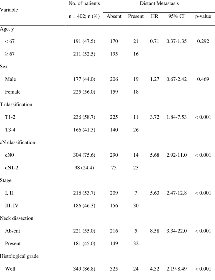
Table 1. Clinicopathological features of 402 patients with primary OSCC
Variable
No. of patients Distant Metastasis
n = 402; n (%) Absent Present HR 95% CI p-value
Age, y < 67 191 (47.5) 170 21 0.71 0.37-1.35 0.292 ≥ 67 211 (52.5) 195 16 Sex Male 177 (44.0) 206 19 1.27 0.67-2.42 0.469 Female 225 (56.0) 159 18 T classification T1-2 236 (58.7) 225 11 3.72 1.84-7.53 < 0.001 T3-4 166 (41.3) 140 26 cN classification cN0 304 (75.6) 290 14 5.68 2.92-11.0 < 0.001 cN1-2 98 (24.4) 75 23 Stage I, II 216 (53.7) 209 7 5.63 2.47-12.8 < 0.001 III, IV 186 (46.3) 156 30 Neck dissection Absent 221 (55.0) 216 5 8.58 3.34-22.0 < 0.001 Present 181 (45.0) 149 32 Histological grade Well 349 (86.8) 325 24 4.32 2.19-8.49 < 0.001
13 Moderate, Poor 53 (13.2) 40 13 INF a, b 344 (85.6) 327 17 9.10 4.75-17.4 < 0.001 c 58 (14.4) 38 20 Vascular/lymphatic invasion Absent 312 (77.6) 299 13 7.67 3.90-15.1 < 0.001 Present 90 (22.4) 66 24 Perineural invasion Absent 361 (89.8) 337 24 5.74 2.92-11.3 < 0.001 Present 41 (10.2) 28 13 Local recurrence Absent 355 (88.3) 330 25 4.03 2.03-8.03 < 0.001 Present 47 (11.7) 35 12 Nodal Metastasis Absent 259 (66.4) 258 1 72.7 10.0-530.2 < 0.001 Present 143 (35.6) 107 36 Mortality Absent 320 (79.6) 315 5 32.3 12.6-83.1 < 0.001 Present 82 (20.4) 50 32
HR, hazard ratio; CI, confidence interval; INF, infiltrative growth pattern.
Univariate analysis was done using the Cox proportional hazards regression model
14
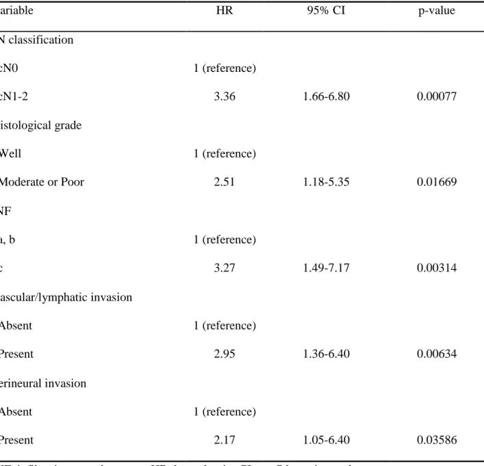
Table 2. Hazard ratios for DM development in the multivariate Cox regression model
Variable HR 95% CI p-value cN classification cN0 cN1-2 1 (reference) 3.36 1.66-6.80 0.00077 Histological grade Well Moderate or Poor 1 (reference) 2.51 1.18-5.35 0.01669 INF a, b c 1 (reference) 3.27 1.49-7.17 0.00314 Vascular/lymphatic invasion Absent Present 1 (reference) 2.95 1.36-6.40 0.00634 Perineural invasion Absent Present 1 (reference) 2.17 1.05-6.40 0.03586
15 References
1. Bray F, Ferlay J, Soerjomataram I, et al. Global cancer statistics 2018: GLOBOCAN estimates of
incidence and mortality worldwide for 36 cancers in 185 countries. CA Cancer J Clin 68:394-424,
2018.
2. Rana M, Kanatas A, Herzberg PY, et al. Prospective study of the influence of psychological and
medical factors on quality of life and severity of symptoms among patients with oral squamous cell
carcinoma. Br J Oral Maxillofac Surg 53:364-70, 2015.
3. Chinn SB, Myers JN. Oral cavity carcinoma: Current management, controversies, and future
directions. J Clin Oncol 33:3269-76, 2015.
4. Krabbe CA, Pruim J, van der Laan BFAM, et al. FDG-PET and detection of distant metastases and
simultaneous tumors in head and neck squamous cell carcinoma: A comparison with chest
radiography and chest CT. Oral Oncol 45:234-40, 2009.
5. Garavello W, Ciardo A, Spreafico R, Gaini RM. Risk factors for distant metastases in head and
neck squamous cell carcinoma. Arch Otolaryngol Head Neck Surg 132:762-6, 2006.
6. Li X, Di B, Shang Y, et al. Clinicopathologic risk factors for distant metastases from head and
neck squamous cell carcinomas. Eur J Surg Oncol 35:1348-53, 2009.
7. Sumioka S, Sawai NY, Kishino M, et al. Risk factors for distant metastasis in squamous cell
carcinoma of the oral cavity. J Oral Maxillofac Surg 71:1291-7, 2013.
8. Sakamoto Y, Matsushita Y, Yamada S, et al. Risk factors of distant metastasis in patients with
squamous cell carcinoma of the oral cavity. Oral Surg Oral Med Oral Pathol Oral Radiol 121:474-80,
2016.
9. Rose BS, Jeong J-H, Nath SK, et al. Population-based study of competing mortality in head and
neck cancer. J Clin Oncol 29:3503-9, 2011.
10. Leon X, Quer M, Orus C, et al. Distant metastases in head and neck cancer patients who
16
11. Myers JN, Greenberg JS, Mo V, Roberts D. Extracapsular spread. A significant predictor of
treatment failure in patients with squamous cell carcinoma of the tongue. Cancer 92:3030-6, 2001.
12. Lim SC, Zhang S, Ishii G, et al. Predictive markers for late cervical metastasis in stage I and II
invasive squamous cell carcinoma of the oral tongue. Clin Cancer Res 10:166-72, 2004.
13. Liao C-T, Wang H-M, Chang JT-C, et al. Analysis of risk factors for distant metastases in
squamous cell carcinoma of the oral cavity. Cancer 110:1501-8, 2007.
14. Amit M, Yen TC, Liao CT, et al. Clinical nodal stage is a significant predictor of outcome in
patients with oral cavity squamous cell carcinoma and pathologically negative neck metastases:
results of the international consortium for outcome research. Ann Surg Oncol 20:3575-81, 2013.
15. Sauerbrei W, Taube SE, McShane LM, et al. Reporting Recommendations for Tumor Marker
Prognostic Studies (REMARK): An Abridged Explanation and Elaboration. J Natl Cancer Inst
110:803-11, 2018.
16. Sobin LH, Gospodarowicz MK, Wittekind C. UICC TNM Classification of Malignant Tumours,
7th ed. New York, Wiley-Blackwell, 2009.
17. National Comprehensive Cancer Network. NCCN Clinical Practice Guidelines in Oncology
Head and Neck Cancers (version 2.2018); 2018. Available at
https://www.nccn.org/professionals/physician_gls/pdf/head-and-neck.pdf. Accessed December 1,
2018.
18. List MA, D'Antonio LL, Cella DF, et al. The performance status scale for head and neck cancer
patients and the functional assessment of cancer therapy-head and neck scale. A study of utility and
validity. Cancer 77:2294-301, 1996.
19. Thomford NR, Woolner LB, Clagett OT. The surgical treatment of metastatic tumors in the lungs.
J Thorac Cardiovasc Surg 49:357-63, 1965.
20. Ebisumoto K, Okami K, Ogura G, et al. The predictive role of infiltrative growth pattern in early
17
21. Kanda Y. Investigation of the freely available easy-to-use software 'EZR' for medical statistics.
Bone Marrow Transplant 48:452-8, 2013.
22. Probert JC, Thompson RW, Bagshaw MA. Patterns of spread of distant metastases in head and
neck cancer. Cancer 33:127-33, 1974.
23. Pietropaoli MP, Damron TA, Vermont AI. Bone metastases from squamous cell carcinoma of the
head and neck. J Surg Oncol 75:136-41, 2000.
24. Al-Othman MO, Morris CG, Hinerman RW, Amdur RJ, Mendenhall WM. Distant metastases
after definitive radiotherapy for squamous cell carcinoma of the head and neck. Head Neck
25:629-33, 2003.
25. Ebrahimi A, Zhang WJ, Gao K, Clark JR. Nodal yield and survival in oral squamous cancer:
Defining the standard of care. Cancer 117:2917-25, 2011.
26. Amit M, Yen TC, Liao CT, et al. The origin of regional failure in oral cavity squamous cell
carcinoma with pathologically negative neck metastases. JAMA Otolaryngol Head Neck Surg
140:1130-7, 2014.
27. D’Cruz AK, Vaish R, Kapre N, et al. Elective versus therapeutic neck dissection in node-negative
oral cancer. N Engl J Med 373:521-9, 2015.
28. Otsuru M, Ota Y, Yanamoto S, et al. A multicenter retrospective study of elective neck dissection
for T1-2N0M0 tongue squamous cell carcinoma: Analysis using propensity score-matching. Ann
Surg Oncol 26:555-63, 2018.
29. Takahashi M, Aoki T, Nakamura N, et al. Clinicopathological analysis of 502 patients with oral
squamous cell carcinoma with special interest to distant metastasis. Tokai J Exp Clin Med 39:178-85,
2014.
30. Mazer TM, Robbins KT, McMurtrey MJ, Byers RM. Resection of pulmonary metastases from
squamous carcinoma of the head and neck. Am J Surg 156:238-42, 1988.
18
neck cancer patients. Head Neck 18:311-6, 1996.
32. Petrella F, Diotti C, Rimessi A, Spaggiari L. Pulmonary metastasectomy: an overview. J Thorac
Dis 9:S1291-8, 2017.
33. Shiono S, Kawamura M, Sato T, et al. Pulmonary metastasectomy for pulmonary metastases of
head and neck squamous cell carcinomas. Ann Thorac Surg 88:856-60, 2009.
34. Fortin A, Couture C, Doucet R, Albert M, Allard J, Tetu B. Does histologic grade have a role in
the management of head and neck cancers? J Clin Oncol 19:4107-16, 2001.
35. Vermorken JB, Specenier P. Optimal treatment for recurrent/metastatic head and neck cancer.
Ann Oncol 21 Suppl 7:vii252-61, 2010.
36. Iovoli AJ, Platek AJ, Degraaff L, et al. Routine surveillance scanning in HNSCC: Lung screening
CT scans have value but head and neck scans do not. Oral Oncol 86:273-7, 2018.
37. Liu JC, Bhayani M, Kuchta K, Galloway T, Fundakowski C. Patterns of distant metastasis in
head and neck cancer at presentation: Implications for initial evaluation. Oral Oncol 88:131-6, 2019.
38. Takes RP, Rinaldo A, Silver CE, et al. Distant metastases from head and neck squamous cell
carcinoma. Part I. Basic aspects. Oral Oncol 48:775-9, 2012.
39. Nekhlyudov L, Lacchetti C, Davis NB, et al. Head and Neck Cancer Survivorship Care
Guideline: American Society of Clinical Oncology Clinical Practice Guideline Endorsement of the
American Cancer Society Guideline. J Clin Oncol 35:1606-21, 2017.
40. Aerle DR, Adams AM, Berg CD, et al. Reduced lung-cancer mortality with low-dose computed
tomographic screening. N Engl J Med 365:395-409, 2011.
41. Agarwal SK, Akali NR, Sarin D. Prospective analysis of 231 elective neck dissections in oral
squamous cell carcinoma with node negative neck - To decide the extent of neck dissection. Auris
Nasus Larynx 45:156-61, 2018.
42. Grigore A, Jolly M, Jia D, et al. Tumor budding: The name is EMT. Partial EMT. J Clin Med
19
43. Li Y, Bai S, Carroll W, et al. Validation of the risk model: high-risk classification and tumor
pattern of invasion predict outcome for patients with low-stage oral cavity squamous cell carcinoma.
Head Neck Pathol 7:211-23, 2013.
44. Kano S, Sakashita T, Tsushima N, et al. Validation of the 8th edition of the AJCC/UICC TNM
20 Figure legends
Figure 1. Primary site of OSCC and the incidence of distant metastases (DM)
The incidence of DM was highest in patients with tongue SCC (16 (8.3%) of 192 patients), followed
by the lower gingiva (12 (12.9%) of 93 patients). There was no significant difference in DM by
primary site (p=0.541, Fisher’s exact test).
Figure 2. Kaplan-Meier survival estimate. (A) 5y-OS of patients with DM and non-DM (p < 0.00001,
log-rank test). (B) 5y-OS of patients with cN0 disease and cN1-2 disease (p = 0.00011). (C) DMFS
of patients with cN0 disease and cN1-2 disease (p = 0.00003). (D) 5y-OS of patients with cN0
disease and cN1-2 disease at the initial diagnosis among patients with DM development (p =
0.02760). (E) 5y-OS of patients whose time to diagnosis of DM ≥ 10.0 months and < 10.0 months (p
< 0.00001). (F) 5y-OS of the surgery group and the non-surgery group (p = 0.00682).
DM, distant metastasis; MST, median survival time.
Figure 3. DM failure rate after definitive surgery. (A) DM failure rates in patients with cN0 disease
and cN1-2 disease (p < 0.00001). (B) DM failure rates in patients who underwent END and did not
undergo END among patients with cN0 disease. (C) DM failure rates of patients with and without
ENE among patients with pathological node-positive disease.