Studies on the functions of Haemaphysalis longicornis
2-Cys peroxiredoxin, and its application for tick control
2-Cys
The United Graduate School of Veterinary Science
Yamaguchi University
Kodai Kusakisako
March 2018
TABLE OF CONTENTS
GENERAL INTRODUCTION
CHAPTER 1: Functional analysis of recombinant 2-Cys peroxiredoxin from the hard tick Haemaphysalis longicornis
1.1 Introduction
1.2 Materials and Methods
1.2.1 Identification and characterization of the cDNA encoding the HlPrx2
protein
1.2.2 Expression and purification of wild-type recombinant HlPrx2 and HlPrx2
mutants
1.2.3 SDS-polyacrylamide gel electrophoresis 1.2.4 Mixed-function oxidation assay
1.2.5 Hydrogen peroxide-scavenging assay 1.2.6 Peroxiredoxin activity assay
1.2.7 Size-exclusion chromatography 1.3 Results
1.3.1 Identification of the 2-Cys peroxiredoxin gene of Haemaphysalis
longicornis
1.3.2 Confirmation of dimer formation by the disulfide bonds via the two
conserved cysteines
1.3.3 Measurement of the antioxidant, hydrogen peroxide-scavenging, and
peroxiredoxin activity of the wild-type and mutant proteins of HlPrx2
1.3.4 Determination of the recombinant HlPrx2
using gel-filtration chromatography
1.4 Discussion Table and Figures
CHAPTER 2: 2-Cys peroxiredoxin is required in successful blood-feeding,
reproduction and antioxidant response in the hard tick Haemaphysalis longicornis 2.1 Introduction
2.2 Materials and Methods 2.2.1 Ticks and animals
2.2.2 Total RNA extraction and cDNA synthesis 2.2.3 Expression analysis of HlPrx2 mRNA
1 4 5 6 6 8 9 9 10 10 11 12 12 13 14 15 16 21 30 31 32 32 33 33
2.2.4 Production of an antiserum against recombinant HlPrx2 2.2.5 Protein extraction and Western blot analysis
2.2.6 Immunostaining
2.2.7 RNA interference (RNAi)
2.2.8 Detection of hydrogen peroxide in adult female ticks during blood-feeding 2.2.9 Statistical analysis
2.3 Results
2.3.1 Transcription profiles of HlPrx2
2.3.2 Localization of Babesia parasites in tick organs 2.3.3 Protein expression profiles of HlPrx2
2.3.4 Effects of HlPrx and/or HlPrx2 gene silencing on the blood-feeding and
reproduction of female ticks
2.3.5 Increasing the concentration of hydrogen peroxide by the double
knockdown of HlPrx genes before and after blood-feeding
2.4 Discussion Tables and Figures
CHAPTER 3: Peroxiredoxins are important for the regulation of hydrogen peroxide concentration induced by paraquat in Ixodes scapularis embryo-derived cell line (ISE6)
3.1 Introduction
3.2 Materials and Methods 3.2.1 Culture of cells
3.2.2 Detection of hydrogen peroxide suing BES-H2O2-Ac in ISE6 cells
3.3.3 Paraquat treatment
3.2.4 RNA interference (RNAi) using double-stranded RNA 3.2.5 Total RNA extraction and cDNA synthesis
3.2.6 In vitro cell proliferation and survival assays
3.2.7 Detection of hydrogen peroxide in peroxiredoxin-silenced ISE6 cells after
treatment with 1-mM paraquat using the BES-H2O2-Ac probe with
Hoechst 33342
3.2.8 Statistical analysis 3.3 Results
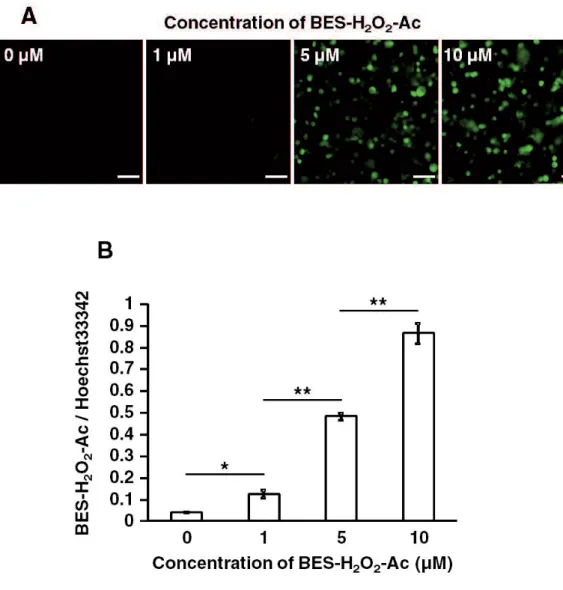
3.3.1 Comparison of the concentration of the BES-H2O2-Ac probe in ISE6 cells
3.3.2 In vitro cell proliferation assay and cell survival assay for ISE6 cells after
treatment with several concentrations of paraquat
34 34 36 37 38 39 39 39 41 43 43 44 45 54 67 68 69 69 69 70 70 73 73 74 75 75 75 76
3.3.3 MTT assay and Trypan blue assay for ISE6 cells after the gene silencing
of IsPrxs using the RNAi method
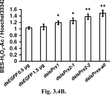
3.3.4 Detection of BES-H2O2-Ac/Hoechst 33342 in IsPrx gene-silenced ISE6
cells after treatment with 1-mM paraquat for 24 hrs
3.4 Discussion Table and Figures
CHAPTER 4: Evaluation of vaccine potential of 2-Cys peroxiredoxin from the hard tick Haemaphysalis longicornis
4.1 Introduction
4.2 Materials and Methods 4.2.1 Ticks and animals
4.2.2 Expression and purification of recombinant H. longicornis 2-Cys
peroxiredoxin (rHlPrx2)
4.2.3 Protein and endotoxin analysis 4.2.4 Immunization protocol of mice 4.2.5 Immune response
4.2.6 Protein extraction and Western blot analysis 4.2.7 Tick infestation on rHlPrx2-caccinated mice 4.2.8 Statistical analysis
4.3 Result
4.3.1 rHlPrx2 protein expression and purification 4.3.2 Antibody titers after immunization
4.3.3 The recognition of native HlPrx2 protein in the crude tick protein sample
using immunized mouse sera
4.3.4 Effects of immunization on H. longicornis nymphal ticks 4.4 Discussion
Tables and Figures
SUMMARY AND CONCLUSION
ACKNOWLEDGEMENTS REFERENCES 77 78 79 83 91 92 93 93 93 94 94 95 96 97 98 98 98 99 101 102 102 107 113 117 120
LIST OF TABLES AND FIGURES
CHAPTER 1
Table 1.1 Primers for the preparation of recombinant and mutant proteins of HlPrx2 Fig. 1.1 Full-length cDNA and deduced amino acid sequences of HlPrx2 from H.
longicornis
Fig. 1.2 Multiple alignment sequence analysis of the deduced amino acid sequence of
HlPrx2 -Cys peroxiredoxin
Fig. 1.3 Confirmation of the dimmerization by disulfide bonds in wild-type, C51S,
C172S, and DM recombinant HlPrx2 prepared using SDS-PAGE analysis
Fig. 1.4 MFO (A), H2O2-scavenging (B), and Peroxiredoxin activity (C) assays for the
recombinant HlPrx2 proteins
Fig. 1.5 Determination of the multimeric recombinant HlPrx2
weight using gel-filtration chromatography
Fig. 1.6 Three-dimensional structure of wild type and three mutant HlPrx2s (C51S,
C172S, and DM)
CHAPTER 2
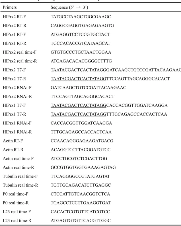
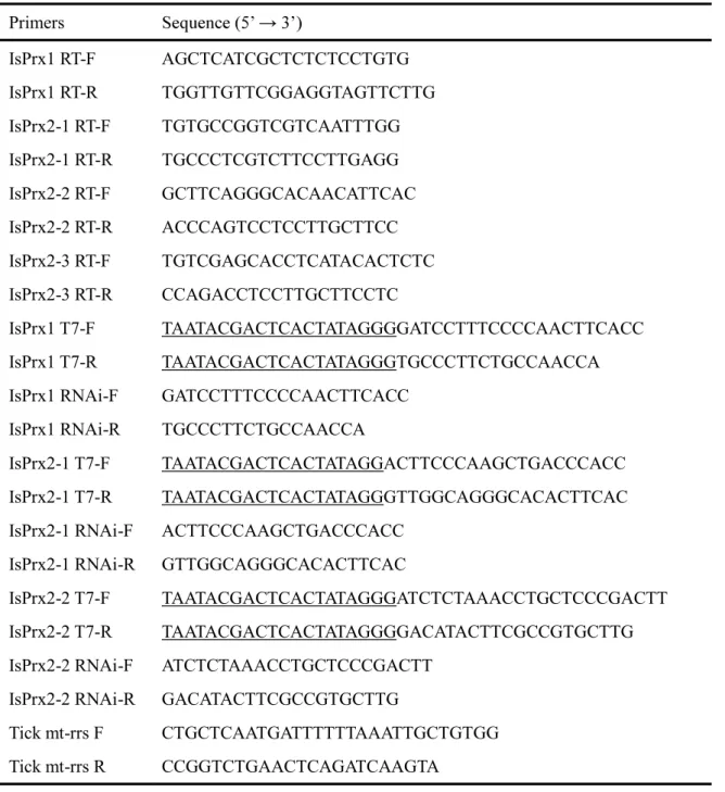
Table 2.1 Gene-specific primers used in Chapter 2
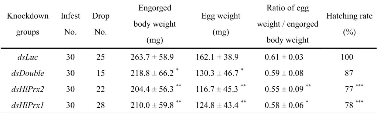
Table 2.2 Effects of HlPrx2 and HlPrx1 genes silencing in ticks
Table 2.3 Candidates for non-specific bands from Japanese white rabbit blood in
Western blot analysis
Fig. 2.1 Transcription profiles of HlPx2 in whole ticks, developmental stages, and
internal organs during blood-feeding
Fig. 2.2 Protein expression profiles of HlPx2 in whole ticks, developmental stages, and
internal organs during blood-feeding
Fig. 2.3 Comparison of normal rabbit blood and engorged-state samples in
developmental stages using Western blot analysis
Fig. 2.4 Confirmation HlPrx and/or HlPrx2 genes-silencing
partially fed adult ticks
Fig. 2.5 Localization of HlPrx2 protein in the internal organs and hemocytes from
engorged adult ticks
Fig. 2.6 Knockdown effects of HlPrx and/or HlPrx2 genes in adult ticks during
blood-feeding
Fig. 2.7 Effects on hydrogen peroxide concentrations from HlPrx and/or HlPrx2
CHAPTER 3
Table 3.1 Gene-specific primers used in Chapter 3
Fig. 3.1 Comparison of the fluorescence of several concentrations of the BES-H2O2-Ac
probe in ISE6 cells
Fig. 3.2 Effects of several concentrations of paraquat on ISE6 cells Fig. 3.3 Effects of IsPrxs gene silencing on ISE6 cells
Fig. 3.4 Effects of 1-mM paraquat treatment on IsPrx gene-silenced ISE6 cells CHAPTER 4
Table 4.1 Endotoxin levels of rHlPrx2
Table 4.2 Effects on nymphal ticks of rHlPrx2 immunization in mice
Fig. 4.1 Purification of recombinant Haemaphysalis longicornis 2-Cys peroxiredoxin
(rHlPrx2)
Fig. 4.2 The trend of the antibody titer in immunized mouse sera Fig. 4.3 Detection of native HlPrx2
ABBREVIATIONS
ROS: reactive oxygen species H2O2: hydrogen peroxide
Prx: peroxiredoxin Prxs: peroxiredoxins
HlPrx2: Haemaphysalis longicornis 2-Cys peroxiredoxin EST: expressed sequenced tag(s)
His: histidine
PBS: phosphate buffered saline
IPTG: isoprop -D-1-thiogalactopyranoside LB: Luria-bertani
rHlPrx2: the recombinant HlPrx2 protein
WT: the wild-type of the recombinant HlPrx2 protein
C51S: the mutant of the recombinant HlPrx2 converting from Cys51 to Ser C172S: the mutant of the recombinant HlPrx2 converting from Cys172 to Ser DM: the double mutant of the recombinant HlPrx2 cited above (C51S and C172S) SDS-PAGE: sodium dodecyl sulfate -polyacrylamide gel electrophoresis
2-ME: 2-mercaptoethanol MFO: mixed-function oxidation FBS: fetal bovine serum
NADPH: nicotinamide adenine dinucleotide phosphate SEC: size-exclusion chromatography
HlPrx: Haemaphysalis longicornis 1-Cys peroxiredoxin qPCR: quantitative polymerase chain reaction
IFAT: indirect immunofluorescent antibody test RNAi: RNA interference
Luc: firefly luciferase gene
ds-: the double-stranded RNA of - mRNA: messenger RNA
ORF: open reading frame IFA: incomplete F
GENERAL INTRODUCTION
Reactive oxygen species (ROS) that include the superoxide anion, hydrogen peroxide (H2O2), and hydroxyl radicals are produced by the incomplete reduction of
oxygen. Among the ROS, hydroxyl radicals are the most reactive and short lived (10-9 sec half-life at 37°C) [1]. In addition, through the Fenton reaction, hydroxyl radicals are produced from H2O2, the most stable ROS. But controlling hydroxyl radicals is very
difficult for organisms because of their short half-lives. Therefore, the removal of H2O2
is biologically advantageous [2].
In high concentrations, H2O2 is known to be a chemical compound harmful
to aerobic organisms due to its ability to seriously damage membrane lipids, nucleic acids, and proteins [3]. To protect against H2O2 toxicity, aerobic organisms have
evolved antioxidant enzymes, such as catalases, peroxidases, and peroxiredoxins (Prxs) [4]. Prxs are ubiquitous antioxidant enzymes that have been investigated in various organisms [5]. Particularly, high levels of Prxs are produced in mammalian cells, including erythrocytes [6]. Erythrocytes are exposed to more oxidative stress than any other mammalian cells, due to the abundance of heme iron and oxygen, which can generate H2O2 [7]. These phenomena indicate that Prxs may play important roles in
Ticks are obligate hematophagous arthropods that need blood feeding at all developmental stages. Blood feeding and the digestion of blood provide nutrition and energy for development, molting, and embryogenesis in ticks [8]. Ticks feed on vertebrate blood that contains high levels of iron, such as ferrous iron, heme, and iron-bound transferrin [9]. Ticks also concentrate iron-containing host blood; this concentration of the blood leads to high levels of iron in ticks. Host-derived iron may react with oxygen in the tick body, resulting in high concentrations of ROS, including H2O2 [10,11]. In addition, ticks have genes of antioxidant enzymes, such as catalases
[12] and Prxs [13-15], to scavenge H2O2. Although the gene silencing of Prxs in ticks
[13], the gene silencing of catalase in ticks is not effective [12]
blood feeding and oviposition through the regulation of the H2O2 concentration in ticks.
In this dissertation, I focused on the 2-Cys Prx in ticks and its application for tick control.
Research Objectives
With following objectives, this dissurtation besides the studies on the functions of
Haemaphysalis longicornis
2-Cys Prx (HlPrx2), and its application for tick control.
AIM 1.
To identify HlPrx2 gene and characterize recombinant HlPrx2 protein AIM 2.
To characterize expression profiles and biological functions of HlPrx2 in the ticks AIM 3.
To clarify the importance of Prxs for antioxidant response using the tick cell line AIM 4.
CHAPTER 1
Functional analysis of recombinant 2-Cys peroxiredoxin from the hard tick Haemaphysalis longicornis
This work has been published as: Kusakisako, K., Masatani, T., Miyata, T., Galay, R.
L., Maeda, H., Talactac, M. R., Tsuji, N., Mochizuki, M., Fujisaki, K., Tanaka, T. (2016). Functional analysis of recombinant 2-Cys peroxiredoxin from the hard tick
1.1 Introduction
Almost all aerobic organisms have developed defense systems to scavenge H2O2. Catalases, peroxidases, and peroxiredoxins (Prxs) are scavengers of H2O2 [4].
Prxs are ubiquitous antioxidant enzymes investigated in various organisms [5]. Particularly, Prxs are produced at high levels in mammalian cells, including erythrocytes [6,16]. The rate constants of catalases, peroxidases, and Prxs were considered to have almost the same rate (107 - 108 M-1 S-1) [17]. These results indicate that Prxs may have important roles in peroxide detoxification in cells [16].
Prxs can be divided into two groups according to the presence of one or two highly conserved cysteines in organisms, 1-Cys or 2-Cys Prx [5]. Structural and mechanistic data showed the further division of the 2-Cys Prxs into two classes, typical and atypical 2-Cys Prxs [16]. Typical 2-Cys Prxs are identified by two conserved cysteines, peroxidatic and resolving cysteines [18]. Deprotonated peroxidatic cysteine reacts with H2O2 via a nucleophilic attack to form cysteine sulfenic acid with the release
of water. Then, the resolving cysteine in another Prx molecule will react with the cysteine sulfenic acid to form homodimers via intermolecular disulfide bonds. These disulfide bonds are reduced by oxidoreductases to revert to the active form [19]. In addition, studies of typical 2-Cys Prxs have showed that dramatic changes in the
oligomeric state (dimers and decamers) are associated with changes in oxidative stress, like those occurring during the catalytic cycle [16]. Atypical 2-Cys Prxs have the same mechanism as do typical 2-Cys Prxs but are functionally monomeric [20]. The 1-Cys Prxs conserve only the peroxidatic cysteine and do not contain a resolving cysteine [21]. The mechanism of the 1-Cys Prxs is considered to be one donor thiol that probably forms a transient mixed disulfide bond with the enzyme, followed by its reduction by a second donor thiol, thus recycling the enzyme [16]. However, there has been only one report on tick Prxs focusing on Haemaphysalis longicornis 1-Cys Prx until now [15]. Thus, the function of tick Prxs is not completely known.
In this study, I identified a 2-Cys Prx gene from H. longicornis (HlPrx2) and prepared some kinds of recombinant proteins of HlPrx2, including three mutants, to clarify the role of HlPrx2 on antioxidant activity produced by H2O2 in ticks.
1.2 Materials and Methods
1.2.1 Identification and characterization of the cDNA encoding the HlPrx2 protein
The putative HlPrx2 gene was identified using an expressed sequence tag (EST) database constructed from the cDNA library of the fat body from H. longicornis as previously described by Boldbaatar et al., [22]. A pGCAP1 plasmid containing an
HlPrx2 gene insert was extracted using a Qiagen® Plasmid Mini Kit (Qiagen, Hilden, Germany). The insert was sequenced using the BigDye® Terminator v3.1 Cycle Sequencing Kit (Applied Biosystems, Tokyo, Japan) and the Applied Biosystems® 3500 XL Genetic Analyzer.
The deduced amino acid translation of the HlPrx2 gene sequence was determined using GENETYX version 7.0 software (GENETYX, Tokyo, Japan). To search homologous genes from GenBank (http://www.ncbi.nlm.nih.gov/genbank), a BLAST server (http://blast.ncbi.nlm.nih.gov/Blast.cgi) was used. The domain structure was determined using the SMART program (http://smart.embl-heidelberg.de/). To predict the three-dimensional structure of HlPrx2, the Phyre2 Protein Fold Recognition Server (http://www.sbg.bio.ic.ac.uk/phyre2/) was used [23]. The theoretical molecular mass and isoelectric point were calculated using a ProtParam tool (http://web.expasy.org/protparam/). Putative signal peptide cleavage sites and N-linked
glycosylation sites were checked by the SignalP 4.1 server
(http://www.cbs.dtu.dk/services/SignalP/) and the NetNGlyc 1.0 server (http://www.cbs.dtu.dk/services/NetNGlyc/), respectively. An alignment analysis was performed using the Clustal Omega (http://www.ebi.ac.uk/Tools/msa/clustalo/).
1.2.2 Expression and purification of wild-type recombinant HlPrx2 and HlPrx2 mutants
Wild-type (WT) recombinant HlPrx2 (rHlPrx2) was expressed as an N-terminus 6-histidine (His) -tagged protein using the expression vector pRSET C (Invitrogen, Carlsbad, CA, USA). The WT open reading frame sequence was amplified by a polymerase chain reaction (PCR) using a forward primer (HlPrx2 F-XhoI) containing a XhoI recognition site and a reverse primer (HlPrx2 R-EcoRI) containing an
EcoRI recognition site (Table 1.1). The amplified PCR product was then purified using
a GENECLEAN® II KIT (MP Biomedical, Solon, OH, USA) and subcloned into the frame of the pRSET C. The WT was expressed in Escherichia coli BL21 (DE3) strain and purified as described previously [24].
To prepare the mutated proteins, the inverse PCR of the plasmid DNA (pRSET C containing HlPrx2 WT) was performed by using mutation primers and KOD-Plus-Neo (Toyobo
single nucleotide substitution (Table 1.1). The self-ligation of PCR products was performed using DNA Ligation Kit (Takara Bio Inc., Shiga, Japan). The self-ligated PCR products were digested by DpnI at 37°C for 4 hrs. The mutated plasmids, HlPrx2 Cys51Ser, HlPrx2 Cys172Ser, and HlPrx2 Cys51Ser/Cys172Ser; double mutant, named
C51S, C172S, and DM, respectively, were transformed into E. coli Stellar strain and subcloned. The mutated plasmids were extracted from E. coli, and the confirmation of mutations was analyzed by DNA sequencing. Finally, the mutant rHlPrx2 proteins were expressed in E. coli BL21 (DE3) strain and purified similarly to the WT protein.
1.2.3 SDS-polyacrylamide gel electrophoresis (SDS-PAGE)
500 ng/lane WT and mutant recombinant proteins were mixed with the same volume of sample buffer with or without 10% 2-mercaptoethanol (2-ME). Consequently, the samples with 2-ME were boiled. The prepared protein samples were resolved in a 12% SDS-PAGE gel under reducing (with 2-ME) or non-reducing (without 2-ME) conditions. After SDS-PAGE, the gel was stained by Coomassie Brilliant Blue and viewed using Gel Doc (Bio-Rad, Hercules, CA, USA).
1.2.4. Mixed-function oxidation assay
Mixed-function oxidation (MFO) assay [25] was performed in accordance with the methods of Masatani et al. [26]. Briefly, a reaction mixture containing 40 µM ferric chloride (FeCl3), 10 mM dithiothreitol (DTT), 20 mM EDTA, and 25 mM Hepes-NaOH
37°C for 1 hr. After pre-incubation, 0.5 µg of pcDNATM 3.1 (+) plasmid DNA (Invitrogen) was added, and the reaction mixture was incubated for another 3 hrs. Nicking of the supercoiled plasmids by MFO assay was evaluated by running electrophoresis on 1% agarose gel stained with ethidium bromide for visualization. Bovine serum albumin (BSA) and recombinant H. longicornis Ferritin 2 (rFer2) were used for negative controls of protein and His-tagged protein, respectively.
1.2.5. Hydrogen peroxide (H2O2)-scavenging assay
H2O2 consumption was measured using the ferrous oxidation of xylenol orange
assay [27], adapted for low peroxide concentrations. The assay reagent consisted of 125 µM xylenol orange, 250 µM ammonium iron (II) sulfate, 100 mM sorbitol, and 25 mM sulfuric acid. After treatment of 50 µg/ml recombinant and mutant proteins with 5 µM H2O2, 100-µl sample solutions were added to a 1-ml assay reagent. The mixture was
vortexed immediately, left at room temperature for 35 min, and measured at 560 nm using spectrophotometer (Ultrospec 2100 pro, GE Healthcare, Pittsburgh, PA, USA).
1.2.6. Peroxiredoxin activity assay
Peroxiredoxin activity assay was performed in accordance with the methods of Kim et al., [28]. A prereaction cocktail that contains 50 mM Hepes-NaOH buffer (pH
rHlPrx2 proteins in 100 µl total volume, was prepared. Then, 100 µM H2O2 was added into each well in the
96-well plate. The reaction was started by adding H2O2, and NADPH oxidation was
monitored for 20 min at 30°C by following absorbance reduction at 340 nm in a microplate reader (SH-9000Lab, Corona Electric, Ibaraki, Japan).
1.2.7. Size-exclusion chromatography
The molecular sizes of the purified WT, C51S, C172S, and DM recombinant proteins were measured by size-exclusion chromatography (SEC). The protein samples were 0.2 µm-filtered before using for the SEC. The 170 µg/ml WT, C51S, C172S, or DM recombinant proteins were loaded into a HiLoad 16/600 Superdex 200 pg column (GE Healthcare) at a flow rate of 0.8 ml/min using the ÄKTAprime plus chromatography system (GE Healthcare) [29]. Phosphate buffered saline (pH 7.4) containing 137 mM NaCl, 27 mM KCl, 81 mM Na2HPO4 12H2O, and 14.7 mM
1.3 Results
1.3.1 Identification of the 2-Cys peroxiredoxin gene of Haemaphysalis longicornis
The 2-Cys Prx gene of H. longicornis (HlPrx2: accession no. LC049075) was isolated from EST clones from the fat body cDNA libraries of the tick. The full-length
HlPrx2 cDNA is 797 bp, with an open reading frame extending from position 97 to 691,
encoding for 197 amino acids (Fig. 1.1). A polyadenylation consensus signal sequence was identified upstream of the poly A tail. The predicted molecular mass of HlPrx2 is 22.3 kDa, and the theoretical isoelectric point is 6.8. The signal peptide and glycoxylation sites were not found by in silico analysis. HlPrx2 has an alkyl hydroperoxide reductase- thiol specific antioxidant (AphC-TSA) domain from positions 8 to 141 and a C-terminal domain of 1-Cys peroxiredoxin (1-cysPrx C) domain from 161 to 196. In addition, BLAST analysis of the deduced amino acid sequences and 2-Cys Prx of other organisms, such as Ixodes scapularis, Ornithodoros parkeri,
Drosophila willistoni, Aedes aegypti, and Homo sapiens (Prx2 and Prx4), revealed that
HlPrx2 shows high similarities of 88%, 86%, 79%, 78%, 77%, and 76%, respectively. Furthermore, the alignment analysis demonstrated that HlPrx2 has two conserved cysteines at positions 51 and 172 and has an YF motif at positions 193 to 194 (Fig. 1.2).
1.3.2 Confirmation of dimer formation by the disulfide bonds via the two conserved cysteines
The scheme of the reaction mechanism of 2-Cys Prx was considered according to Lu & Holmgren [19]. Briefly, deprotonated peroxidatic cysteine reacts with H2O2 to
form cysteine sulfenic acid. Then, the resolving cysteine in another molecule will react with the cysteine sulfenic acid and form homodimers via intermolecular disulfide bonds. To determine whether the wild-type protein of rHlPrx2 (WT) can form homodimers by disulfide bonds via the two conserved cysteines, I performed SDS-PAGE analysis with or without 2-mercaptoethanol. SDS-PAGE analysis demonstrated that WT formed a dimer by disulfide bonds under non-reducing conditions (Fig. 1.3). In addition, to investigate whether the conserved cysteines play an important role for the dimerization of rHlPrx2, I carried out the same experiment using mutant proteins (rHlPrx2 Cys51Ser, rHlPrx2 Cys172Ser, and rHlPrx2 Cys51Ser/Cys172Ser; double mutant, named C51S, C172S, and DM, respectively). However, the mutant proteins of rHlPrx2, C51S, C172S, and DM could not form a dimer under non-reducing conditions. These results indicate that either Cys51 or Cys172 is necessary for the dimerization of rHlPrx2.
1.3.3 Measurement of the antioxidant, hydrogen peroxide-scavenging, and peroxiredoxin activity of the wild-type and mutant proteins of rHlPrx2
To demonstrate the antioxidant activity of the WT protein, I performed an mixed-function oxidation (MFO) assay using recombinant proteins [25]. Through agarose gel electrophoresis using plasmid DNA, a band of nicked plasmid DNA, injured by ROS, appears in a higher position as compared to a normal coiled plasmid DNA. In other words, the nicked and coiled forms indicate oxidized and anti-oxidized forms, respectively. The MFO assay showed that the WT had antioxidant activity from a 4A). Additionally, to investigate the roles of the conserved cysteines in the antioxidant activity of rHlPrx2, I performed an MFO assay using the mutant proteins of rHlPrx2, C51S, C172S, and DM. C51S and DM mutants did not show antioxidant activity at any concentration. Nevertheless, the C172S mutant r
scavenge H2O2 [19]. Accordingly, I performed an H2O2-scavenging assay for the WT
rHlPrx2. These results showed that the WT has antioxidant activity, indicating that it can scavenge H2O2 (Fig. 1.4B). Moreover, the H2O2-removal ratio of WT was 67%
when comparing 0 and 30 min of the calculated H2O2 concentration. In contrast, the
H2O2-scavenging assay for the mutant proteins C51S, C172S, and DM to check whether
the conserved cysteines contribute to H2O2 scavenging. The assay revealed that C51S
and DM mutants could not scavenge H2O2. Importantly, the C172S mutant was able to
scavenge H2O2, and the H2O2
-2-Cys Prxs are known to be reduced by the thioredoxin system including thioredoxin and thioredoxin reductase [19]. Thus, to clarify whether rHlPrx2 protein can scavenge H2O2 with the thioredoxin system, I performed a peroxiredoxin activity assay. The
assay revealed that the WT rHlPrx2 with the thioredoxin system catalyzed the H2O2-dependent oxidation of NADPH (Fig. 1.4C), however, mutant proteins of rHlPrx2,
including C172S mutant, and BSA did not. These results suggest that Cys51 of rHlPrx2 is necessary for antioxidant activity, and also, both Cys51 and Cys172 are essential for thioredoxin-dependent activity.
1.3.4 Determination of the recombinant HlPrx2 gel-filtration chromatography
Previous studies show that 2-Cys Prxs form an oligomer [16,30]. To investigate the exact molecular size of the rHlPrx2, gel-filtration chromatography was performed. The molecular weight of the rHlPrx2, WT, C51S, C172S, and DM were 399 kDa, 388
kDa, 448 kDa and 377 kDa, respectively. Notably, only the WT retained the other peak of the 57 kDa molecular mass (Fig. 1.5). This result paralleled that in Fig. 2, strongly suggesting that either Cys51 or Cy172 is essential for the dimerization of rHlPrx2.
1.4 Discussion
In the present study, I identified the HlPrx2 gene, and the sequence analysis of the HlPrx2 amino acids revealed that HlPrx2 has the AphC-TSA domain and the 1-cysPrx C domain. The AphC-TSA domain is conserved in the Prxs family [16]. In addition, the 1-cysPrx C domain is recognized as a member of the Prxs superfamily that protects cells from membrane oxidation [21]. Alignment analysis of the deduced amino
acid sequences of the HlPrx2 -Cys Prxs revealed that HlPrx2 is
similar to 2-Cys Prxs, and their similarities with HsPrx2 and HsPrx4 are 77% and 76%, respectively. HsPrx2 is located in the cytosol or cell membrane of erythrocytes [31]. Using an indirect immunofluorescence antibody test, I found HlPrx2 along the basal lamina or cell membrane of some H. longicornis tick tissues, including the midgut and ovary, as well as in the cell membrane of hemocytes (unpublished data). These results suggest the possibility that HlPrx2 functions to protect cells against membrane oxidation at the midgut, ovary, or hemocyte of the H. longicornis tick. Some
reports showed that the decamers of some peroxiredoxins associated with membrane [32,33]. HsPrx2, that is similar to HlPrx2 with amino acid sequence similarity of 77%, is also lacks a signal peptide and associated with plasma membrane of erythrocytes, however, in the case of patient lack of band 3 gene, HsPrx2 cannot be associated with the plasma membrane in the erythrocytes [31]. In addition, HsPrx2 does not have the large hydrophobic region as well as HlPrx2. These results suggest that HsPrx2 needs [31] without the signal peptide and the large hydrophobic region. Therefore, HlPrx2 may also have some partners to associate with plasma membrane without the signal peptide and the large hydrophobic region.
According to Lu & Holmgren [19], the deprotonated peroxidatic cysteine (Cys51 of HlPrx2) reacts with H2O2 to form cysteine sulfenic acid with the release of
H2O. Then, the resolving cysteine (Cys172 of HlPrx2) of another HlPrx2 molecule will
react with the cysteine sulfenic acid to form homodimers via intermolecular disulfide bonds (Fig. 1.6). To demonstrate the dimerization of rHlPrx2, I performed SDS-PAGE analysis and found that only the WT formed a dimer under a non-reducing conditions but C51S, C172S, and DM mutants did not. Additionally, I performed MFO and H2O2-scavenging assays to demonstrate the antioxidant activity for scavenging H2O2 of
the recombinant and mutant proteins of HlPrx2; the assays demonstrated that WT and C172S possessed antioxidant activity. In the MFO assay, ROS such as superoxide, H2O2,
and hydroxyl radicals may be generated [34]. Hydroxyl radicals are primary oxidants that injure the plasmid DNA. However, hydroxyl radicals may be generated by a Fenton reaction. These results indicate that rHlPrx2 has almost the same molecular dynamism of the catalytic cycle of typical 2-Cys Prxs as mentioned by Lu & Holmgren [19], and may function as the antioxidant enzyme for the H2O2 scavenger in the tick body.
There have been some reports about a mutation of the resolving cysteine in 2-Cys Prxs, indicating that the mutation results in lowered catalytic rates of the wild type [35,36]. Additionally, reports of truncating several C-terminal residues, including the YF motif of 2-Cys Prxs (see in Fig. 1.2, indicated by right and left arrow), suggested that the antioxidant activity of the mutant was increased [30,37]. The YF motif covers the peroxidatic cysteine of another 2-Cys Prx to protect from overoxidation [5] and limits the dynamics of active site including the peroxidatic cysteine [37]. Based on MFO and H2O2-scavenging assays, the WT and the C172S mutant had antioxidant 2O2-removal
ratio of the WT and the C172S mutant of 67% and 79%, respectively. In my study, when the resolving cysteine of rHlPrx2 was mutated, the antioxidant activity seems to
be increased. Same observation was seen when the YF motif was truncated [30,37]. Additionally, the Prx activity assay demonstrated that WT with the thioredoxin system could scavenge H2O2 but C172S mutant could not. These results indicate that a
positional clash between Cys51 and the YF motif of the C172S mutant of rHlPrx2 may occur, leading to the malfunction of the YF motif. This hypothesis suggests that the YF motif of rHlPrx2 might be strongly related to Cys172.
Prxs are known to form oligomers as shown in some reports [16,30]. Through gel-filtration chromatography of the WT and mutant proteins of rHlPrx2, I obtained high molecular weight fractions of the WT, C51S, C172S, and DM. However, mutants of rHlPrx2 could not form a dimer in SDS-PAGE or gel-filtration chromatography. In
[5,30,38]. Although the antioxidant activity was decreased conversely, the chaperone functions of the native HsPrx2 increased significantly upon exposure to increasing concentrations of H2O2 [30]. In addition, the HsPrx2 mutants retained their chaperon
activity [30]. These results and a large amount of H2O2 that can be generated in the
[11] might indicate that HlPrx2 is important to the function as a chaperone protecting the stability of the basal lamina or cell membrane of the midgut and ovary in tick. Moreover, I estimated the three-dimensional structures of
the wild-type and mutant proteins of HlPrx2 by using the Phyre2 Protein Fold Recognition Server. As predicted by using HsPrx4 [PDB ID: 3TKP [37] ] as template, the Phyre2 server revealed a three-dimensional structure of HlPrx2 WT and prepared HlPrx2 decamer and dimer structures by using WinCoot software [39]. The three-dimensional structures of mutant HlPrx2 proteins, C51S, C172S, and DM, were prepared by the same method as for the WT. However, the three-dimensional structures of those proteins are almost the same as the structures of the decamer and dimer forms (Fig. 1.6). These results indicate that Cys51 and Cys172 may not influence the interface of decamers for chaperon activity.
In summary, I identified a 2-Cys Prx gene from H. longicornis (HlPrx2) and prepared some kinds of rHlPrx2 proteins, including three mutants. I determined that Cys51 is necessary for the antioxidant activity of rHlPrx2 for removing H2O2, and both
Cys51 and Cys172 are essential for the thioredoxin-dependent activity. Moreover, either Cys51 or Cys172 is critical for the dimerization of rHlPrx2 but not for oligomerization. Therefore, HlPrx2 may play an important role in the antioxidant activity, including chaperone activity, in ticks.
Table and Figures in CHAPTER 1
Table. 1.1. Primers for the preparation of recombinant and mutant proteins of HlPrx2 HlPrx2 F-XhoI
-CCGCTCGAGTAATGGACGTGGTTATG-HlPrx2 R-EcoRI -CGGAATTCCAGATCTATTGTTTGGCG-HlPrx2 Cys51Ser F -CGTCTCC
CCGACGGAAATCATCGCC-HlPrx2 Cys51Ser R -GGGA GACGAAAGTGAAGTCAAGCGGATAG-HlPrx2 Cys172Ser F -TGGCGAAGTGTCC CCTGCTAACTGGAAGCCTGGC-HlPrx2 Cys172Ser R -AGTTAGCAGGGGA CACTTCGCCATGCTTGTCAGTG-Single underline indicates XhoI recognition site. Broken underline indicates EcoRI recognition site. Serine codons converting from cysteine codons are highlighted in black, and double underlines indicate that the nucleotides are changed by a single nucleotide substitution.
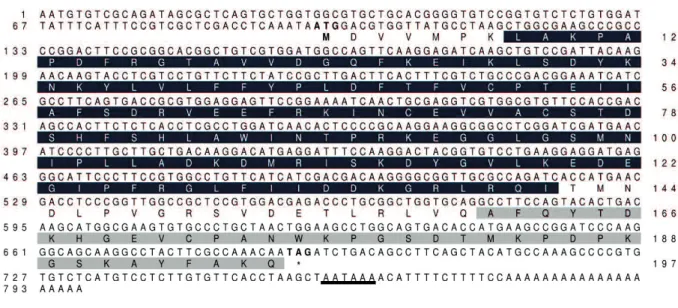
Fig. 1.1. Full-length cDNA and deduced amino acid sequences of HlPrx2 from H.
longicornis. AphC-TSA and 1-cysPrx C domains are highlighted in black and gray,
respectively (AphC-TSA domain: 8 to 141, 1-cysPrx C domain: 161 to 196). The putative polyadenylation signal after the stop codon (TAG) is underlined. The start and stop codons are boldfaced.
Fig. 1.2. Multiple alignment sequence analysis of the deduced amino acid sequence of
HlPrx2 -Cys peroxiredoxin. Asterisks indicate the position of
conserved cysteines (amino acid numbers 51 and 172) of 2-Cys peroxiredoxins. The two-directional arrow indicates the YF motif of typical 2-Cys Prxs (amino acid number 193 to 194). Similar and homologous amino acids are highlighted in black and gray, respectively. HlPrx2 (Hl), alkyl hydroperoxide reductase of Ixodes scapularis (Is; XP_002405466.1), thioredoxin peroxidase of Ornithodoros parkeri (Op; ABR23404.1), peroxiredoxin of Drosophila willistoni (Dw; XP_002071309.1), 2-Cys thioredoxin peroxidase of Aedes aegypti (Aa; AAL37254.1), peroxiredoxin-2 of Homo sapiens (Hs2; NP_005800.3), and peroxiredoxin-4 of H. sapiens (Hs4; CAG46469.1).
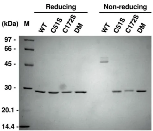
Fig. 1.3. Confirmation of the dimmerizetion by disulfide bonds in wild-type, C51S,
C172S, and DM rHlPrx2 prepared using SDS-PAGE analysis. 500 ng of rHlPrx2 (WT, C51S, C172S, and DM) were loaded per lane. WT, wild type; C51S, Cys51Ser; C172S, Cys172Ser; DM, double mutant, Cys51Ser/Cys172Ser; M, marker.
Fig. 1.4. (A) Measurement of the antioxidant activity of the rHlPrx2 proteins using
MFO assay. The left column indicates the names of recombinant proteins. Lane 1 is the 1,000 b DNA ladder marker. Lanes 2 to 4 represent treatments with or without FeCl3 or
DTT. Lanes 5 to 12 are plasmids with doubling serial dil
of recombinant proteins. Lanes 13 and 14 are bovine serum albumin (BSA) and recombinant H. longicornis ferritin 2 (rFer
column indicates the status of plasmid, the nicked form (NF), and the coiled form (CF). NF and CF indicate oxidized and non-oxidized status, respectively. BSA and rFer2 are negative controls for proteins and His-tagged proteins, respectively. WT, wild type; C51S, Cys51Ser; C172S, Cys172Ser; DM, double mutant, Cys51Ser/Cys172Ser. (B) Hydrogen peroxide-scavenging assay for the rHlPrx2 proteins treated with 5 µM H2O2.
The concentration of H2O2 was measured at 0, 2, 4, 8, 10, 15, 20, and 30 min after
adding H2O2. BSA indicates bovine serum albumin as negative control and sampling
was done at 0, 10, 20 and 30 min after adding H2O2. (C) The peroxiredoxin activity
assay for the rHlPrx2 proteins and the thioredoxin system with 100 µM H2O2. The
absorbance of 340 nm was measured at every minute until 20 min after adding H2O2.
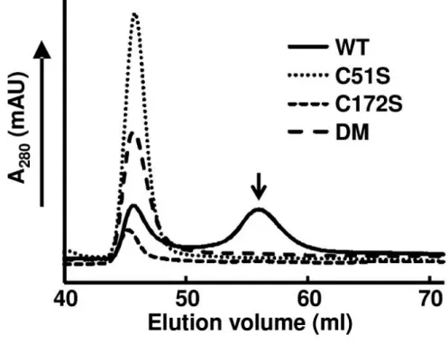
Fig. 1.5. Determination of the multimeric recombinant HlPrx2
weight using gel-filtration chromatography. 170 µg/ml rHlPrx2s (WT, C51S, C172S, and DM) were loaded. The molecular weight of WT, C51S, C172S, and DM are 399 kDa, 388 kDa, 448 kDa and 377 kDa, respectively. The second peak of rHlPrx2 WT shown with the arrow indicates a 57 kDa molecular mass. WT, wild type; C51S, Cys51Ser; C172S, Cys172Ser; DM, double mutant, Cys51Ser/Cys172Ser.
Fig. 1.6. One or both of the conserved cysteines replaced by serines that generate three
mutant HlPrx2s (C51S, C172S, and DM) are shown in three-dimensional structure {model based on template 3TKB [37]}, constructed using Phyre2 software [23]. Moreover, the decamer and dimer structures of HlPrx2 are prepared by using WinCoot software [39]. Yellow and red spheres indicate cysteines and serines, respectively. For each protein, the model on the left is the predicted decamer, while the model on the right is the predicted dimer model. The amino acid sequences are enclosed in gray boxes.
CHAPTER 2
2-Cys peroxiredoxin is required in successful blood-feeding, reproduction and
antioxidant response in the hard tick Haemaphysalis longicornis
This work has been published as: Kusakisako, K., Galay, R. L., Umemiya-Shirafuji,
R., Hernandez, E. P., Maeda, H., Talactac, M. R., Tsuji, N., Mochizuki, M., Fujisaki, K., Tanaka, T. (2016). 2-Cys peroxiredoxin is required in successful blood-feeding, reproduction, and antioxidant response in the hard tick Haemaphysalis longicornis.
2.1 Introduction
Prxs can be divided into two groups according to the presence of one or two highly conserved cysteines in organisms, 1-Cys or 2-Cys Prx [5]. 2-Cys Prxs are identified by two conserved cysteines, peroxidatic and resolving cysteines [18]. On the other hand, the 1-Cys Prxs have a conserved peroxidatic cysteine and do not contain a resolving cysteine [21]. Enzymes of the Prx family exhibit antioxidant activity that catalyzes the reduction of H2O2 into water (H2O), with thioredoxin as an immediate
hydrogen donor or donor thiol, respectively [21].
In some endoparasites, such as Plasmodium and Fasciola parasites, Prxs have been characterized as antigens or secreted proteins, suggesting that endoparasite Prxs may participate in interactions between the parasites and their hosts [40,41]. Therefore, to evaluate the efficacy of antigens for these endoparasites, basic biological and bio-histological analyses such as mRNA and protein expression profiles, and the localization of proteins in these parasites have been studied.
Ticks need blood meals to develop from one stage to the next and for reproduction. Blood-feeding and the digestion of blood provide nutrition and energy for molting, development, and the vitellogenesis of ticks [8]. Ticks feed on vertebrate blood that contains iron, such as heme, ferrous iron, and other pro-oxidants. Ticks also
concentrate host blood with iron; this concentration of the blood leads to high levels of iron in ticks. Host-derived iron may react with oxygen in the tick body, and then high levels of reactive oxygen species (ROS), including H2O2, may be generated [11].
Haemaphysalis longicornis 1-Cys Prx (HlPrx) has been reported previously [15];
however, there is still little knowledge about the biological functions of Prxs in ticks. In Chapter 2, I analyzed mRNA and protein expression profiles and the localization of proteins in tick tissues of H. longicornis 2-Cys Prx (HlPrx2). Moreover,
HlPrx and/or HlPrx2 gene silencing was performed to clarify their functions in ticks
using RNA interference. Finally, I demonstrated that the double knockdown of HlPrx and HlPrx2 led to increased oxidative stress in ticks.
2.2 Materials and Methods
2.2.1 Ticks and animals
The parthenogenetic Okayama strain of H. longicornis has been maintained by blood-feeding on the ears of Japanese white rabbits (KBT Oriental Co. Ltd, Saga, Japan) in the Laboratory of Infectious Diseases, Joint Faculty of Veterinary Medicine, Kagoshima University [42]. Rabbits were cared for in accordance with the guidelines
approved by the Animal Care and Use Committee of Kagoshima University (Approval no. VM13007) and maintained under regulated conditions throughout the experiments.
2.2.2 Total RNA extraction and cDNA synthesis
To extract total RNA, whole ticks were homogenized using an Automill (Tokken, Chiba, Japan), while dissected organs were disrupted using a pellet pestle motor (Sigma-Aldrich, St. Louis, MO, USA). The extracted RNA was purified using TRI® Reagent (Sigma-Aldrich), and then treated with an RQ1 RNase-Free DNase (Promega, Madison, WI, USA). cDNA synthesis was performed with ReverTra Ace- -®
RNA.
2.2.3 Expression analysis of HlPrx2 mRNA
The expression analysis of the HlPrx2 mRNA was performed with real-time PCR using THUNDERBIRDTM SYBR® qPCR Mix (Toyobo) with a 7300 real-time PCR system (Applied Biosystems, Foster City, CA, USA). Gene-specific primers were designed to target HlPrx2 and the internal control genes, as shown in Table 2.1. Standard curves were made from four-fold serial dilutions of the cDNA of adult ticks fed for three days. The PCR cycle profile was as follows: initial denaturation at 95°C for 10 min, 40 cycles of a denaturation step at 95°C for 15 s, and an annealing/extension
step at 60°C for 60 s. The data was analyzed with 7300 system SDS software (Applied Biosystems). At the first step of real-time PCR, actin, tubulin, P0, and L23 genes were evaluated for standardization and L23 was selected as the tick reference in the current study.
2.2.4 Production of an antiserum against recombinant HlPrx2
To prepare mouse anti-HlPrx2 HlPrx2 (rHlPrx2;
WT rHlPrx2 in Chapter 1 [14]
(Sigma-Aldrich) and intraperitoneally injected into ddY female mice (four weeks old, HlPrx2
with Fre -Aldrich) twice at two-week intervals to
boost the generation of antibodies against rHlPrx2. Their blood was collected two weeks after the third immunization to obtain specific antisera to rHlPrx2.
2.2.5 Protein extraction and Western blot analysis
Homogenized ticks were suspended in phosphate buffered saline (PBS) and ultrasonicated three times, two minutes each (Vibra-CellTM; Sonics and Materials, Newtown, CT, USA) on ice and finally centrifuged at 500×g. The supernatant was
resolved in a 12% SDS-PAGE gel under reducing conditions. After sodium dodecyl sulfate-polyacrylamide gel electrophoresis (SDS-PAGE), the proteins were transferred onto a polyvinylidene difluoride membrane (Immobilon®-P; Millipore, Danvers, MA, USA). The membrane was blocked overnight at 4°C with 3% skim milk in PBS (pH 7.4) (blocking solution); it was incubated with a 1:500 dilution of anti-rHlPrx2 mouse sera in blocking solution at 37°C for 1 h. For a loading control, tubulin was detected using antiserum against recombinant H. longicornis tubulin [43]. After washing five times in PBS containing 0.05% Tween 20 (PBS-T), the membrane was incubated with a 1:50,000 dilution of horseradish peroxidase (HRP)-conjugated sheep anti-mouse IgG (Dako, Glostrup, Denmark) in blocking solution at 37oC for 1 h. After washing five times in PBS-T, bands were detected using AmershamTM ECLTM Prime Western Blotting Detection Reagent (GE Healthcare, Buckinghamshire, UK) and viewed using FluorChem®FC2 software (Alpha Innotech, San Leandro, CA, USA). To accurately determine differences in the protein expression, band densitometry analysis was performed using Alpha View Software (Alpha Innotech). The band densitometry analysis results shown in this study represent the mean of three trials of Western blot analysis.
2.2.6 Immunostaining
To confirm the localization of HlPrx2 in tick tissues, indirect immunofluorescent antibody tests (IFAT) were performed. Engorged ticks were dissected under a stereomicroscope (SZX10, Olympus, Tokyo, Japan) for collecting tick internal organs. Dissected organs were fixed in a 4% paraformaldehyde phosphate buffer solution (pH 7.4) at 4°C overnight. After washing with a sucrose series, organs were embedded in a Tissue-Tec® O.C.T Compound (Sakura Finetek, Torrance, CA,
Kawamoto's film method (Leica Microsystems, Tokyo, Japan) and a cryostat (Leica CM 1850, Leica Microsystems, Wetzlar, Germany). The films were blocked with 5 % skim milk in PBS (pH 7.4) (blocking solution) at 37°C for 1 hr, and then incubated with 1:50 dilution in a blocking solution of anti-HlPrx2 mouse serum at 37°C for 1 hr. For the negative control, normal mouse serum (1:50) was used. After washing three times in PBS, the slides were incubated at 37°C for 1 h with Alexa Fluor® 594 goat anti-mouse IgG (Invitrogen, Carlsbad, CA, USA) with 1:1,000 dilution in blocking solution. After removing the antibody by washing three times with PBS, the films placed on a slide glass and mounted with DAPI (VECTASHIELD®; Vector Laboratories, Burlingame, CA, USA), and then covered with a cover glass. The images were recorded using a
confocal laser scanning microscope (LSM700, Carl Zeiss, Jena, Germany). Hemocytes were prepared using a slide glass instead of film as described previously by Galay et al. (2016) [44]. Briefly, hemolymph collected from ticks by amputating the legs was smeared directly on glass slides and air-dried. After drying, hemocyte smears were fixed with 4% paraformaldehyde in PBS at room temperature for 10 min. Thereafter, the
2.2.7 RNA interference (RNAi)
Two separate PCR reactions of approximately 469 bp with a single T7 promoter were generated using the following primer sets: a T7-attached gene-specific forward primer (HlPrx2 T7-F) and gene-specific reverse primer (HlPrx2 RNAi-R) and a T7-attached gene-specific reverse primer (HlPrx2 T7-R) and gene-specific forward primer (HlPrx2 RNAi-F) (Table 2.1). After gel purification of PCR products using a GENECLEAN® II KIT (MP Biomedicals, Irvine, CA, USA), double-stranded RNA of
H. longicornis 2-Cys peroxiredoxin (dsHlPrx2) was synthesized using the T7
RiboMAXTM Express RNAi System (Promega) with two separate single-promoter -stranded RNA of H.
gene-specific primers (Table 2.1). The firefly luciferase (Luc) gene [45] was used for a control (dsLuc group). One microgram of dsLuc, dsHlPrx, dsHlPrx2, or dsDouble (dsHlPrx and dsHlPrx2
unfed adult female ticks in each experimental group through the fourth coxae into the hemocoel. Injected ticks were observed for one day at 25°C and subsequently transferred to rabbits with each group infesting separate ears. Three to four days after attachment, three ticks were manually detached to confirm gene silencing using RT-PCR. The remaining ticks were allowed to feed until engorgement, and the total number of engorged ticks, the engorged body weight, the oviposition, and the hatching rate were assessed.
2.2.8 Detection of hydrogen peroxide (H2O2) in adult female ticks during
blood-feeding
The H2O2 concentration in ticks was measured using the ferrous oxidation of
xylenol orange assay [27]. Briefly, homogenized unfed and partially fed ticks were suspended in 200 µl of Milli-Q H2O, while homogenized engorged ticks were
suspended in 900 µl of Milli-Q H2O. The samples were centrifuged at 500×g, and the
supernatant was collected. The supernatant from the engorged ticks was further diluted 10 times in Milli-Q H2O. Ninety microliters of the supernatant from unfed and partially
fed ticks or the diluted supernatant from engorged ticks was used for a sample solution as described later. The assay reagent consisted of 125 µM xylenol orange, 250 µM ammonium iron (II) sulfate, 100 mM sorbitol, and 25 mM sulfuric acid. One hundred microliters of the sample solutions was added to a 1-ml assay reagent. The mixture was vortexed immediately, left at room temperature for 30 min, and measured at 560 nm using a spectrophotometer (Ultrospec 2100 pro; GE Healthcare, Pittsburgh, PA, USA). Finally, the ratio of the H2O2
weight (mg) was calculated.
2.2.9 Statistical analysis
All experiments were conducted in two or three separate trials. Data except for
t-test. Hatching rate analysis was
done using the chi-square test. P< 0.05 and P< 0.01 were considered to be statistically significant vs control.
2.3 Results
2.3.1 Transcription profiles of HlPrx2
The mRNA levels of HlPrx2 in whole female ticks and internal organs during blood-feeding and in different developmental stages (egg, larval, nymphal and adult
stages) were investigated using real-time PCR. HlPrx2 mRNA was upregulated in whole female ticks, developmental stages, and all internal organs (salivary glands, midgut, ovaries, fat bodies, synganglia and hemocytes) during blood-feeding (Fig. 2.1). In the whole body, mRNA was upregulated at day 1 and, in spite of higher expression levels as compared to those of unfed stage, gradually decreased thereafter (Fig. 2.1A). Upregulation of the mRNA level was also observed in the developmental stages from unfed to engorgement, and the immature stages, including the egg, showed higher expression levels as compared to the adult stage (Fig. 2.1B). In the midgut, mRNA was drastically increased at day 1 and decreased thereafter (Fig. 2.1C, Midgut). In the ovary, the expression level gradually increased until day 2, drastically increased at day 3, and decreased thereafter (Fig. 2.1C, Ovary). In the hemocytes, the expression level increased at day 1 and remained almost the same at day 2, drastically increased from day 3 to day 4, and then slightly decreased at the engorged state (Fig. 2.1C, Hemocytes). The expression levels of HlPrx2 gene in ovaries and hemocytes were higher than those of other internal organs. In other tissues, such as the salivary glands, fat bodies and synganglia, mRNA was upregulated from unfed to day 1 and remained at a high level until engorgement (Fig. 2.1C). These results indicate that the mRNA of HlPrx2 gene was upregulated in ticks by blood-feeding. The high levels of mRNA expression in the
ovaries and hemocytes suggest that HlPrx2 gene may be related to the reproduction and immune response of ticks.
2.3.2 Protein expression profiles of HlPrx2
The protein expression levels of HlPrx2 in whole female ticks and internal organs during blood-feeding and in different developmental stages were investigated by Western blot analysis using HlPrx2-specific antisera. The predicted molecular mass of HlPrx2 protein is approximately 22 kDa, and the theoretical isoelectric point (pI) is 6.8; the signal peptide and glycoxylation sites were not found in silico analysis [14]. However, the calculated molecular mass in Western blot analysis was approximately 26 kDa. The mobility of native HlPrx2 protein in Western blot analysis decreased because the pI= 6.8 is slightly low. HlPrx2 expression was generally upregulated during blood-feeding in the whole body, the developmental stages, and the midgut (Fig. 2.2). In the whole body and the developmental stages, the HlPrx2 expression level was upregulated from unfed to engorgement (Fig. 2.2A, B). Notably, in the developmental stages, protein expression levels seemed to be almost the same, although immature stages, including the egg, showed higher mRNA expression levels as compared to those of the adult stage (Figs. 2.1B and 2.2B). In Fig. 2.2B, other bands under HlPrx2 band at
the engorged state of all stages can be seen. These bands are considered to be non-specific bands derived from the blood of the host rabbit (Fig. 2.3). These non-specific bands in the rabbit blood are cross-reacted with rHlPrx2 antisera; thus, these are considered to be a candidate for the cross-reacted protein related to 2-Cys peroxiredoxin of rabbit (Figs. 2.3 and 2.4). Moreover, in the knockdown ticks, the band of HlPrx2 protein was decreased as compared to control group (Fig. 2.4). Therefore, the anti-rHlPrx2 mouse serum used in this study was considered as specifically working.
In the midgut, although the protein expression level was very low in unfed stage, it significantly increased from unfed to partially fed states and significantly decreased to engorged state (Fig. 2.2C, Midgut). In the salivary glands, ovaries, and fat bodies, the protein expression levels of HlPrx2 were constant during blood-feeding (Fig. 2.2C). These results indicate that the protein expression of HlPrx2 is strongly upregulated in the whole body, especially the midgut, by blood-feeding; however, the expression levels of HlPrx2 protein in the other tissues, such as the salivary glands, ovaries and fat bodies, were constant during blood-feeding. The drastic increase of HlPrx2 protein expression in the midgut during blood-feeding suggests that HlPrx2
2.3.3 Localization of HlPrx2 in the salivary glands, midgut, ovaries and hemocytes from engorged adult female ticks
Western blot analysis showed the high expression of HlPrx2 protein in the whole body and internal organs. To determine localization in the cells of internal organs, IFAT was performed using some internal organs of engorged female ticks. In the salivary glands, positive fluorescence was detected in the cell membrane of the acinar cells (SA) and granular cells (SGG) and in the basal lamina of the salivary duct (SD) (Fig. 2.5 Salivary glands). In the midgut, positive fluorescence was detected in the basal lamina of the digestive cells (Fig. 2.5 Midgut). In the ovary, positive fluorescence was detected in the cell membrane of the oocytes and basal lamina of the oviduct (Fig. 2.5, Ovary), whereas in the hemocytes, positive fluorescence was detected in the cell membrane (Fig. 2.5, Hemocytes). These results demonstrate that the HlPrx2 protein was associated to the tissue membranes.
2.3.4 Effects of HlPrx and/or HlPrx2 gene silencing on the blood-feeding and reproduction of female ticks
To clarify the functions of the HlPrx and HlPrx2 genes, gene silencing using the RNAi method was conducted. Gene silencing was confirmed by semi-quantitative RT-PCR and Western blot analysis (Fig. 2.6A). The knockdown of HlPrx and/or
HlPrx2
significantly decreased (Fig. 2.6B, C). Notably, double knockdowns, wherein both
HlPrx and HlPrx2 were silenced, showed almost the same results as HlPrx silencing. HlPrx2 silencing resulted in a greater decrease in engorged body weight and egg weight
when compared to those of dsDouble and dsHlPrx silencing. However, the hatching rates of dsHlPrx and dsHlPrx2 groups were similar (Table 2.2). These results suggest that the knockdown of HlPrx and/or HlPrx2 genes significantly decreased engorged body weight, egg weight and hatching rate as compared to the dsLuc group.
2.3.5 Increasing the concentration of H2O2 by the double knockdown of HlPrx genes
before and after blood-feeding
To elucidate the observed effects of HlPrx and/or HlPrx2 gene silencing during blood-feeding, H2O2 concentrations were measured in female ticks. Gene
silencing was also confirmed by semi-quantitative RT-PCR (data not shown). In the unfed and engorged states, the dsDouble group showed significantly higher concentrations of H2O2 as compared to the dsLuc group (Fig. 2.7, Unfed and Engorged).
HlPrx or HlPrx2 gene-silenced groups only showed slightly higher concentrations of
the other hand, in the engorged state, the HlPrx2 gene-silenced group showed a slightly higher concentration of H2O2, whereas the HlPrx gene-silenced group showed a slightly
lower concentration of H2O2 as compared to the dsLuc-injected group (Fig. 2.5,
Engorged). These results demonstrate that the knockdown of both HlPrx and HlPrx2 genes leads to a high concentration of H2O2 in ticks before and after blood-feeding.
2.4 Discussion
To protect against the toxicity of H2O2, aerobic organisms have evolved
antioxidant enzymes, such as catalases, peroxidases and Prxs [17]. Moreover, ticks lack heme synthesis and catabolism pathways because they are unable to prepare -aminolevulinic acid, a heme precursor, even at genomic levels [46,47]. Therefore, they rely on heme from their host and store heme in hemosomes of the midgut without digestion [11,48]. These facts suggest that ticks might face difficulties in producing proteins that contain heme, such as catalase and peroxidase, which are both H2O2-scavenging enzymes [16]. Moreover, ticks must acquire nutrients from the host
blood meal and metabolize these nutrients via catabolism and anabolism [49].
Plasmodium parasites also take in nourishment from host blood and are likely to utilize
H2O2, because they lack catalase and peroxidase [40]. Therefore, Prxs might be
similarly essential to the regulation of the H2O2 concentration for ticks.
In this study, I found that HlPrx2 mRNA expression was upregulated by blood-feeding (Fig. 2.1). On the other hand, HlPrx2 protein expression was almost stable during blood-feeding, except in the midgut (Fig. 2.2). In the whole body, although mRNA expression was upregulated by blood-feeding when compared to the unfed state (Fig. 2.1A, B), protein expression seemed to be constant in all states of blood-feeding except for the engorged state, where it showed an increased expression level (Fig. 2.2A, B). Fasciola parasites secrete Prxs into their hosts to regulate their environment for survival in the host body [41]. My results suggest that ticks may also secrete HlPrx2 protein into hosts as Fasciola parasites do, and the inconsistency of protein expression in comparison with mRNA expression may be due to the release of HlPrx2 proteins. Protein expression in the whole body increased according to the state of engorgement (Fig. 2.2C). This drastic change seems to be related to body size, because tick body weight notably increases from day 4 to engorgement, and the increase in body weight is about 100-fold compared to unfed ticks [50]. It may be also in response to the very large amounts of blood ingested during the rapid engorgement stage, which may expose ticks to higher levels of ROS. Although other developmental
stages (larval and nymphal stages) also showed similar tendencies in HlPrx2 protein expression (Fig. 2.2B), HlPrx2 mRNA expression in larval and nymphal stages was higher than in the adult stage (Fig. 2.1B). This result suggests that HlPrx2 protein might have an important role in the molting and survival of immature stages during blood-feeding and after engorgement.
In the internal organs, especially the midgut, HlPrx2 mRNA and protein expression was consistent (Figs. 2.1C and 2.2C). The mRNA and protein expression levels were negligible in the unfed midgut (Figs. 2.1C and 2.2C). Blood-feeding acts as a trigger, inducing the upregulation of HlPrx2 mRNA and protein expression. In IFAT examination of the midgut, HlPrx2-specific fluorescence was detected in the basal lamina (Fig. 2.5). There have been some reports that the multimer of 2-Cys Prxs are associated with membranes, such as red blood cells [32,33]. My results, along with those of previous reports, suggest that HlPrx2 protects digestive cells against membrane oxidation and suppresses unnecessary diffusion of H2O2 from midgut lumen and
digestive cells. On the other hand, the midgut, ovaries, and fat bodies are known to produce vitellogenin, a phospholipoglycoprotein and a member of the lipid transfer protein superfamily that is the precursor of major yolk proteins in all oviparous organisms [22,51]. During blood-feeding, the expression patterns of tick vitellogenin
are upregulated from day 3 to engorgement; the highest expression of mRNA and protein is observed upon engorgement [22]. Vitellogenin also has a positive effect on oxidative stress resistance in bees and is a preferred target of oxidative carbonylation in comparison with hemolymph proteins in adult bees [52]. In addition, in the ovaries and fat bodies, HlPrx2 mRNA expression was upregulated from around day 3, and protein expression was present stably (Figs. 2.1C, 2.2C). This indicates HlPrx2 protein could protect vitellogenin and the organs synthesizing vitellogenin, such as the midgut, the fat bodies, and the ovaries, from the oxidative stress that occurs during blood-feeding. In the salivary glands, HlPrx2 mRNA expression was upregulated during blood-feeding (Fig. 2.1C), while protein expression was upregulated from unfed to partially fed states (Fig. 2.2C). Moreover, in the case of HlPrx, the other known peroxiredoxin of H.
longicornis, mRNA is upregulated in the salivary glands, and HlPrx protein is also
highly expressed in the salivary glands [15]. Anti-HlPrx antibodies were detected in the host serum after several repeated tick infestations [15], suggesting that the HlPrx was released from ticks into the host eliciting to produce anti-HlPrx on immune response. In
Fasciola parasites, infective parasites excyst from a dormant state following ingestion
and penetrate the intestinal wall before migrating to the liver; in this nutrient- and oxygen-rich environment, the parasites undergo rapid growth and development, and
energy is supplied by aerobic respiration [53]. This developmental situation of Fasciola parasites is similar to the development of ticks during blood-feeding. In addition,
Fasciola parasites secrete Prxs into their host to regulate their environment for survival
in the host body [41]. These findings strongly suggest that tick Prxs may be also
Fasciola parasites.
In hemocytes, HlPrx2 mRNA expression was upregulated during blood-feeding, and a specific fluorescence was also detected in cell membranes of the hemocytes (Figs. 2.1C and 2.3). In Ixodes ricinus, two Prx homologous genes (Accession nos. AY333958 and AY333959) were strongly induced in the hemolymph after Borrelia burgdorferi infection [54]. Furthermore, Borrelia exploits the salivary Salp25D, a protein homologous to Prx in Ixodes scapularis, for protection against reactive oxygen intermediates generated by the mammalian neutrophils at the vector-host interface [55]. These results indicate that HlPrx2 might be related to immune response, e.g. digestion of foreign bodies such as Borrelia and Babesia parasites in hemocytes. In the mosquito Anopheles stephensi, 2-Cys Prx (AsPrx-4783) expression induced in the midgut was two to seven times higher in malaria parasite-infected insects than in uninfected mosquitoes [56]. Two Prx genes of I. ricinus
were also induced in the midgut by B. burgdorferi infection [54]. HlPrx2 in the midgut may also be involved in immune response; however, further investigation is necessary.
Knockdown experiments of HlPrx and/or HlPrx2 genes were also performed and the H2O2 concentrations after the knockdown of these genes were measured (Table
2.2 and Figs. 2.6 and 2.7). The H2O2 concentration of no injection group in the unfed
state was about 3 µM (data not shown). In comparison to insects, the H2O2
concentration in normal state silkworms was also reported at about 3 µM [57]. These observations may suggest that at a normal state, tick and silkworm H2O2 concentrations
might have the same range. Therefore, this detection method of H2O2 concentration was
considered as functionally acceptable. In the unfed and engorged states, the dsDouble group showed significantly higher concentrations of H2O2 as compared to the dsLuc
group. These results suggest a synergistic regulation of H2O2 by both HlPrx and HlPrx2.
In addition, phenotype evaluation after the knockdown of HlPrx and/or HlPrx2 demonstrated significant decreases in the engorged body weight, egg weight and hatching rate, particularly after HlPrx2 knockdown. The antioxidant activities evaluated by a metal-catalyzed oxidation system seemed to be almost the same comparing 1-Cys Prx and 2-Cys Prx from the bumblebee Bombus ignites [58]. The donors of 1-Cys Prxs and 2-Cys Prxs are thiol and thioredoxin, respectively [21]. Thioredoxin is a major
disulfide reductase system which can provide electrons to a large range of enzymes and is found to be critical for DNA synthesis and defense against oxidative stress [19]. Taken together, the 1-Cys and 2-Cys Prxs seemed to have almost the same antioxidant activity and their donors are different. These data indicate that 2-Cys Prx is more related to cell metabolism through the antioxidant activity because of its utilization of thioredoxin as donor, thus, HlPrx2 knockdown in the ticks led to the significant decrease in engorged body weight, egg weight, and hatching rate in spite of no significant effect to H2O2 concentrations in the knockdowned ticks. Therefore, these
findings suggest that HlPrxs play an important role in successful blood-feeding and reproduction, with HlPrx2 being apparently more significant. Additionally, the observed effects in the dsDouble group were milder than those of the dsHlPrx2 group. H2O2 can
activate signaling pathways to stimulate cell proliferation, differentiation and migration in multicellular organisms [59]. These results suggest that the dsDouble group, but not the dsHlPrx2 group, was exposed to a high concentration of H2O2, leading to higher
engorged body weight, egg weight and hatching rate as compared to the dsHlPrx2 group.
In endoparasites, Prx has been shown to be the most important detoxifying enzyme for their survival [40,41] making it a candidate for use in vaccine development