Establishment of proven molecular evolution
method PLM through the acquisition of Aβ42
aggregation-inhibitory/cytotoxicity-preventive
peptides
(
Aβ42 重合阻害/細胞毒性阻止ペプチドの取得などを通じ
ての分子進化戦略 PLM 法の確立)
2 0 1 4 年 3 月
埼玉大学大学院理工学研究科(博士後期課程)
理工学専攻(主指導教員 西垣 功―)
Establishment of proven molecular evolution method
PLM through the acquisition of Aβ42 aggregation
inhibitory/cytotoxicity-preventive peptides
By
Sunita Ghimire Gautam
A dissertation submitted in partial fulfillment of requirements for
the degree of Doctor of Philosophy
Department of Functional Materials Science
Graduate School of Science and Engineering
Saitama University
March2014
i
To
ii
Acknowledgements
First of all, I would like to extend my heartfelt gratitude to my respected supervisor Professor Koichi Nishigaki for his invaluable guidance thorough out this study. He is an outstanding supervisor who always inspired me to learn new things while providing a unique research environment.
I would like to acknowledge Dr. Kang Cheng of RIKEN Braine Science Institute. It would not be possible to continue my PhD study without his guidance during my enrollment in PhD program while I was working in RIKEN Brain Science Institute. I am very indebted for his continuous support thorough out my PhD study.
I would like to thank Associate Prof. Naoto Nemoto and Assistant Prof. Miho Suzuki for their excellent advices. I wish to thank my examination committee members including Prof. Junichi Nakai and Associate Prof. Masamichi Ohkura for their constructive comments.
I am very thankful to my parents and family members for their continuous support during my study. My special acknowledgement goes to my husband Dr. Jhabindra Prasad Ghimire for his unconditional support and encouragement to achieve my PhD degree. I am thankful to my son Avash Kei Ghimire whose arrival brought happiness in my life during a stressful period of PhD study.
At last but not the least, I would like to remember and convey my sincere gratitude to all my seniors and juniors of Nishigaki and Nemoto Lab, all my collogues of Lab for Neuronal Circuit Dynamics of RIKEN Brain Science Institute, my dearest friends and relatives who are always there for me to give advice and support during my study.
iii
Abstract
There is a great demand for the development of novel therapeutic molecules that combine the high specificity and affinity shown by biologics with the bioavailability and lower cost of small molecules. Since peptides (typically less than 100 amino acids in size) possess high binding affinity and specificity comparative to antibodies and have the high possibility to penetrate cells due to their smallness, peptides are ideal therapeutic candidates to bridge the gap between small molecules and biologics. Although many research studies are going on to develop the therapeutic peptides, there is a great deal of room to develop much better therapeutic peptides. This situation motivated us to develop the novel peptides with high binding affinity, specificity and functional activity towards different therapeutic targets. So, the objectives of this study are i) to develop novel peptides which have a high binding affinity and specificity towards beta amyloid 42 (Aβ42); ii) to screen the peptide-based inhibitors of Aβ42 aggregation and cytotoxicity so that these peptides can be therapeutic candidates for Alzheimer`s disease (AD) iii) to prove that progressive library method (PLM) can be an established molecular evolution method to engineer the peptides with high binding affinity, specificity and potential functional activity against different therapeutic targets, e.g. Aβ42, cathepsin E and etc.
Several decades of cumulated research evidence has proven that Aβ42 is the main cause of neuronal death in the brain of the patient with AD. Therefore, inhibition of Aβ42 aggregation holds great promise for the prevention and treatment of AD. Although several drugs based on Aβ immunotherapy and small molecule have been developed
iv
and recently much effort has been given to develop drugs based on the peptides/peptide aptamers, none of them has yet translated into new medicines. To this end, it was aimed to develop the peptides which have high binding affinity and specificity to Aβ42.
To achieve the first two objectives of this study, the mRNA (cDNA) display technique and the progressive library method (PLM) were used. Once evolved peptides by the second stage all-steps-all-combinations (ASCS) method were further evolved by the third stage paired peptide (PP) method. The PP library generated from the second library selection products was subjected to in vitro selection against Aβ42 by using cDNA display method and then cloning and sequence analysis was performed to select the most frequently occurred PPs in the library.
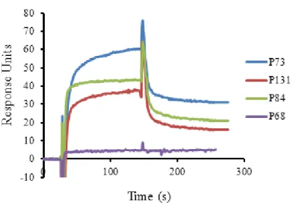
By using surface plasmon resonance experiment, it was found that two PPs, P84 and P131 had higher binding affinity for the Aβ42 peptide (Kd value of 20 nM and 12 nM,
respectively) than the previously reported Aβ42 binding peptides. To the best of our knowledge, this is the first report showing the development of randomly evolved peptide aptamers with the highest binding affinity to Aβ42. Then the functional characterization of P84 and P131 was performed to check their potential of inhibiting aggregation and cytotoxicity of Aβ42 peptide by using thioflavin T assay, atomic force microscopy assay and PC12 cells based cytotoxicity assay. It was found that both P84 and P131inhibited the aggregation of Aβ42, leading to the reduction of the cytotoxic effect of Aβ42 on PC12 cells. Therefore, it is believed that these PPs can be the potential therapeutic seeds for the AD.
v
As of now, PLM consists of the evolution of the first, second and third library and each library selection can be assigned as module finding, module shuffling and module pairing, quite similar phenomena occurring in natural evolution of proteins. As the last step in PLM, in this study the fourth library was introduced based on the point mutation together with the DNA shuffling method to further improve the affinity and functional activity of the most improved paired peptide evolved from the third library. So, the P109 peptide, which showed the strongest binding affinity to cathepsin E at neutral pH (Kd value of 2 nM), was selected and a randomly base-substituted library (fourth
library) was generated by inserting point mutations in the sequence of P109. One mutant containing single point mutation of P109, here after named SK1, showed 1.6 fold improved binding affinity to cathepsin E at neutral pH compared to the affinity of non-mutated P109. SK1 also showed significantly improved functional activity (5% increased functional activity compared to that of P109).
In this study, highly functional peptides with a strong binding affinity towards the respective peptides were successfully developed from the third and fourth library, demonstrating the effectiveness of the progressive library method (PLM) in the evolution of peptides/proteins.
Since the natural evolution of proteins is based on point mutation and recombination, the evolution strategy for proteins, i.e., PLM seems to be completed by the introduction of the fourth library of point mutation-based one following the recombination-based second and third libraries.
vi
List of figures
Figure 1.1. Progressive library method. Functional peptides were identified from the
first random peptide library………..……....8
Figure 2.1. Schematic representation of generation of paired peptide (PP) library by Y-ligation-based block shuffling (YLBS) method………..…....21
Figure 2.2. Scheme for in vitro peptide selection……….……….…..24
Figure 2.3. Random combination of peptide blocks selected from the first and resulted PP second library library………...………...…..…..31
Figure 2.4. Determination of the binding affinity of the PPs to Aβ42 by surface plasmon resonance (SPR)………...……….……....32
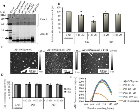
Figure 2.5. P84 and P131 inhibit the fibrillization of Aβ42………..….……...34
Figure 2.6. AFM images of Aβ42 incubated under modified conditions for fibrillization………..………..………..35
Figure 2.7. Effect of the PPs on the oligomerization of Aβ42……….…….….38
Figure 2.8. PPs inhibit the cytotoxicity of Aβ42……….………...40
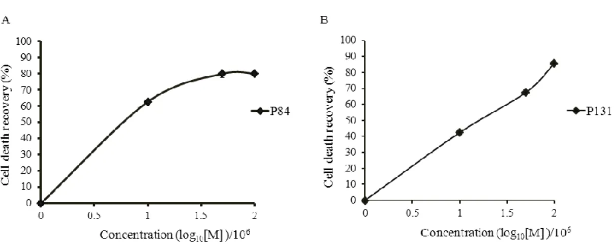
Figure 2.9. Tentative EC50 values of P84 and P131……….41
Figure 2.10. PPs inhibited the induction of caspase-3/7 activity by Aβ42………...42
Figure 2.11. Schematic representation to show the effectiveness of PLM to develop novel peptides with stronger binding affinity to the Aβ42………….…………..…..44
vii
Figure 2.12. A hypothetical model for the aggregation inhibition by the PPs……..47
Figure 3.1. Schematic representation of generation of the fourth library and in vitro
selection of cathepsin E binding peptides………..………….56
Figure 3.2. Determination of binding affinity of SK1 to cathepsin E at
Physiological pH………..………...….60
Figure 3.3. Comparison of cathepsin E activity enhanced by chemically
Synthesized P109 and its mutant SK1………..………...……61
Figure 3.4. Demonstration of methodological effectiveness of complete
viii
List of tables
Table 2.1. Peptide blocks and spacers used for constructing the third library (paired
peptide library)………....20
Table 2.2 . Paired peptides (PPs) obtained from in vitro selection of the thirdly
generated library………...………...30
Table 2.3. Paired peptides selected and their properties………...…..31
Table 3.1. Cathepsin E binding peptides obtained from in vitro selection
ix
List of abbreviations
ASAC all-steps-all-combinations
AD Alzheimer`s disease
APP amyloid precursor protein
ANS 8-Anilinonaphthalene-1-sulfonate
AFM atomic force microscopy
Aβ beta amyloid
Aβ40 beta amyloid 40
Aβ42 beta amyloid 42
BBB blood brain barrier
CCK-8 cell counting kit-8
DMSO Dimethyl Sulphoxide
DMEM Dulbeco`s modified eagle medium
D-PBS Dulbeco`s phosphate buffer saline
eRAPANSY evolutionary rapid panning and analysis system
x
Kd dissociation constant
µM micro molar
nM nano molar
PAGE poly acrylamide gel electrophoresis
PLM progressive library method
PP (s) paired peptide (s)
PS1/2 presenilin 1/2
SDS sodium dodecyl sulphonate
SPR surface plasmon resonance
ThT Thioflavin T
WST-8 water-soluble tetrazolium salt-8
xi
Table of contents
Dedication
………..…..…...…..i
Acknowledgements
……… .……….……..……..ii
Abstract
………..…………..……..iii
List of figures
………...…………...……..………...vi
List of tables
………...……...………..…………..viii
List of abbreviations
………...…………...ix
Table of contents
………...………...………xi
Chapter One: Introduction and literature review………...…...
11.1 Background……….…...….…….……1
1.2 Peptides as therapeutic drugs………..…….2
1.3 Directed evolution………..……….…………..…..3
1.4 mRNA/cDNA display technology………..……...…...………...5
1.5 Evolutionary rapid panning and analysis system (eRPANSY)………..……….6
1.6 Progressive library method (PLM)……….………....….7
1.7 Research objectives and methodology………...……..……...…..9
xii
Chapter Two: Acquisition of Aβ42 aggregation inhibitory/
cytotoxicity-preventive peptides using PLM………...
132.1 Abstract
………..……….…
132.2 Introduction
………..
142.2.1 Alzheimer`s Disease
………...…..……
142.2.2 Beta amyloid 42 (Aβ42) as a therapeutic target for AD
………...…..
152.2.3 Objectives relevant to this study
………...……...
172.3 Materials and Methods
………..……..
182.3.1 Generation of a paired peptide library and cDNA-peptide fusion product ……...18
2.3.2 In vitro selection of novel peptides ………...………….…...….23
2.3.3 Preparation of Aβ42 aggregates………...………..…24
2.3.4 Measurement of binding affinity………...………...……..25
2.3.5 Thioflavin T (ThT) binding assay……….………..……..…….26
2.3.6 Atomic force microscopy………..……….…….……….….…….26
2.3.7 Tricine-SDS-PAGE………...………..……...…..……..27
2.3.8 8-Anilinonaphthalene-1-sulfonate (ANS) fluorescence analysis……..………27
xiii
2.4 Results………...………29
2.4.1 In vitro selection of novel peptides from the PP library……….….…...29
2.4.2 Improved binding affinity of the novel peptides to Aβ42………..32
2.4.3 P84 and P131 inhibit the fibrillization of Aβ42………..….………..33
2.4.4 Effect of P84 and P131 on the oligomerization of Aβ42………..….…....36
2.4.5 Effect of P84 and P131 on the cytotoxicity of Aβ42………...……….…..37
2.5 Discussion………...………....43
2.6 Conclusions………...……….….…..49
Chapter Three. Improvement of binding affinity and functional
activity of cathepsin E activator P109 peptide by the fourth library
method of PLM………..51
3.1 Abstract……….……….………..51
3.2 Introduction………...………...………...52
3.3 Materials and Methods………...54
3.3.1 Preparation of protease cathepsin E and its substrate……….……54
3.3.2 Generation of the fourth library (Mutated library)……….55
3.3.3 In vitro selection of cathepsin E activating peptides……….………….55
xiv
3.3.5 Cathepsin E protease activity assay………..……..58
3.4 Results………...…………58
3.4.1 In vitro selection of cathepsin E binding peptides from the fourth library...…..…58
3.4.2 Improved binding affinity and functional activity of SK1 to cathepsin E……..…59
3.5 Discussion………..……….………….…….…61
3.6 Conclusions………...………..63
Chapter Four: Overall conclusions and prospects to the future
study...65
4.1 Overall conclusions………..…...……….65
4.2 Prospects to the future study………..…………66
1
Chapter 1
Introduction and literature review
1. Background
Current drug industry has been dominated by the drugs based on small molecules and biological macromolecules (biologics) (Verdine et al., 2007). The small molecules are generally synthetic organic compounds with a simple chemical structure and molecular size less than 1000 Dalton (less than 100 atoms). Small size, low cost and low price, oral bioavailability, ready synthesis, membrane-penetrating ability and stability are the advantages of small molecules. However the drugs based on small molecules have the major disadvantage of low specificity and higher attrition rate in the clinic (Craik et al., 2013). On the other hand, biologics are mainly protein or carbohydrate based (like antibodies, insulins etc). Unlike small molecules, biologics have a very high affinity and specificity to the targets so there is increasing demand of drugs based on biological macromolecules (Reichert et al., 2005). The main hurdle with biologic drugs is their very poor cell permeability due to the very big size of biologics (50 KDa -150 KDa). Due to this nature, 90% of drug targets are not accessible for targeting (Verdine et al., 2007). In addition, only 10% of proteins encoded in the genome are estimated to be amenable to small-molecule targeting and another 10% by biologic drugs, and thus 80% of proteins are currently termed as `undruggable` (Schlippe et al., 2012). Moreover, biologic drugs are very costly due to the complexity of production in large quantities. Furthermore, biologics are very sensitive to various environmental factors like heat, light, agitation etc. From the above mentioned facts about small drugs and biologic
2
drugs, it is clear that there exist a big gap between the small molecules and biologic drugs. Therefore, the time has now come to discover new drug leads that fit between small molecules and biologics with the goal of combining advantages of small molecules and biologics.
1.2 Peptide as a therapeutic drug
An important goal of drug discovery is to develop nonmacromolecular species that retain the favorable molecular properties of antibodies and small molecules. Since peptides (usually less than 50 amino acids in size) possess high binding affinity and specificity comparable to antibodies and have the high possibility of cells/tissue penetration due to their smallness, these peptides are the ideal therapeutic candidates to bridge the gap between small molecules and biologic drugs (Zhang et al., 2000). Since peptides are much smaller than antibodies, peptides can be readily synthesized, optimized, evaluated and do not cause serious immune responses. Moreover, peptides are potent and could be metabolically cleaved and readily cleared from the body minimizing their toxic side effects.
Traditionally, therapeutic peptides can be obtained from three different sources such as i) natural or bioactive peptides produced by plants, animal or human (naturally occurring peptide hormones or from fragments of larger proteins); ii) peptides discovered from chemical libraries and iii) peptides isolated from genetic/recombinant libraries. Since bioactive peptides are selected by nature to have a stable fold, they have key contact residues to impact function and they have increased in vivo stability against proteases. However, there are only limited bioactive peptides against the specific target proteins. Until few years back, the peptide based drugs were mainly dominated by the
3
bioactive peptides due to the lack of the technology to develop the peptides as potent as bioactive peptides (Sato et al., 2006). However the development of the recombinant display technologies with the ever-increasing levels of diversity, it has been possible to find novel peptides as rival of those found in nature (Sato et al., 2006; Landon et al., 2005). Over the past decade, recombinant display technologies have been essential tools in the discovery of peptide and protein ligands and have played a vital role in the directed molecular evolution of therapeutic peptides.
1.3 Directed molecular evolution
Directed molecular evolution mimics natural evolution of proteins/ peptides in the laboratory (in vitro) but operates on molecular level and focuses on specific molecular properties. Directed molecular evolution results novel peptides/proteins through generation and selection of combinatorial libraries of nucleic acids and peptides. Molecular selection technologies involve the construction of large, diverse DNA molecules and the establishment of associated selection mechanisms for identifying and isolating those that encode the relevant peptides, which bind with high affinity and specificity to a given target molecule (McGregor et al., 2008). These methods depend on the physical linkage of each nucleic acid with its encoded peptide. Moreover, the molecular selection methods depend on different display technologies for the development of novel peptides.
Based on the mode of selection of the library, there are in vivo and in vitro display technologies. The in vivo display technologies include phage display, bacterial display and yeast display. Phage display was developed by George P. Smith in 1985. Bacterial display systems were first introduced by Freudl et al. and Charbit et al. in 1986 where
4
as yeast display was developed by Borden and Wittrup in 1997. Since the invention, these display methods have been successfully applied to many different areas of research, including immunology, cancer research, drug discovery, epitope mapping, protein-protein interactions, plant science and infectious diseases, targeting a broad range of protein families. Though phage display has shown great potential in the discovery of new therapeutics and in recent years, the best known peptide display is the phage display technology itself (McGregor et al., 2008; Huang et al., 2012).
Phage display technology is based on the construction of a polypeptide library fused to a bacteriophage coated protein. In the phage display technique, a DNA library is genetically fused to a filamentous bacteriophage (commonly used bacteriophage is M13) so that co-infection by the bacteriophage, newly emerging phage particles encase individual gene sequences while displaying the corresponding gene-encoded polypeptides on their outer surfaces (Huang et al., 2012). In this way phage particles provide the physical link between genes and their encoded polypeptides. Usually phage display results about 1010 different polypeptides in the library which is incubated with an immobilized target molecule. All the non-binding library members are removed by washing and those rare species of the library which bind to the target are eluted. Some of the peptides derived from phage display are currently in clinical and preclinical trials (Giuliani et al., 2007).
Although phage display is now playing a significant role for the discovery of novel therapeutic peptides (Giuliani et al., 2007), there are some limitations of this technology. As already mentioned that Phage display has the peptides displayed on the surface of the bacteriophage, it allows only the screening and enrichment of peptides based on
5
natural amino acids, resulting the reduced diversity of the selected sequences. Furthermore, the library size is limited by the transformation efficacy of the bacteriophage and also the displayed peptide size is small.
1.4 mRNA/cDNA display technology
In order to overcome the limitations of phage display technology, two display technologies, mRNA (cDNA) display and ribosome display technologies, were developed at the end of the 1990s. The mRNA display technology was first described by Roberts and Szostak in 1997 and the same principle was reported concomitantly under the name of in vitro virus by Nemoto et al. in 1997 whereas ribosome display technology was first described by Hanes and Pluckthun in 1997. Since both mRNA and ribosome display technologies are based on the in vitro selection of the library, the library size is as large as 1013 differentvariants. Both display technologies begin with the library of DNA sequences coding for the random peptides. In case of mRNA display, the translated peptide is associated with the progenitor mRNA via a puromycin linker (Roberts et al., 1997 and Nemoto et al., 1997). Puromycin is an analog of the 3’ end of a tyrosyl-tRNA with a part of its structure mimicking a molecule of adenosine, and the other part, a molecule of tyrosine. Since puromycin has a non-hydrolysable amide bond, it interferes with translation resulting the release of translation products (Roberts et al., 1997). In case of ribosome display, the translated peptide is associated with the progenitor mRNA and the stalled ribosome.
Although both mRNA display and ribosome display technologies have the great merit of very high diversity of the library and synthesis of proteins/peptides by using both natural and unnatural amino acids, mRNA display is superior over ribosome display
6
technology (Roberts et al., 1999). In case of ribosome display, selection stringency is limited to keep ribosome-mRNA-polypeptide in a complex because of the noncovalent ribosome-mRNA-polypeptide complex. Furthermore, ribosome is very large in size so that there might be unwanted interaction between selection target and the ribosome, and this may lead to a loss of potential binders during the selection cycle. On the other hand, puromycin linker used in mRNA display system is much smaller comparing to ribosome so that this linker may have less chance to interact with an immobilized selection target. Therefore, the mRNA display technology is more likely to give less biased results.
Although the mRNA display system was originally developed for evolutionary protein engineering based on in vitro translation system, they were subsequently used for various purposes like for the discovery of therapeutic peptides from natural and/or non-natural and cyclic and/or linear amino acid libraries, for functional analysis including, protein-protein, drug-protein, peptide-protein and antigen-antibody interactions (Takahashi et al., 2003; Wada et al., 2013; Schlippe et al., 2012). Previous studies have revealed that in vitro selection of peptides from wide variety of libraries is a powerful approach for the development of new therapeutics against target molecules like GPCR which is a major drug target (Wada et al., 2013).
1.5 Evolutionary rapid panning and analysis system (eRAPANSY)
cDNA display (Tabuchi et al., 2001), a modified version of the mRNA display has been successfully applied in evolutionary rapid panning and analysis system (eRAPANSY) to develop novel peptide drug candidates against different targets like cathepsin E (acidic protease) (Kitamura et al., 2009). The eRAPANSY consists of three
7
primary steps; i) generation of huge diversity (1013) library of molecules in the form of cDNA display by employing a Y-ligation based block shuffling (YLBS) technique (Kitamura et al., 2002), ii) selection of peptides either based on selection-by-function or selection-by-affinity and iii) generation of progressive libraries by progressive library method (PLM), which allows the generation and selection of clones with greater activity and affinity than in the previous library, through the use of the YLBS. Novel peptides with a strong binding affinity (Kd in the pM range) has been successfully applied against
the protease cathepsin E proving the effectiveness of this technique in vitro molecular evolution of therapeutic peptides (Kitamura et al., 2012; Madhu et al., 2011; Komatsu et al., 2012).
1.6 Progressive library method (PLM)
PLM is one of the important strategies for systemic in vitro evolution method to develop advanced peptides against a specific target protein (Kitamura et al., 2012). PLM was first introduced by Kitamura et al. in 2009, which consists of generation of progressive libraries through the use of the Y-ligation-based-block shuffling (YLBS) method (Kitamura et al., 2002). In PLM, the successfully selected peptides from the preceding library are used to build the successive library. The successfully selected peptides (8 amino acids in length) from the secondary library by using in vitro cDNA display method showed improved functional activity and binding affinity against the protease cathepsin E at acidic pH compared to that of the peptides selected from the primary library. The peptides thus obtained from the secondary library were randomly shuffled to generate the third library (paired peptide (PP) library). The library was then used for in vitro selection by affinity or by function. The successfully selected paired
8
peptides (PPs) exhibited highest functional activity and binding affinity to cathepsin E at physiological pH and acidic pH (Biyani et al., 2011, Kitamura et al., 2012 and Komatsu et al., 2012). The general scheme of progressive library method is summarized in Fig.1.1 (Komatsu et al., 2012).
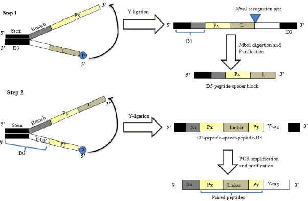
Figure 1.1. Progressive library method. Functional peptides were identified from the
first random peptide library. The second library was generated by combining peptide blocks obtained from the primary library and subjecting them to the next round of selection. The tertiary library was constructed by pairing two peptides selected from the second library (Komatsu et al., 2012).
The primary, secondary and tertiary library selections used in PLM can be regarded as module finding, module shuffling and module pairing respectively, quite similar phenomena occurring in natural evolution of proteins. The functional activity and affinity of PPs against cathepsin E under different conditions proved the methodological
9
effectiveness of PLM in the in vitro evolution of therapeutic peptides. These previous studies on cathepsin E also demonstrated the proof of principal that PLM can be used to develop the therapeutic peptides against a vast array of targets like Aβ42 which is proven to be a main pathological hallmark of Alzheimer`s disease.
1.7 Research objectives and methodology
The plethora of research evidence has proven that Aβ42 plays a key role in Alzheimer`s disease pathogenesis (Selkoe DJ, 2001). Moreover, several lines of research evidences have proven that aggregation of Aβ42 is a main pathological hallmark of AD (Hardy and Selkoe, 2002). However, as of now there is no cure of AD. So, development of nM affinity peptides which can inhibit or reverse the aggregation and cytotoxicity of Aβ42 holds a great potential of therapeutic value for the treatment of AD. Although several peptides and small molecules as Aβ42 aggregation inhibitors have been reported by many research groups, none of them have been developed as a drug. To this end, PLM was used in previous study to find out the novel peptides against Aβ42 (Ueno-Tsuji et al., 2011) through in vitro selection of primary and secondary library. However, the best peptide from secondary library showed the moderate level of binding affinity to Aβ42 (Kd value in µM range) which is far below the binding affinity needed to be used
as a potential therapeutic candidate for Aβ42. Since the novel PPs evolved from tertiary library of PLM showed strongest binding affinity and functional activity against cathepsin E (Komatsu et al., 2012; Kitamura et al., 2012), this fact motivated us to generate and screen the paired peptide (PP) library which is the third library of PLM to develop novel peptides with improved binding affinity to Aβ42. Therefore, the objectives of this study are listed below.
10
i) Development of novel peptides with higher binding affinity and specificity to Aβ42.
ii) Functional screening of novel peptides which can inhibit Aβ42 aggregation and cytotoxicity so that these peptides can be the therapeutic candidates for Aβ42
iii) Establishment of the progressive library method (PLM) as an effective in vitro molecular evolution method to engineer novel peptides with high binding affinity, specificity and functional activity against different therapeutic targets, e.g. Aβ42, cathepsin E and etc.
In order to fulfill the first objective of this study, the third library (paired peptide library) was generated by random pairing of peptides selected from the second library using YLBS method. Once evolved peptides by the all-steps-all-combinations (ASCS) method introduced in secondary library were further evolved by the paired peptide (PP) method. The PP library generated from the third library selection products was subjected to in vitro selection against Aβ42 by using cDNA display method and then cloning and sequence analysis was performed to select the most frequently occurred PPs in the library. After 4th round of successive in vitro selection by cDNA display method, the potential PPs were selected from the cloning and sequence analysis. The surface plasmon resonance (SPR) analysis was performed to confirm the binding affinity of the selected PPs to Aβ42. In order to fulfill the second objective, different biophysical characterizations based on thioflavin T assay, atomic force microscopy assay, polyacrylamide gel electrophoresis and in vitro bioassay were performed. Indeed, the introduction of PP approach in case of Aβ42 can provide strong support that PLM is an effective in vitro evolution method. Furthermore, in order to fulfill the third objective of this study, the fourth library was introduced by generating random mutagenesis library
11
of cathepsin E activating paired peptide P109 (Kd value of 2 nM) based on single point
mutation together with DNA shuffling approach.
1.8 Thesis organization
The whole thesis has been divided into four chapters. Chapter 1 is about the introduction and literature review of this study. The objectives of this study are mentioned at the end of this chapter.
Chapter 2 is about the establishment of proven molecular evolution method PLM through acquisition of Aβ42 aggregation-inhibitory/cytotoxicity-inhibitory peptides. This chapter consists of the major research findings of this study. Identification of novel PPs with highest binding affinity to Aβ42 has been described in this chapter. Similarly, inhibition of Aβ42 aggregation and cytotoxicity by PPs has been shown by performing different biophysical characterizations like thioflavin T assay, atomic force microscopy and PC12-cells based bioassay.
Chapter 3 is about the improvement of binding affinity of cathepsin E activator P109 peptide. A fourth library was introduced based on the point mutation together with the DNA shuffling method. A randomly base-substituted library (fourth library) was generated by inserting point mutations in the sequence of P109. The library was used for affinity-based in vitro selection using cDNA display method. The results mentioned in this chapter suggested that fourth library is the last step of PLM, supporting that PLM is an established in vitro molecular evolution method to develop new therapeutic peptides against different target proteins/peptides.
12
Chapter 4 is about the overall conclusions and prospects to the future study. The major findings of this study and future prospects of the study have been described in this chapter.
13
Chapter 2
Acquisition of Aβ42 aggregation-inhibitory/cytotoxicity-preventive
peptides using PLM
2.1 Abstract
Several decades of cumulated research evidence has proven that aggregation of beta amyloid 42 (Aβ42) is the main cause of neuronal death in the brain of patient with Alzheimer’s disease (AD). So, Inhibition of Aβ42 aggregation holds great promise for the prevention and treatment of AD. Although several drugs based on Aβ immunotherapy and small molecule have been developed, none of them has yet translated into new medicines. The main problem with Aβ immunotherapy drugs is their failure to cross the blood brain barrier. On the other hand, small molecule drugs have the potential of off target specificity and high clinical attrition rate. Since peptides have the high affinity and specificity comparable to the immunotherapy drugs and also peptides have the high possibility of crossing the blood brain barrier due to their small size compared to immunotherapy drugs, recently much effort has been given to develop drugs based on the peptides/peptide aptamers. Despite intensive research studies on the peptide candidates for AD therapeutics, there is a great deal of room for improvement in the design of much better peptides. So, in this study, systematic in vitro evolution method called progressive library method (PLM) was used and novel peptides with nano-molar affinity to Aβ42 were developed. Particularly, two peptides, P84 and P131 showed about 26 fold and 48 fold, respectively, higher binding affinity to Aβ42 compared to the peptide with strongest binding affinity to Aβ42 developed from
14
previous library. Furthermore, both paired peptides showed strong inhibition of Aβ42 aggregation, resulting the recovery of PC12 cells from the cytotoxic effect of aggregated Aβ42. Our results suggest that these novel peptides can be the potential therapeutic seeds for Alzheimer`s disease.
2.2 Introduction
2.2.1 Alzheimer`s Disease
Alzheimer`s disease (AD) is an age-related neurodegenerative disorder and is the most common cause of dementia among aged people (Brookmeyer et al., 2007). AD was named after the name of German neuropathologist Alois Alzheimer who first described this disease in 1906 (Berchtold et al., 1998). Clinically, the symptoms of AD include the loss of memory, thinking ability and eventually leading to the loss of ability to carry out the simplest tasks (Selkoe DJ, 2001). The main affected area of the brain by AD is the hippocampal area which is the main region responsible for memory formation. It has been predicted that AD will affect 1 in 85 people worldwide by 2050 (Brookmeyer et al., 2007). The histopathological features of AD include the presence of amyloid beta plaque, neurofibrillary tangles and neuronal atrophy (Hardy and Selkoe, 2002). Although the exact cause of AD remains unknown and most of the reported evidences are rather sporadic, a small fraction of AD patients (less than 1%) genetically inherit AD through autosomal dominant inheritance which results disease onset before the age of 65. Mutations that increase the accumulation of beta amyloid (Aβ) deposition are associated with familial AD (FAD). Mutations in amyloid precursor protein (APP), presenilin 1 (PS 1) and presenilin 2 (PS 2) are proven to be responsible for the onset of FAD (Nunan and Small 2002). These mutations all result in an increased production of
15
Aβ42 and lead to AD neuropathology, suggesting that Aβ42 deposition is one of the pivotal steps in the pathogenesis of AD. This supports the amyloid cascade hypothesis which proposes that the increased production or decreased clearance of Aβ peptides causes the AD (Hardy and Selkoe, 2002).
2.2.2 Beta amyloid 42 (Aβ42) as a therapeutic target for AD
The Aβ peptide, which consists of 40 to 42 amino acids, is generated from the β – amyloid precursor protein (APP) by the subsequent proteolytic activities of β- and ϒ- secretases (Haass et al., 1992; Seubert et al., 1992 and Shoji et al., 1992). Extensive research studies strongly support the central role of amyloid beta (Aβ) in the pathogenesis of Alzheimer`s disease (Hardy and Selkoe, 2002). Based on molecular genetics, exploration of mutations in APP and direct alteration of Aβ production in AD experimental model animals further supports the key role of Aβ in AD pathogenesis (St. George-Hyslop et al., 2000 and Jonsson et al., 2012). At the early days of AD research, the extracellular amyloid fibril formation was regarded as the main cause of AD (Hardy et al., 1992) however the recent studies have proven that progressive accumulation of soluble Aβ oligomers play a vital role in the pathogenesis of AD by causing long term potentiation impairment and synaptic dysfunction (Lue et al., 1999; McLean et al., 1999; Haroutunian et al., 2000; Walsh et al., 2007; Shankar et al., 2008 and Benilova et al., 2012).
Aβ40 and Aβ42 peptides are the two major peptides present in AD patients. Although Aβ40 is produced in significantly large amount compared to Aβ42 by enzymatic cleavage from the APP, Aβ42 has been found more neurotoxic due to its higher
16
hydrophobicity, which leads to faster oligomerization and aggregation (Walsh et al., 2007 and Blennow et al., 2006). Furthermore, it has been reported that Aβ42 oligomers are more toxic than protofibrils or fibrils (Ahmed et al., 2010). Thus, Aβ42 has become a major therapeutic target with different anti- Aβ strategies being accompanied. One of the strategies includes lowering the production of the Aβ peptide by inhibiting the enzymes responsible for Aβ generation (Jasvinder et al., 2011). Similarly, another strategy is to prevent the formation of Aβ aggregates and to increase the rate of Aβ clearance from the brain. This strategy can be accomplished by Aβ immunotherapy which uses anti- Aβ antibodies generated following vaccination or introduced passively and over the last one decade, Aβ immunotherapy has entered for clinical trials in humans (Lemere et al., 2010).
Until today, there is no cure of AD and the available medications can temporarily slow the worsening of symptoms. Although great effort and cost has been invested to understand the pathophysiology of AD and to develop the Aβ immunotherapy drugs, this process has not yet translated into new medicines due to the failure of drugs in different phases of clinical trials (Barry et al., 2012 and Salkoe et al., 2011). Similarly, despite the invaluable role of antibodies in medical diagnostics and biomedical research, antibodies are tedious and expensive to generate, and it is very difficult to produce them in very large quantities. To this end, much effort has been devoted to design small molecules and peptides capable of inhibiting Aβ aggregation and to neutralize Aβ cytotoxicity. Most of these small molecules have been identified by screening thousands of already available small molecules (Angela FM, 2012) whereas small peptides were generated as a partial fragment of the Aβ42 peptide by rational design or by
17
combinatorial libraries (Takahashi et al., 2010; and Baine et al., 2010). Despite the intensive research studies on such small molecules and peptides as a potential candidate for AD therapeutics, there is lot of space to design much better small peptides with high binding affinity and specificity to Aβ42 peptides. This fact motivated us to perform this study.
2.2.3 Objectives relevant to this study
The objectives of the study described in this chapter are i) to develop novel peptides with high binding affinity and specificity to Aβ42 and ii) to develop potential therapeutic seeds to inhibit and reverse the misfolding and aggregation of Aβ42.
In order to fulfil these objectives, it was aimed to develop high-affinity Aβ42 binding peptide aptamers based on evidence supporting the use of peptide aptamers for biological and therapeutic applications (Li et al., 2011). Here, the evolution of novel peptide aptamers using the mRNA (cDNA) display technique [Nemoto et al., 1997; Robert et al., 1997] and paired peptide (PP) method (Kitamura et al., 2012; Komatsu et al., 2012) is described. In the previous study, peptides with a moderate binding affinity to Aβ42 (Kd in µM) were explored
from the preceding libraries by the all-steps-all-combinations (ASAC) method (Tsuji-Ueno et al., 2011). In the present study, thus evolved peptides were further evolved by the PP method. The evolved peptide aptamers, hereafter called paired peptides (PPs), contained two peptide moieties which were arbitrarily combined by linking through a definite length of a linker sequence. The selected two peptides had higher binding affinity for the Aβ42 peptide than previously reported Aβ42 binding peptides (Tsuji-Ueno et al., 2011). Furthermore, different
18
biophysical experiments were performed to show that the novel peptides with improved binding affinity can inhibit the aggregation of Aβ42 leading to the reduction of the cytotoxic effect of Aβ42 in PC12 cells.
2.3 Materials and Methods
Chemicals were obtained from Wako (Japan), unless otherwise stated.
2.3.1 Generation of a paired peptide library and cDNA-peptide fusion product
A paired peptide (PP) library was generated by following the Y-ligation-based block shuffling (YLBS) method (Kitamura et al 2002) with slight modifications. In brief, cDNAs coding for 8 different types of peptide blocks selected from the first and second libraries (Tsuji-Ueno et al., 2011) and two types of linkers (Table 2.1) were synthesized and purified (> 95%) by JBios, Japan. Before starting the first round of YLBS, phosphorylation of 5` end of 3`-half spacer and 3`-half Y-Tag was performed by incubating the mixture of 100 µM 3`-half spacer mixture or 3`-half Y-Tag (1 µl), 10× PNK buffer (1 µl), 10 mM ATP (1 µl), 1 µl of PNK (10,000 U/ml) (NEB) and water up to 10 µl at 37oC for 1 h. Then, the reaction mixture was heat inactivated at 85oC for 15 min.
After phosphorylation reaction, the first round of YLBS was performed. For this purpose, 10 pmol/µl 5`-half peptide blocks mixture was mixed with 10 pmol/µl 3`-half phosphorylated mixture, 5µl of 2× ligation buffer (specially prepared for YLBS, Kitamura et al., 2002) and 1 mM ATP (1 µl) and then the mixed reagents were used for annealing reaction by incubating the reaction mixture for 2 min at 94oC, for 10 min at
19
60oC and then for 10 min at 40oC. The lowering of temperature should be very gradual (0.1oC/sec). After that, as shown in Figure 2.1 below, the first round of YLBS was performed with biotin-labeled 5′-half peptide blocks and 3′-half linker blocks by adding 2 µl T4 DNA ligase (Takara) to the annealing reaction and incubating at 25oC for overnight (up to 16 hours). The YLBS product was then digested with the MboI restriction enzyme (NEB) and then purified using Dynabeads MyOne Streptavidin C1 (Invitrogen) to obtain single-stranded 5′-half DNA sequence blocks coding for “peptide-spacer blocks.” Then, a second round of YLBS was performed with D5-peptide-spacer blocks and a 3′-half peptide blocks to obtain DNA sequences that code for PPs composed of “peptide-spacer-peptide” blocks by following the protocols as mentioned above. The required cDNA construct to use for cDNA display (Xa-paired peptide-Y-Tag) was prepared by PCR using forward primer b-5`-half-stem (5`-GGCTCGCGAATA CTGCGATTGAAGGTCGT-3`) and reverse primer 3`-stem rev (5`-GGCTCGCGAATACTGCGAAGG AGTGAG-3`). In this experiment, at first two PP libraries were generated by using short and long linker separately and only the final product obtained after PCR was mixed together resulting the PP library. The reason of doing so was to minimize the possible biased diversity of PPs during YLBS which is supposed to be more efficient with short linker compared to that with long linker. Theoretically, the PP library consisted of 128 different variants (8 different types of 5'-half peptide blocks x 2 types of linkers x 8 different types of 3'-half peptide blocks). However, the actual library diversity was assumed to be much higher due to the possible substitution and indel mutations during library generation process (Komatsu et al., 2012).
20
Table2.1. Peptide blocks and spacers used for constructing the
third library (Paired peptide library)
Name (5` -> 3`) Nucleotide sequence Length Amino acid sequences (bp)
5-half peptide blocks
P5001 D5-GGC AGC AGC CCT CCT GGC CTG TGC 53 GSSPPGLC P5002 D5-TGC TGC AAG CCT TCC TGT TGC TGT 53 CCKPSCCC P5105 D5-TGC GGC ATT CTC GAT CCC ATC CCT
TGG
56 CGILDPIPW P5109 D5-GGC TGC CCA TGC ATT GGC ATT ATT 53 GCPCIGII P5128 D5-CCA AAC CCA CCA GAT CAA ATC CCC
ACC
56 PNPPDQIPT P5226 D5-CCA AAC CCA CCA GAT CCC ATC CAT
TGG
56 PNPPDPIHW P5241 D5-GAC GGC AAA TCC ATT GGC CCA ATT 53 DGKSIGPI P5268 D5-GAC TGC TCC TCC GAT CTC ACT CCT
TCG
56 DCSSDLTPS 3-half linker blocks
L1 D3-GGA GGC TCC GGA GGC AGC 48 GGSGGS L2 D3-GGC GGA GGC TCC GGT GGA GGG AGC
GGA GGC GGT TCT
66 GGGSGGGG GGS 3-half peptide blocks
P5001 D3`-GGC AGC AGC CCT CCT GGC CTG TGC 76 GSSPPGLC P5002 D3`-TGC TGC AAG CCT TCC TGT TGC TGT 76 CCKPSCCC P5105 D3`-TGC GGC ATT CTC GAT CCC ATC CCT
TGG
79 CGILDPIPW P5109 D3`-GGC TGC CCA TGC ATT GGC ATT ATT 76 GCPCIGII P5128 D3`-CCA AAC CCA CCA GAT CAA ATC CCC
ACC
79 PNPPDQIPT P5226 D3`-CCA AAC CCA CCA GAT CCC ATC CAT
TGG
79 PNPPDPIHW P5241 D3`-GAC GGC AAA TCC ATT GGC CCA ATT 76 DGKSIGPI P5268 D3`-GAC TGC TCC TCC GAT CTC ACT CCT
TCG
79 DCSSDLTPS
D5 : GGC TCG CGA ATA CTG CG ATT GAA GGT CGT (29 bp) D3 : GAT CTC ACT CCT T CGC AGT ATT CGC GAG CC (30 bp)
D3`:AGG ACG GGG GGC GGC GGG GAA A GAT CTC ACT CCT T CGC AGT ATT CGC GAG CC (52 bp)
The underlined sequences are complementary to eachother. Sequences in bold letters in D5 code for Xa factor recognition sequence (IEGR). The sequences written in italic in D3` (Y-Tag sequences) are the sequences for puromycin linker binding site. LI: Liker 1; L2: Linker 2
21
Figure 2.1. Schematic representation of generation of paired peptide (PP) library by Y-ligation-based block shuffling (YLBS) method. Note that 8
different types of peptide blocks (Px as 5`-half peptide blocks and Py as 3`-peptide blocks) and two types of linkers (L) were used in this study. The first step YLBS was performed with Px and two types of linkers (L). The YLBS product was digested with the MboI restriction enzyme and then purified using Dynabeads MyOne Streptavidin C1 to obtain single-stranded 5′-half DNA sequence blocks coding for “D5-peptide-spacer blocks”. Then, second step YLBS was performed with 5`-half “D5-peptide-spacer blocks” and Py to obtain DNA sequences that code for paired peptides composed of “D5-peptide-spacer-peptide-D3`” blocks. Here, Xa represents the Xa factor recognition site (IEGR which is a part of D5). D5 represnts the device sequences of 5` half (stem and brach part) which consists of cDNA sequences coding for Xa factor recognition site; D3 represents the device sequences of 3` half of first step YLBS; D3` represents the device sequences including Y-Tag of 3` half for second step YLBS; Y-Tag represents the cDNA sequences for puromycin-linker binding site. The black box of each device represents the complementary cDNA sequences.
This library was then used to design the full cDNA construct by using overlap extension PCR. The purified cDNA construct containing paired peptides thus obtained by overlap
22
extension PCR was ready to use for in vitro transcription, puromycin linker ligation, and in vitro translation to generate cDNA-peptide fusion constructs as shown in Figure 2.2. For in vitro transcription reaction, 1 pmol of cDNA construct was mixed with 3 µl of 25 mM each rA, U, C and GTP mixture, 2 µl of 5× T7 transcription and 1 µl of T7 enzyme mixture (Ribomax Large Scale RNA production system, Promega). The total mixture was gently mixed and incubated at 37oC for 2 h. After that, 1 µl of RQ1 RNase free DNase (1 U/µl) was added and incubated at 37oC for 15 min.Then, RNA purification was performed by using RNase minielute purification kit by following the manufacturer`s protocol (Promega).
The mRNA thus obtained was used for puromycin linker ligation. For this purpose, 50 pmol mRNA procuct was mixed with 60 pmol of streptavidin puromycin liker, 2.5 µl of 10× T4 RNA ligase buffer (Takara) and RNase free water up to total volume of 25 µl. All the reagents were mixed gently and incubated for ligation reaction (the ligation conditions were 2 min incubation at 90oC followed by 2 min incubation at 72oC, 30 s incubation at 25oC and holding at 4oC). Then 1 µl of T4 PNK enzyme (9 U/µl, Toyobo) and 1 µl of T4 RNA ligase (40 U/µl, Takara) were added to the ligation mixture and then incubated at 25oC for 2 h.
The ligation product was then used for in vitro translation reaction. Retic Lysate IVT Kit from Ambion was used for in vitro translation. 10 pmol of puromycin linker ligation product was mixed with 1.25 µl of 20× Master Mix-Met deficience reagent, 1.25 µl of 20× Master Mix-Leu deficiene reagent, 34 µl of reticulocyte lysate and RNase free water was added for up to 50 µl. The total reaction mixture was mixed gently and incubated at 30oC for 30 min. Then, 20 µl of 3 M KCl (RNase free) and 6 µl of 1 M MgCl2 was added, mixed gently and incubated at 37oC for 1 and half hour.
23
Finally, the in vitro translation product was checked by using SDS PAGE. The in vitro translation product (mRNA-peptide fusion construct) was then used for reverse transcription.
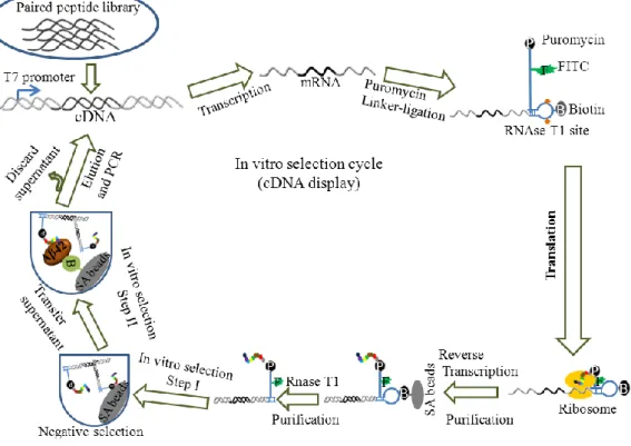
The reverse transcription (RT) of the mRNA-peptide fusions was performed on Dynabeads MyOne Streptavidin C1 by using RT kit ((ReverTraAce –alpha, Toyobo) following manufacturer`s protocol. The RT product was digested with 500 U RNase T1 (Ambion) in order to cleave the biotin-site on the SBP linker. Then, the cDNA-peptide fusions released from the beads were isolated using His-Tag purification kit (HiS Mag Sepharose Ni, GE healthcare). Finally, cDNA-peptide fusion constructs were generated by trimming unwanted peptide portion of this construct using factor Xa protease treatment (1 U/ µl, Invitrogen) followed by Xa removal using BioSpin columns (Qiagen). The cDNA-peptide fusion constructs thus obtained were used for in vitro selection process as shown in Figure 2.2.
2.3.2 In vitro selection of novel peptides
The cDNA-peptide fusion constructs obtained from the PP library (Figure 2.2) were mixed with Dynabeads MyOne Streptavidin C1 and incubated at room temperature for 30 min to remove any non-specific binding peptides. The supernatant was collected in a new tube containing biotin-LC-Aβ42 (AnaSpec Inc., U.S.A) and incubated at room temperature for 20 min and 10 min for first and second rounds of selection, respectively, while the incubation was done for 5 min at room temperature and at 37oC for the third and fourth rounds of selection, respectively. The used biotin-LC-Aβ42 was monomerized and stored at -80oC prior to use according to the method of Tsuji-Ueno et al (2011). Then, Dynabeads MyOne Streptavidin C1 were added and incubated for
24
Figure 2.2. Scheme for in vitro peptide selection. A paired peptide library was
used for this purpose.
30 min to collect the Aβ42-binding PPs.
The beads were washed with D-PBS (-) buffer containing 0.5% tween-20. The Aβ42-binding peptides were eluted with a 25% ammonia solution at 65oC for 15 min. The eluted solution was ethanol precipitated, and the pellet was resuspended in double distilled water. The cDNAs coding for the PPs were cloned and sequenced to select the most readily available novel peptides for chemical synthesis and further testing.
2.3.3 Preparation of Aβ42 aggregates
Aβ42 peptide was purchased from AnaSpec Inc., U.S.A. Monomerized Aβ42 peptide was prepared as described previously (Tsuji-Ueno et al., 2011). Just before use, the
25
monomerized Aβ42 peptide that was stored at -80o
C was thawed and dissolved in 50 mM NaOH to obtain a 0.5 mM Aβ42 peptide solution that was further diluted with 1× D-PBS (-) to a final concentration of 200 µM to neutralize the NaOH. The peptide solution was sonicated for 3 min and then ultra-centrifuged at 15,000 rpm for 10 min. Finally, the supernatant of the peptide solution was collected to obtain a completely dissolved peptide solution. This solution was stored at -80oC and it was considered a monomerized peptide solution. For the surface plasmon resonance (SPR) experiment, the Aβ42 samples were prepared by following the protocol described by Tsuji-Ueno et al. (2011). Aβ42 fibrillization was performed by incubating 100 µM monomerized Aβ42 peptide solutions in the presence or absence of different concentrations of PPs at 37oC with shaking at 350 rpm for up to 48 h. The chemically synthesized PPs with purity more than 95% were obtained from SCRUM, Japan. Similarly, Aβ42 oligomerization was performed by incubating 50 µM Aβ42 peptide solutions in the presence or absence of different concentrations of PPs at 37oC without shaking. After incubation, the samples were then diluted to the concentrations required for the different experiments.
2.3.4 Measurement of binding affinity
The binding affinity (the dissociation constant [Kd]) of each peptide for Aβ42 was
determined at 25oC by SPR using the Biacore 2000 (GE Healthcare). PPs were immobilized on different lanes of a CM5 Biacore sensor chip (GE Healthcare, UK) by a general amine coupling method using immobilization buffer (pH 8.5) containing 10 mM borate and 1 M NaCl. The reference lane was prepared without PPs. Then, different concentrations of monomerized Aβ42 (2.5 µM, 5 µM, 10 µM, 20 µM, and 40 µM) were
26
added to determine the binding affinity of the PPs for Aβ42. For all samples, PBS (-) buffer (pH 7.4) was used as the running buffer and a 50 µM NaOH solution was used to remove Aβ42. The sensograms obtained were fit to a 1:1 Langmuir binding model and the Kd values were determined using BIA evaluation software (GE Healthcare).
2.3.5 Thioflavin T (ThT) binding assay
At various time points, an aliquot of the 100 µM Aβ42 peptide solution prepared for fibrillization and a 50 µM Aβ42 peptide solution prepared for oligomerization were taken and diluted to 10 µM. 50 µL of the diluted peptide solutions was placed in each well of a 96-well plate (Black with clear, flat bottom; Costar) along with 50 µL of a 100 µM ThT (Sigma-Aldrich, India) solution (prepared in 50 mM glycine buffer, pH 8.5). The same concentration of ThT solution without Aβ42 samples was used for background fluorescence measurement. Fluorescence was measured at 490 nm using a Tecan Infinite M200 microplate reader. At first, the background ThT fluorescence was subtracted from the ThT fluorescence of samples. Then, the ThT data in the absence and presence of PPs were normalized to the ThT data of fibrillized Aβ42 sample in the absence of PPs and the values were expressed in percentage. So, at each time point, ThT data of Aβ42 sample in the absence of PPs was set to 100% in order to calculate the percentage of inhibition of ThT fluorescence (i.e., in order to calculate the percentage of inhibition of fibrillization) of Aβ42 sample in the presence of varied concentrations of PPs.
2.3.6 Atomic force microscopy
Aβ42 peptide fibrillization and oligomerization solutions were diluted to 25 µM. Then 20 µL of the diluted sample was deposited on freshly cleaved mica. The mica was
27
incubated at room temperature for 30 min and then rinsed with ultra-pure water and any remaining water was removed under inert gas. Then, the sample was dried in a desiccator overnight. Atomic force microscopy (AFM) images were obtained using a NanoWizard3 ultra (JPK Instruments AG). A Cantilever AC 160TS (Olympus) was used for AC-mode imaging.
2.3.7TRICINE-SDS-PAGE
FAM-labeled Aβ42 peptide (AnaSpec Inc., U.S.A) was mixed with unlabeled Aβ42 peptide at a molar ratio of 1:1 at a final concentration of 50 µM Aβ42 peptide, and was then incubated under the oligomerization conditions as described above. An aliquot of the peptide solutions from these oligomerization reactions was diluted with 1× PBS (-) at different time intervals prior to tricine-SDS-PAGE. A 16% running gel and a 4% stacking gel were prepared following a previously established protocol (Schägger, 2006). A 0.1 µM Aβ42 peptide solution was applied in the wells of the stacking gel. A protein marker (Precision Plus protein standard; Biorad) was added to one well. Electrophoresis was carried out at 30 V for approximately 30 min until the sample completely entered the stacking gel. Then, the voltage was increased to 90 V and the electrophoresis was run at this voltage until it was finished. Gel images were obtained with a Scanner (Biorad) and the fluorescence intensity was quantified by using Quantity One software (Biorad).
2.3.8 8-Anilinonaphthalene-1-sulfonate (ANS) fluorescence analysis
A 50 µM solution of Aβ42 with or without 10 µM or 100 µM PPs in PBS (-) was incubated at 37oC without agitation for up to 48 h. Then, 50 µM ANS (Sigma-Aldrich,
28
Switzerland) was added to assess the conformation of 10 µM Aβ42. The ANS fluorescence spectra (λex = 380 nm, bandpass 10 nm) was measured from 420 nm to 570 nm emission using a Tecan Infinite M200 microplate reader.
2.3.9 Cell –based assay
Rat pheochromocytoma (PC12) cells were purchased from the RIKEN BioResource Center, Japan and the cells were cultured in DMEM medium supplemented with 10% horse serum, 10% fetal bovine serum, and 1% penicillin-streptomycin. All cell culture reagents were purchased from GIBCO, U.S.A. unless otherwise stated. PC12 cells (2,500 cells/well in 25 µL of medium) were cultured in 384-well tissue culture-treated plates for 24 h at 37°C. Aβ42 peptide fibrillization and oligomerization samples incubated in the absence or presence of PPs were added to the cells at a final concentration of 10 µM and mixtures were cultured for up to 48 h at 37°C. The NaOH carryover from the Aβ peptide solution was 0.6 mM per well. Cells treated with 0.6 mM NaOH without the peptide samples were used as a control. Cell viability was measured using the cell counting kit-8 (CCK-8) cell proliferation and cytotoxicity assay (Dojindo, Japan) following the manufacturer’s instructions. Briefly, CKK-8 solution was diluted 1:2 in PBS (-) buffer and 5 µL of the diluted solution was added to the cells. The cells were incubated at 37°C for 2 h. Then, the absorbance of water-soluble tetrazolium salt-8 (WST-8) was measured at 450 nm using the Tecan Infinite M200 microplate reader. The absorbance values were normalized to the control.
To detect caspase-3/7 activity induced by Aβ42 sample in the absence or presence of different concentrations of PPs, an Apo-ONE Homogeneous Caspase-3/7 Assay Kit (Promega, USA) was used according to the manufacturer`s protocol. Caspase activity
29
was detected by fluorescent emissions with excitation at 485 nm and emission at 530 nM. The PC12 cell culture and cell treatment with Aβ42 samples was same as that of cell viability assay. The Aβ42 concentration used for cell viability and caspase assay was the same. However, only 10 µM and 100 µM PPs were used in case of caspase assay.
2.4 Results
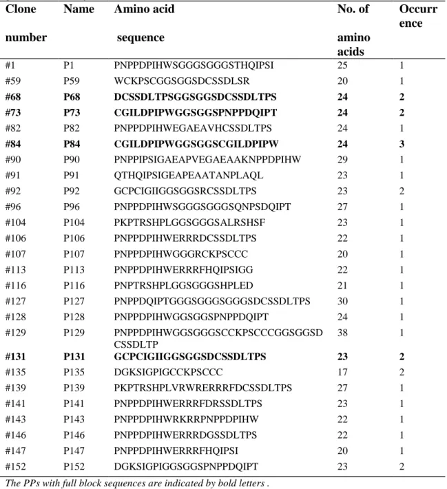
2.4.1 In vitro selection of novel peptides from the PP library
The PP library was constructed by random shuffling of eight peptide blocks selected from the previous libraries (Table 2.1) using YLBS (Kitamura et al., 2009 and 2012; Komatsu et al., 2012) as shown in Figure 2.1. The PPs were then used to construct the mRNA ready to be used for in vitro transcription followed by puromycin-linker ligation, in vitro translation and in vitro reverse transcription process to make the mRNA (cDNA) display product as shown in Figure 2.2. Then, in vitro selection using the mRNA display (Figure 2.2) provided PPs containing peptide blocks linked via a linker. The PPs had different length and block-diversity (Table 2.2). The combination of PPs was completely random (Table 2.2 and Figure 2.3), and in addition some PPs had indel and frame shift mutations due to the relaxed nature of the restriction enzyme (MboI) used, which expanded the diversity of the mutant library. Furthermore, most of the PPs were linked via a short linker (6 amino acids in length). Although the theoretical size of the PP library was 128 variants, the actual diversity of the library was found to be much bigger. One of the reason is that the designed molecular diversity of the paired peptides is very small (in this case, 128 variants) due to the limited number of blocks, which is
30
far smaller than the number of molecules used for the selection ( usually about 1012) (Kitamura et al., 2012)
Table 2.2. Paired peptides (PPs) obtained from in vitro selection of the thirdly
generated library
Clone Name Amino acid No. of Occurr ence
number sequence amino
acids #1 P1 PNPPDPIHWSGGGSGGGSTHQIPSI 25 1 #59 P59 WCKPSCGGSGGSDCSSDLSR 20 1 #68 P68 DCSSDLTPSGGSGGSDCSSDLTPS 24 2 #73 P73 CGILDPIPWGGSGGSPNPPDQIPT 24 2 #82 P82 PNPPDPIHWEGAEAVHCSSDLTPS 24 1 #84 P84 CGILDPIPWGGSGGSCGILDPIPW 24 3 #90 P90 PNPPIPSIGAEAPVEGAEAAKNPPDPIHW 29 1 #91 P91 QTHQIPSIGEAPEAATANPLAQL 23 1 #92 P92 GCPCIGIIGGSGGSRCSSDLTPS 23 2 #96 P96 PNPPDPIHWSGGGSGGGSQNPSDQIPT 27 1 #104 P104 PKPTRSHPLGGSGGGSALRSHSF 23 1 #106 P106 PNPPDPIHWERRRDCSSDLTPS 22 1 #107 P107 PNPPDPIHWGGGRCKPSCCC 20 1 #113 P113 PNPPDPIHWERRRFHQIPSIGG 22 1 #116 P116 PNPTRSHPLGGSGGGSHPLED 21 1 #127 P127 PNPPDQIPTGGGSGGGSGGGSDCSSDLTPS 30 1 #128 P128 PNPPDPIHWGGSGGSPNPPDQIPT 24 1 #129 P129 PNPPDPIHWGGSGGGSCCKPSCCCGGSGGSD CSSDLTP 38 1 #131 P131 GCPCIGIIGGSGGSDCSSDLTPS 23 2 #135 P135 DGKSIGPIGCCKPSCCC 17 2 #139 P139 PKPTRSHPLVRWRERRRFDCSSDLTPS 27 1 #141 P141 PNPPDPIHWERRRFDRSSDLTPS 23 1 #143 P143 PNPPDPIHWRKRRPNPPDPIHW 22 1 #146 P146 PNPPDPIHWERRRDGSSDLTPS 22 1 #147 P147 PNPPDPIHWERRRFHQIPSI 20 1 #152 P152 DGKSIGPIGGSGGSPNPPDQIPT 23 2
The PPs with full block sequences are indicated by bold letters . Furthermore, these four PPs were selected for chemical synthesis.
31
It was also observed the deletion of long linker (12 amino acids in length), resulting 6 to 8 amino acids in length. Based on the full block sequence information of parent blocks used for YLBS and based on the frequency of occurrence, four PPs were selected for further characterization (Table 2.3).
Figure 2.3. Random combination of peptide blocks selected from the first and second library resulted PP library. Single straight arrow head shows that two peptides
(N -> C) are connected via a linker. Double curve arrow head shows the self-paired peptides via a linker. Note that dotted arrow head means the linker is partially deleted; black arrow head for short linker and red arrow head for long linker.
Table 2.3. Paired peptides selected and their properties
a The GRAVY value is calculated by dividing the sum of the hydropathy values for each residue by the length of the sequence (Kyle and Doolittle; 1982).
Name Systematic Amino acid Source peptide No. of Kd (M) code name sequence blocks representation amino acids
P68 (pp(Aβ42)7068) DCSSDLTPSGGSGGS DCSSDLTPS P5268-L1-P5268 24 No binding P73 (pp(Aβ42)7073) CGILDPIPWGGSGGS PNPPDQIPT P5105-L1-P5128 24 3.7×10⁻⁶ P84 (pp(Aβ42)7084) CGILDPIPWGGSGGSCGILDPIPW P5105-L1-P5105 24 2.0×10⁻⁸ P131 (pp(Aβ42)7131) GCPCIGIIGGSGGS DCSSDLTPS P5109-L1-P5268 23 1.2×10⁻⁸