原 著
糖尿病患者における下肢切断部位および再切断率の 23 年間にわたる経時的変化
東京女子医科大学糖尿病センター(糖尿病・代謝内科) オ ダ ユ リ イ ク ラ カ ズ キ ハ ナ イ コウ カ ト ウ ウチガタ ヤ ス コ 小田 友里・井倉 和紀・花井 豪・加藤 ゆか・内潟 安子 (受理 平成 29 年 9 月 7 日)Changes in Level of Lower Extremity Amputation and Re-amputation Rate in Diabetic Patients Yuri ODA, Kazuki IKURA, Ko HANAI,
Yuka KATO and Yasuko UCHIGATA
Diabetes Center, Tokyo Women s Medical University School of Medicine
Objective: To examine the time-dependent changes in the incidence of major amputation and re-amputation in diabetic patients who underwent lower extremity amputation (LEA).
Method: We recruited 188 consecutive patients who underwent LEA due to diabetic foot ulcers at the foot care unit in the Diabetes Center of Tokyo Women s Medical University Hospital between 1993 and 2016. Patients were classified into three groups according to the amputation date: group 1: 1993-2000 (n=37), group 2: 2001-2008 (n=64) and group 3: 2009-2016 (n=87). Major amputation was defined as an amputation above the ankle, and re-amputation was defined as LEA of the same limb within 6 months after the first LEA. The time-dependent changes in each group were compared by Jonckheere-Terpstra test.
Result: The ratio of the major amputation at the final LEA during each group were 81.1 % in group 1, 70.3 % in group 2 and 34.5 % in group 3, and it was significantly reduced over time (p for trend < 0.001).The ratio of the re-amputations during each group were 29.7 % in group 1, 26.6 % in group 2 and 33.3 % in group 3, and there were not any significant difference between 3 groups (p for trend = 0.510). As for the amputation parts of re-amputation patients, major re-amputations were performed 90.9 % in group 1, 82.4 % in group 2 and 41.4 % in group 3, and it was significantly reduced over time (p for trend = 0.001).
Conclusion: The incidence of major amputation in the diabetic patient who underwent LEA was reduced over time. Although there were not any significant change with the incidence of re-amputation, but the incidence of major amputation at the re-amputation parts was reduced.
Key Words: diabetes, diabetic foot ulcers, lower extremity amputation, re-amputation
緒 言 下肢切断となった糖尿病患者は,著しい quality of life(QOL)の低下のみならず,きわめて高い死亡 率を有することが知られている1)∼3) .下肢切断のなか でも特に足関節より上位での大切断は,足関節より 下位での小切断と比較して生命予後が不良である4) . したがって糖尿病患者において下肢切断,特に大切 断の回避は予後の改善に繋がる可能性がある重要な :井倉和紀 〒162―8666 東京都新宿区河田町 8 番 1 号 東京女子医科大学糖尿病センター Email: [email protected] doi: 10.24488/jtwmu.87.Extra2_E234
Copyright Ⓒ 2017 Society of Tokyo Women s Medical University
! # $ 東女医大誌 第 87 巻 臨時増刊 2 号 頁 E234∼E238 平成 29 年11月 " # %
Table 1 Comparison of clinical characteristics among each group Group 1 (n=37) Group 2 (n=64) Group 3 (n=87) p value Age (years) 61±9 64±13 63±12 0.536 Male (%) 86 80 76 0.420 Type 2 diabetes (%) 92 94 92 0.876 Duration of diabetes (years) 20±9 21±11 22±11 0.582 HbA1c (%) 7.7±2.0 7.5±1.9 7.7±2.1 0.838 Dialysis (%) 65 53 46 0.165 History of cardiovascular disease (%) 68 56 57 0.512 History of leg revascularization (%) 0 9 57 <0.001 Smoking (current or ever) 68 55 66 0.310 Wagner grade 4 or 5 (%) 89 91 90 1.000 Group 1: 1993-2000, Group 2: 2001-2008, Group 3: 2009-2016.
Data are mean±SD or percent of patients. HbA1c: hemoglobin A1c, SD: standard deviation.
課題である. 近年,糖尿病足潰瘍に対する集学的治療の進歩に より,多くの国や地域で下肢切断,特に大切断の発 生率が減少しているが,本邦からの報告はいまだ少 ないのが現状である5)∼11) .一方,糖尿病患者における 下肢切断は高い手術リスクを伴うにもかかわらず, 切断後,治癒に至らず再切断となる例も少なくな い12) .これまで,再切断率の経時的変化に関して一定 の見解は得られていない13) . そこで本研究は,東京女子医科大学糖尿病セン ターで糖尿病足潰瘍により下肢切断を施行された患 者における下肢切断部位および再切断率の 23 年間 にわたる経時的変化を検討した. 対象および方法 1.研究デザイン 本研究は一施設のヒストリカルコホートを用いた 観察研究であり,東京女子医科大学倫理委員会で承 認され(承認番号 3905), ヘルシンキ宣言に基づき, 特に対象患者のプライバシーの保護などに最大限の 注意を払ったうえで行った. 2.対象 1993 年 1 月から 2016 年 3 月までの間に,東京女 子医科大学糖尿病センターフットケア外来に通院歴 があり,糖尿病足潰瘍から下肢切断に至った患者 204 名を抽出した.このうち,下肢切断時の採血デー タが不明であった 16 名を除外し,最終的に 188 名を 本研究の対象とした.対象者を切断日により 1 期: 1993 年∼2000 年(37 名),2 期:2001 年∼2008 年 (64 名),3 期:2009 年∼2016 年(87 名)に分類した. 3.アウトカム 足関節より上位(膝上切断または膝下切断)での 切断を大切断,足趾切断も含めた足関節より下位で の切断を小切断と定義し,各群の初回切断部位およ び最終切断部位を調査した.初回の切断後 6 か月以 内における同肢での切断を再切断と定義した. 4.統計解析 連続量の表記は平均値±標準偏差とした.HbA1c は,National Glycohemoglobin Standardization
Pro-gram:NGSP 値に換算して表記した14) .3 群間の連 続量の比較は一元配置分散分析,離散量の比較は Fisher の正確検定を用いて行った.3 群間の切断率 の比較は,ヨンクヒール・タプストラ検定および多 変量ロジスティック回帰分析による傾向検定を用い て解析した.オッズ比は多変量ロジスティック回帰 分析を用い算出した.なお,多変量ロジスティック 回帰分析では,年齢,性別,HbA1c,糖尿病罹病期 間,心血管病の既往,透析療法の有無,および Wag-ner 分類 4 度以上を共変量とした.p 値<0.05 を統 計学的に有意とした.以上の統計解析は,SAS Ver-sion 9.4 を用いて行われた. 結 果 1.各群における切断時の患者背景 切断日により分類した 3 群における各群の患者背 景を Table 1 に示す.対象患者 188 名の平均年齢は 63±12 歳,女性は 39 名(20.7 %)であった.年齢, 性別,糖尿病型,糖尿病罹病期間,HbA1c,透析療 法の有無,心血管病の既往,喫煙歴,Wagner 分類 4 度以上に関して,いずれも 3 群間に差を認めな かった. 2.全対象者の下肢切断部位と再切断率 全対象者における初回切断部位は,大切断が 74 名(39.4 %),小切断が 114 名(60.6 %)であった.
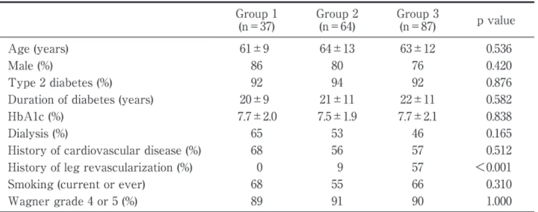
Fig. 1 Amputation level of each group
a: First amputation level, b: Final amputation level.
Major amputation was defined as an amputation above the ankle, and minor amputation was defined as an amputation under the ankle. The ratio of the major amputation during each group was significantly reduced over time both first and final (p for trend<0.001) .
81.1% 70.3% 34.5% 18.9% 29.7% 65.5%
Group 1 Group 2 Group 3 0% 20% 40% 60% 80% 100% 59.5% 51.6% 21.8% 40.5% 48.4% 78.2%
Group 1 Group 2 Group 3 0% 20% 40% 60% 80% 100% a b Rate (%) 䕕Minor 䕔Major (n=37) (n=64) (n=87) (n=37) (n=64) (n=87)
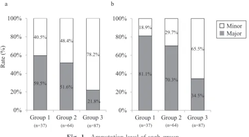
Fig. 2 Re-amputation rate of each group
Re-amputation was defined as LEA of the same limb within 6 months after the first LEA. The ratio of the re-amputations during each group were not any signif-icant difference between 3 groups (p for trend=0.510).
29.7% 26.6% 33.3%
Group 1 Group 2 Group 3 0% 20% 40% 60% 80% 100% Rate (%) (n=37) (n=64) (n=87) 全対象者における最終切断部位は,大切断が 105 名 (55.9 %),小切断が 83 名(44.1 %)であった.最終 的に大切断に至った 105 名のうち,膝上切断は 22 名,膝下切断は 83 名であった.最終的に小切断で治 癒に至った 83 名のうち,中足骨レベルでの切断は 30 名,足趾切断は 53 名であった. 再切断を施行したのは 57 名(30.3 %)であった. 最終的な再切断部位は大切断が 36 名,小切断が 21 名であった.小切断で再切断したが治癒に至らず最 終的に大切断となったのは 7 名であった. 3.大切断率の経時的変化 各群の初回切断部位における大切断率は,1 期: 59.5 %(22 名),2 期:51.6 %(33 名),3 期:21.8 % (19 名)で,有意に減少した(p for trend < 0.001). 各群の最終切断部位における大切断の割合は,1 期:81.1 %(30 名),2 期:70.3 %(45 名),3 期: 34.5 %(30 名)であり,経時的に有意な低下を認め た(p for trend < 0.001)(Fig. 1).多 変 量 ロ ジ ス ティック回帰分析において,1 期を対照とした最終 的に大切断に至るオッズ比は,2 期:0.65(95 %CI: 0.21 ― 1.95,p = 0.437),3 期:0.11(95 %CI:0.04 ― 0.31,p < 0.001)であり,経時的に有意な低下(p for trend < 0.001)を認めた. 4.再切断率の経時的変化 各群の再切断の割合は,1 期:29.7 %(11 名),2 期:26.6 %(17 名),3 期:33.3 %(29 名)であり,
3 群間に差を認めなかった(p for trend= 0.510)(Fig.
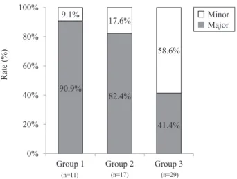
2).再切断患者の初回切断部位は,1 期 11 名中 2 名(18.2 %),2 期 17 名中 2 名(11.8 %),3 期 29 名 中 1 名(3.4 %)が大切断であった.多変量ロジス ティック回帰分析において,1 期を対照とした再切 断 に 至 る オ ッ ズ 比 は,2 期:0.92(95 %CI:0.37 ― 2.30,p = 0.853),3 期:1.31(95 %CI:0.55 ― 3.11,p = 0.539)であり,これも経時的な変化を認めなかっ た(p for trend = 0.539). 次に,再切断患者においてその最終切断部位を検 討したところ,1 期 11 名中 10 名(90.9 %),2 期 17
Fig. 3 Final re-amputation level of each group
The ratio of major amputations in final re-amputation was significantly reduced over time (p for trend= 0.001). 90.9% 82.4% 41.4% 9.1% 17.6% 58.6%
Group 1 Group 2 Group 3 0% 20% 40% 60% 80% 100% 䕕Minor 䕔Major Rate (%) (n=29) (n=17) (n=11) 名中 14 名(82.4 %),3 期 29 名中 12 名(41.4 %)が 最終的に大切断となり,経時的に有意な低下を認め た(p for trend = 0.001)(Fig. 3).多 変 量 ロ ジ ス ティック回帰分析において,1 期を対照とした最終 的に大切断に至るオッズ比は,2 期:0.66(95 %CI: 0.05 ― 8.75,p = 0.754),3 期:0.07(95 %CI:0.01 ― 0.74,p = 0.028)であり,経時的に有意な低下(p for trend = 0.028)を認めた. 考 察 東京女子医科大学糖尿病センター(以下当セン ター)における 1993 年から 2016 年までの 23 年間に わたる調査を行ったところ,糖尿病足潰瘍を原因と した下肢切断患者のうち,初回切断部位および最終 切断部位ともに,大切断に至る割合は経時的に有意 に減少していた.一方,再切断率は経時的変化を認 めなかったが,再切断部位の検討で大切断の割合が 減少していた. 1990 年代から 2010 年までに行われた欧米におけ る糖尿病患者を対象とした調査では,年代や地域に よって多少異なるが,多くの報告で糖尿病患者にお ける大切断の発生率の経時的な減少が報告されてい る5)∼11) .本研究は下肢切断糖尿病患者のみを対象と した調査ではあるが,足壊疽を示す Wagner 分類 4 度以上の割合を含む患者背景は,各年代で差を認め ず,さらに,患者背景で調整した多変量モデルでも, 経時的に最終切断部位における大切断患者の割合は 減少していた.このことから,既報同様,当センター においても糖尿病足潰瘍から大切断に至る割合は減 少している可能性が示唆される.本研究からは大切 断の割合が減少した原因を明らかにすることはでき ないが,近年の治療の進歩,特に末梢動脈疾患(pe-ripheral arterial disease;PAD)に対する血行再建 術の進歩が今回の結果に影響した可能性が推測され る.糖尿病患者に合併する PAD は,下 動脈以下に 多く15) , 血管内治療後の再狭窄率が問題であった16) . しかし,近年,技術と器具の進歩により膝下動脈領 域の重症虚血肢に対する血管内治療の有用性も拡大 してきている17) .当院では 2001 年以降より下肢血行 再建術を施行するようになり,2 期が 9.4 %(6 名), 3 期が 57.5 %(50 名)と徐々に症例数は増加し,現 在では膝下動脈領域の重症虚血肢でも早期から積極 的に下肢血行再建術を施行している.このことが今 回の結果につながった可能性が考えられる.また 3 期は 1 期,2 期と比較すると,初回切断部位が大切断 で あ る 割 合 が 少 な く,activity of daily living (ADL)・QOL 維持のため,救肢に対する意識が高 まり,可能な限り切断部位の縮小を目指した治療が 行われていることも大切断の割合が減少している原 因のひとつと考えられる. 本研究における再切断率は,経時的な変化を認め なかった.Izumi らが行った 1993 年から 1997 年に かけての米国からの報告では,下肢切断を施行した 糖尿病患者 277 名において,切断後 1 年での再切断 率は 26.7 %であった12) .おおよそ同年代である本研 究の 1 期における再切断率は 29.7 %であり,ほぼ同 等であった.Larsson らは,経時的に再切断の割合が 減少していることを報告している13) .本研究では,再 切断の経時的変化は認めなかったが,再切断が大切 断となる割合は減少していた.小切断と比較して, 大切断は ADL・QOL の低下のみならず,出血など 明らかに手術リスクが高くなる.さらに,下肢切断 となる糖尿病患者は心血管合併症を複数持つことが 多く18) ,その点において今回の結果の意義は小さく ないと考えられる. 最後に本研究の限界について述べる.まず,本研 究は一施設,特に大学病院に通院中の患者を対象と したため外的妥当性がない,すなわち,本研究対象 患者が必ずしも日本人における下肢切断糖尿病患者 を代表するコホートではない可能性がある.また, 対象患者が少なく,統計学的検出力が不十分であっ た可能性は否定できない.さらに,下肢切断部位の 決定や再切断に影響すると考えられる切断時におけ る下肢血流評価や炎症反応,患者の臨床像として脂 質,血圧,糖尿病網膜症,腎症,神経障害,飲酒歴
などが不明であった点が挙げられる. 結 語 東京女子医科大学糖尿病センターにおいて,糖尿 病足潰瘍から下肢切断に至った患者を 23 年間にわ たり調査したところ,大切断となる患者は経時的な 減少を認めた.一方,再切断を必要とする患者の割 合は変化を認めなかった.しかし,再切断が必要と なる患者においても,それが大切断である傾向は減 少していた.今後,下肢切断に至った患者のみなら ず,全糖尿病患者を対象とした大規模な多施設研究 において,本邦における下肢切断糖尿病患者の実態 を明らかにする必要がある. 開示すべき利益相反はない. 文 献
1)Faglia E, Favales F, Morabito A: New ulceration, new major amputation, and survival rates in dia-betic subjects hospitalized for foot ulceration from 1990 to 1993: a 6.5-year follow-up. Diabetes Care 24: 78―83, 2001
2)Moulik PK, Mtonga R, Gill GV: Amputation and mortality in new-onset diabetic foot ulcers strati-fied by etiology. Diabetes Care 26: 491―494, 2003 3)Boulton AJM, Vileikyte L, Ragnarson-Tennvall
G et al: The global burden of diabetic foot disease.
Lancet 366: 1719―1724, 2005
4)Hambleton IR, Jonnalagadda R, Davis CR et al: All-cause mortality after diabetes-related amputa-tion in Barbados: a prospective case-control study. Diabetes Care 32: 306―307, 2009
5)Gregg EW, Li Y, Wang J et al: Changes in diabetes-related complications in the United States, 1990-2010. N Engl J Med 370: 1514―1523, 2014 6)Moxey PW, Gogalniceanu P, Hinchliffe RJ et al:
Lower extremity amputations―a review of global variability in incidence. Diabet Med 28: 1144―1153, 2011
7)Rasmussen BS, Yderstraede KB, Carstensen B et
al: Substantial reduction in the number of
amputa-tions among patients with diabetes: a cohort study over 16 years. Diabetologia 59: 121―129, 2016 8)Jørgensen ME, Almdal TP, Faerch K: Reduced
in-cidence of lower-extremity amputations in a Danish diabetes population from 2000 to 2011. Diabet Med
31: 443―447, 2014
9)Vamos EP, Bottle A, Edmonds ME et al: Changes in the incidence of lower extremity amputations in individuals with and without diabetes in England between 2004 and 2008. Diabetes Care 33 : 2592 ― 2597, 2010
10)Holman N, Young RJ, Jeffcoate WJ: Variation in the recorded incidence of amputation of the lower limb in England. Diabetologia 55: 1919―1925, 2012 11)The Global Lower Extremity Amputation Study
Group: Epidemiology of lower extremity
amputa-tion in centres in Europe, North America and East Asia. The Global Lower Extremity Amputation Study Group. Br J Surg 87: 328―337, 2000
12)Izumi Y, Satterfield K, Lee S et al: Risk of ream-putation in diabetic patients stratified by limb and level of amputation: a 10-year observation. Diabetes Care 29: 566―570, 2006
13)Larsson J, Eneroth M, Apelqvist J et al: Sustained reduction in major amputations in diabetic patients: 628 amputations in 461 patients in a defined popula-tion over a 20-year period. Acta Orthop 79: 665―673, 2008
14)Kashiwagi A, Kasuga M, Araki E et al: Interna-tional clinical harmonization of glycated hemoglo-bin in Japan: From Japan Diabetes Society to Na-tional Glycohemoglobin Standardization Program values. J Diabetes Investig 3: 39―40, 2012
15)Jude EB, Oyibo SO, Chalmers N et al: Peripheral arterial disease in diabetic and nondiabetic patients: a comparison of severity and outcome. Diabetes Care 24: 1433―1437, 2001
16)Liistro F, Porto I, Angioli P et al: Drug-eluting balloon in peripheral intervention for below the knee angioplasty evaluation ( DEBATE-BTK ) : a randomized trial in diabetic patients with critical limb ischemia. Circulation 128: 615―621, 2013 17)Adam DJ, Beard JD, Cleveland T et al: Bypass
versus angioplasty in severe ischaemia of the leg (BASIL): multicentre, randomised controlled trial. Lancet 366: 1925―1934, 2005
18)Resnick HE, Carter EA, Lindsay R et al: Relation of lower-extremity amputation to all-cause and car-diovascular disease mortality in American Indians: the Strong Heart Study. Diabetes Care 27 : 1286 ― 1293, 2004