Acta Med. Nagasaki 38:265 - 269
Changes of Concentrations of Free Amino Acids in HeLa Cells Induced by Antitumor Agent
Yutaka Tagawa,1) Yoshikazu Mine, Toru Yasutake, Satoshi Matsuo and Masao Tomita
First Department of Surgery, Nagasaki University School of Medicine, 7-1 Sakamoto 1-chome, Nagasaki-city, Nagasaki 852, Japan 1) To whom requests for reprints should be addressed.
In this report we have investigated that the amino acid is most actively metabolized in the tumor cells damaged by antitumor agents, and discussed imbalanced amino acid procedure to be combined with cancer chemotherapy.
The concentrations of 24 free amino acids in medium and in cells were determined in HeLa cells treated with Adriamycin. The free amino acids that decreased in the medium but increased in cells were glutamine and arginine. From this result, we treated HeLa cells with Adriamycin in medium of glutamine-deficiency and obtained a marked cytocidal effect.
It would be difficult to make tumor tissues of glutamine-depletion in the whole body. For clinical application, local administration such as intraarterial infusion of L-glutamine antagonists combined with antitumor agents is expected as a practical administration method.
Key Words: Glutamine, Free amino acid, Adriamycin, Amino acid imbalance
not yet been clarified in tumor tissues or tumor cells treated with antitumor agents.
The author' has previously reported that the cultured cancer cells treated with antitumor agents were signifi- cantly enlarged in size, suggesting abnormal increase in
intracellular protein. The increase of intracellular protein has been observed for 42K, 45K and 270K with Carvocon, an antitumor agent, by Maehara et al.') These studies suggest the possibility that cancer chemotherapy might have induced some changes in the levels of amino acids participating in protein synthesis.
In this study, the authors have examined the changes of free amino acids in HeLa cells treated with Adriamycin (ADM) as a fundamental study for amino acid imbalance therapy to be combined with cancer chemotherapy.
Introduction
The recent progress in total parenteral nutrition has facili- tated the desired combination of amino acids to be admin- istered, enabling strict nutritional control. With this improvement, amino acid imbalance therapy for cancer is being reviewed.
The first clinical application of amino acid imbalance was reported by Allan et al ') in 1965 with phenylalanine and tyrosine imbalance. However, the amino acid imbal- ance state would be intolerable for human body if as potent effect of amino acid imbalance as that obtained in animal experiments is expected. Efforts are, therefore, being made to find a way out in its use as an adjunct to cancer chemo- therapy. An example of the efforts was methionine- and cystine-free amino acid solution reported by Goseki et al.") The combination of this solution and chemotherapy was effective in the patients of terminal digestive cancer with- out developing mortal side effects. Clinical studies of amino acid imbalance therapy are steadily progressing with the aim to potentate antitumor agents. These studies have mainly focussed on amino acid imbalance so far. On the other hand, changes of concentrations of amino acids have
Materials and Methods
HeLa cells in logarithmic growth phase were used for this experiment. The HeLa cells were grown in monolayer culture in CO, incubator in RPMI1640 (GIBCO, LTI) medium supplemented with 10% fetal calf serum (FCS). 2 X 10⁶/10ml of HeLa cells were plated in a 100mm dish and allowed to stand in an incubator for 20hr before use.
The antitumor agent used in this study was ADM (KYO- WA HAKKO Co., Ltd), which was added to the RPMI1640 medium in 100mm dish to make the final concentration of 0.5,ug/ml. After making the antitumor agent contact with the culture for 2hr, it was washed twice with PBS, and then the culture was replaced by fresh RPMI1640 medium supplemented with 10% FCS.
After the exchange of medium, changes with time of the concentrations of free amino acids in medium up to 72hr and the concentrations of free amino acids in cells at 72hr were determined. Furthermore, treatment with Colcemide at the final concentration of 0.I,ug/ml was performed for 24hr to analyze free amino acids in cells of M phase. The floating cells were harvested to use for the sample in determination of free amino acids in M phase cells.
Growth curves of HeLa cells treated with ADM were
compared in RPM11640 medium and RPM11640 medium of glutamine-deficiency (NIKKEN Biomedical Laboratory Co., Ltd).
The medium on HeLa cells treated and not treated with ADM were deproteinized with 2.5% sulfosalicylic acid at 0, 6, 12, 24 and 72hr of culture, and the supernatant after centrifugation at 1000rpm was used as the sample for analysis of free amino acids in medium. For the determi- nation of free amino acids in cells, HeLa cells treated and not treated with ADM were harvested with 0.12% Trypsin and 0.01% EDTA at 72hr of culture. Thus obtained single cells were centrifugally precipitated and washed with PBS.
The pellet was deproteinized with 2.5% sulfosalicylic acid and sonicated. It was further centrifuged for 15min at 3000rpm, and the supernatant was filtered to use for the analysis of free amino acid.
Twenty-four amino acids were determined in samples of 500,ag/ml each by ion exchange chromatography and ninhydrin reaction with a high performance amino acid analyzer (HITACHI 835, Type 50).
Results
1) Free Amino Acids in Medium
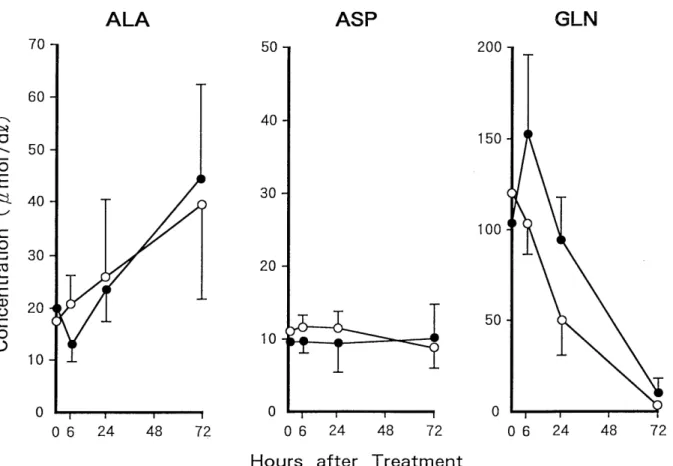
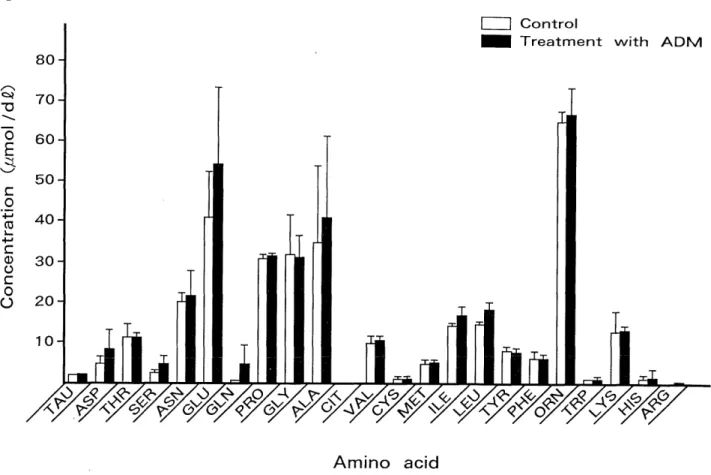
Concentrations of the 24 amino acids changed virtually in the same pattern in groups treated and not treated with ADM up to 72hr, though a slight difference was observed between the two groups at 6hr. Among the 24 amino acids, proline (PRO), alanine (ALA), ornithine (ORN) and glu- tamic acid (GLU) increased with time, and taurine (TAU), asparaginic asid (ASP) and glycine (GLY), showed no change in concentration, whereas threonine (THE), serine (SER), asparagine (ASN), glutamine (GLN), isoleucine (ILE), citrulline (CIT), cystine (CYS), methionine (MET), leucine (LEU), tyrosine (TYR), phenylalanine (PHE), tryp- tophan (TRP), lysine (LYS), histidine (HIS) and arginine (ARG) decreased. Figure 1 shows changes with time for 72hr of concentrations of ALA, ASP and GLN as respec- tive examples. Any of the amino acids showed higher concentration at 72hr in the sample treated with ADM than that not treated (Fig. 2)
2) Free Amino Acids in Cells
The concentration of each amino acid at 72hr was com- pared between samples treated with ADM and not treated.
Fig. 1. Changes of concentrations of free amino acids in medium. 0, control. ®, treatment with ADM. (n = 3, Bars indicate mean + SD)
Fig. 2. Concentrations of free amino acids in medium at 72 hour. (n = 3, Bars indicate mean ± SD)
All the 22 amino acids other than CYS and ARG showed higher concentration in the sample treated with ADM.
Furthermore, SER, ASN, GLN, TRP and ALA, among others, showed twice or more increase by the treatment with ADM as compared with the non-treated control (Fig.
3).
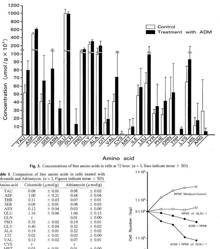
Concentrations of amino acids at 72hr in the cells treated with ADM were compared with those in M phase cells treated with Colcemide to evaluate the effect of cell cycle.
GLN, ALA and ARG showed higher concentrations in ADM-treated cells than Colcemide-treated ones (Table 1).
3) Growth Curve of HeLa Cells
The amino acids that decreased in the culture medium of HeLa cells treated with ADM and more increased in the cells treated with ADM than in M phase cells were GLN and ARG. Therefore, the growth curve of HeLa cells in RPMI1640 medium of GLN-deficiency was investigated. A marked cytocidal effect was obtained at 72 to 96hr by adding ADM to medium of GLN-deficiency. The effect of this treatment was greater than the inhibitory effects of RPMI1640 medium of GLN-deficiency alone and RPMI1640 medium containing ADM on the growth of HeLa cells (Fig. 4).
Fig. 3. Concentrations of free amino acids in cells at 72 hour. (n = 3, Bars indicate mean ± SD) Table 1. Comparison of free amino acids in cells treated with
Colcemide and Adriamycin. (n = 3, Figures indicate mean ± SD) Amino acid Colcemide (,umol/g) Adriamycin (,amol/g)
TAU 0.08 ± 0.01 0.08 ± 0.02
ASP 1.00 ± 0.21 0.68 ± 0.04
THR 0.11 ± 0.03 0.07 ±0.01
SER 0.08 ± 0.01 0.08 ± 0.03
ASN 0.12 ± 0.04 0.03 ± 0.00
GLU 1.16 ± 0.06 1.06 ± 0.15
GLN t 0.01 ± 0.00
PRO 0.35 ± 0.02 0.19 ± 0.01
GLY 0.40 ± 0.04 0.32 ± 0.02
ALA 0.19 ± 0.01 0.22 ± 0.02
CIT 0.02 ± 0.01 0.02 ± 0.00
VAL 0.12 ± 0.02 0.07 ± 0.01
CYS t t
MET 0.03 ± 0.01 0.01 ±0.00
ILE 0.17 ± 0.03 0.06 ± 0.01
LEU 0.19 ± 0.02 0.10 ± 0.06
TYR 0.08 ± 0.01 0.04 ± 0.00
PHE 0.07 ± 0.02 0.04 +-0.00
ORN 0.25 ± 0.07 0.05 ± 0.01
TRP 0.02 ± 0.00 0.01 ± 0.00
LYS 0.10 ± 0.01 0.01 ±0.00
HIS 0.07 ± 0.01 0.02 ± 0.01
ARG t 0.01 ± 0.00
t: trace Fig. 4. Growth curve of HeLa cells in each condition.
Discussion
In the present study, depletion of which amino acid in imbalanced amino acid solution should most effectively damage tumor cells when combined with chemotherapy or when used after chemotherapy was investigated in vitro.
Thirteen amino acids out of 24 decreased in the culture medium of HeLa cells and increased in cells by the treat- ment with ADM. There are two possibilities for the 13 amino acids in the situation which cell proliferation stopped by the treatment with ADM: One possibility is that they furiously participate in some intracellular metabolism and the other is that the half life of disintegration of these amino acids is extended.
Attention should also be given to the fact that ADM has an effect of cell accumulation in G2M phase on cell cycle.
This raises concern that the present study might have evaluated only concentrations of free amino acids in G2M cells. Concentrations of free amino acids in ADM-treated M phase cells were compared with those treated with Colcemide to exclude the possibility. In this comparison, amino acids that showed higher concentrations by the treatment with ADM than with Colcemide were GLN, ALA and ARG. GLN and ARG, above all, were found to have decreased in medium by the treatment with ADM. On the basis of the finding, a marked cytocidal effect was obtained in the medium deficient in GLN, which is a non-essential amino acid, combined with ADM. This suggests an import- nat role of GLN in the intracellular metabolism of HeLa cells damaged by ADM.
Report on the relation between GLN metabolism and growth kinetics of tumor cells''' indicate that a large part of energy production of HeLa cells depends on GLN, and GLN in HeLa cells, in the presence of fructose, supplies about 95% of the whole energy. HeLa cell, as suggested by these reports, may be an exceptional cell. Woolly et al 6) report that single-amino acid depletion affected cell growth kinetics potently in the order of GLN, TRP and ASN.
Dranoff et al $' report on their experiment with the cell lines obtained from anaplastic glioma and medulloblastoma that anaplastic glioma failed to proliferate without the supply of GLN, but medulloblastoma well proliferated. As a result, they point out that the dependency on GLN differs between cell lines. Matsuno et al 9' and many investi- gators,"' 10-13) on the other hand, have concluded in their reports about the GLN metabolism in tumor cells that GLN may be an essential amino acid for highly GLN-requiring tumor cells based on their observations: 1) Many tumor cells tend to need much GLN for growth and 2) GLN cannot be synthesized sufficiently in tumor cells because they are with poorly active GLN synthetase.
The amino acid imbalance therapy is clinically applied in some hospitals as an adjunct to cancer chemotherapy. It might, however, perform metabolism of GLN on the liver or other important organs and furthermore GLN would be
extensively distributed in the body,` `') making its clinical application very difficult. This might be a reason why GLN-depleted amino acid imbalance therapy is not so common. On the other hand, Wiss et al 16) have reported that responses were obtained in 10 out of 22 patients of colon cancer who received a regimen with Azotomycin, an L-glutamine antagonist, combined with 5-fluorouracil. L- glutamine antagonists, as suggested by this report, can inhibit glutamine metabolism and are well-available for clinical application. It would be more practical if amino acids' concentrations could be locally controlled. One expectable administration method for that purpose would be to use an intraarterial catheter or other devices to administer antitumor agents and L-glutamine antagonists selectively to the tumor-controlling artery.
References
1) Allan, J. D., Ireland, J. T., Milner, J. and Moss, A. D.: Treatment of leukemia by amino acid imbalance. Lancet 1:302-303, 1965.
2) Goseki, N., Onodera, S. and Menjo, M.: Clinical study of amino acid imbalance as an adjunct to cancer therapy. J. Jpn. Soc. Cancer Ther.
17:1908-1916, 1982.
3) Goseki, N., Mori, S., Habu, H. and Menjo, M.: Effect of intravenous methionine-free hyperalimentation combined with anticancer drugs
(RT-Therapy) on adenocarcinoma of gastrointestinal tract. JJPEN
2:265-271, 1980.
4) Tagawa, Y.: Relationship between G2 accumulation and RNA content of PC1 cells in unbalanced growth induced by antitumor agents. J.
Jpn. Soc. Cancer. Ther. 22:590-602, 1987.
5) Maehara, H., Ania, H., Kusumoto, H. and Sugimachi, K.: Effect of antitumor drug on protein synthesis chinese hamster V79 cells. Pro- ceedings of the 45th Annual Meeting of the Japanese Cancer Associ-
ation, Sapporo, Hokkaido (Abstr.) 1110, 307, 1986.
6) Woolly, P. V., Colt, R. and Magno, M.: Possible L-glutamine antago- nists in the treatment of gastrointestinal cancer. Cancer Treatment
Reports 63:1039-1040, 1979.
7) Reitzer, L. J., Wice, B. M. and Kennell, D.: Evidence that glutamine, no sugar, is the major energy source for cultured HeLa cells. J. Biol.
Chemo. 254:2669-2679, 1979.
8) Dranoff, G., Elion, G. B., Friedman, H. S., Campbell, G. L. and Bigner, D. D.: Influence of glutamine on growth human glioma and
medulloblastoma in cultures. Cancer Res. 45:4077-4081, 1985.
9) Matsuno, T. and Satoh, T.: Glutamine metabolism in the avian host bearing transplantable hepatomatous growth induced by MC-29 virus.
Inst. J. Biochem. 18:187-189, 1986.
10) Sebot, J. S. and Weber, G.: Negative correlation of L-glutamine
concentration with proliferation rate in rat hepatoma. Lite Sci.
34:341-306, 1984.
11) Pine, M. J.: Depletions in glutamine, asparagine, isoleucine and ornithine pools of rodent and human tumor. Proceedings of the 74th
Annual Meeting of the American Association for Cancer Research,
San Diego, CA (Abstr.) 24, 47, 1983.
12) Pilkington, G. J. and Lantos, P. L.: The role of glutamine synthetase in the diagnosis of cerebral tumors. Neuropathol. Appl. Neurobiol.
8:227-236, 1982.
13) Abou-Khalil, W. H., Yunis, A. A. and Abou-Khalil, S.: Prominent glutamine oxidation activity in mitochondria of hematopoietic tumors.
Cancer Res. 43:1990-1993, 1983.
14) Yoshimura, K. and Mochizuki, H.: Infusion of amino acids supplem- ented with glutamine a key amino acids gut mucosal metabolism in
stressed cases. Igalu no Ayumi 149:385-392, 1989.
15) Ishikawa, E.: Dynamic aspects of free amino acids in the blood. Eiyo to shokuryou 30:241-248, 1977.
16) Wiss, A. J. and Mastranglo, M. J.: Phase I study of a combination of azotomycine (NCC-19893) in malignant disease. Cancer Chemother.
Res. 54:109-110, 1970.