Acta Med. Nagasaki 43 : 92-96
- Case Report -
Adenocarcinoma of the Colon Presenting as Intussusception in Adult:
Report of Two Cases
Hirofumi KAJIYAMA1), Kunihiko MURASE2), Akirou ITOH1), Shinichiro KANZAKI1), Kazufumi YAMASAKI1), Koichi TAURA1), Hajime ISOMOTO2), Fuminao TAKESHIMA2), Yohei MIZUTA2), Shigeru KOHNO2)
1) Department of Internal Medicine, Nagasaki Municipal Medical Center
2) Second Department of Internal Medicine, Nagasaki University School of Medicine
We present two adult cases of intussusception caused by adenocarcinoma of the colon. These cases represent typical cases of adult intussusception, a rare disease characterized by insidious onset, vague abdominal symptoms, and elusive diagnosis. On physical examination, the masses were palpa- ble in the right hypochondrium. Colonic intussusception were diagnosed by characteristic finding on ultrasonography (US) and computer tomography (CT). Endoscopic examina- tion showed pedunculated tumors with central ulceration, in the cecum, which caused colonic intussusception, and endoscopic biopsies showed adenocarcinoma of the colon. US and CT are useful non-invasive diagnostic tools that allow early detection of intussusception. Colonoscopy is recom- mended for differential diagnosis and possibly a direct re- duction of intussusception.
Key words: intussusception, adenocarcinoma, colon
Introduction
Intussusception is a condition characterized by invagination of one part of the intestine into the adja- cent proximal segment. The blood supply of the inner layers of the intussusception could be potentially im- paired, resulting in severe ischemia and gangrene.
Several forms of intussusception may be encountered but the most common are the ileo-cecal and ileo-colic.
Address Correspondence : Kunihiko Murase, M.D.
Second Department of Internal Medicine, 7-1 Sakamoto 1-Chome, Nagasaki 852-8501, Japan
In adults, the presenting symptoms of intussusception are those of partial intestinal obstruction. Among the
causes of mechanical bowel obstruction, adhesions are the most common, followed by strangulated hernia and volvulus (1-3). Intussusception is very rare in adults and is rather a disease of infants and children;
representing the presenting symptoms in 3-16% of all the above diseases (4-6). Both benign and malignant neoplasms of the intestine might also present as intussusception (7-9).
The diagnosis of intussusception in adults is difficult due to the variability of clinical presentation and pre- operative diagnosis of intussusception is infrequent in adult population. The preoperative diagnosis in the majority of patients is bowel obstruction (10, 11). In patients with a vague clinical picture, ultrasonography (US) and computed tomography (CT) have more re- cently been used to facilitate diagnosis of intussusception (2, 12-17). Several reports have also described the use of endoscopy in the diagnosis and management of colonic intussusception (18, 19).
We describe the diagnosis of intussusception of the cecum and ascending colon with malignant growths.
The diagnosis was established in both cases using US, CT and colonoscopy.
CASE REPORT
Case 1
A 87-year-old male presented in May 1996 with one month history of a progressive worsening of lower ab- dominal dull pain and abdominal fullness. The patient
was admitted to our hospital for further evaluation.
On physical examination, the abdomen was slightly distended with no tenderness or hyperactive bowel
sound. A funicular mass was palpable in the right hypochondrium. Laboratory data demonstrated mild anemia (hemoglobin 11.5 g/dl).
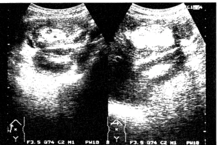
Ultrasonography demonstrated mixed hypoechoic- echogenic structure with a hyperechoic center, so
called target-like appearance, compatible with the diag- nosis of intussusception (Fig. 1). On abdominal CT, in- testinal intussusception was suspected as it produced a layer pattern secondary to mesenteric fat, intestinal fluid, and bowel wall (Fig. 2). Colonoscopy identified an
Fig 1. Case 1. Ultrasonography demonstrating the layering of intestinal intussusception.
Fig 2. Case 1. Abdominal CT demonstrating a complex colonic mass composed of a fat layer, intestinal fluid, and bowel wall.
Fig 3. Case 1. Colonoscopy showing the ulcerated tumor with regular surface in the cecum.
Fig 4. Case 1. Resected specimens showing ulcerated mass, 40 X 35 mm in the cecum.
Fig 5. Case 1. Histopathologic examination of the biopsy specimen showing a poorly differentiated adenocarcinoma
(magnification X 100).
ulcerated tumor with regular surface in cecum (Fig. 3), and biopsy established the diagnosis of adenocarcinoma.
The patient underwent right hemicolectomy. Gross ex- amination of the specimens showed 40 X 35 mm ulcer- ated tumor in the cecum (Fig 4). Histopathological ex- amination of the biopsy material showed a poorly differentiated adenocarcinoma with serosal invasion (Fig. 5). The postoperative course was satisfactory, and the patient is in good health two years after the operation without any evidence of tumor recurrence.
Case 2
A 61-year-old female presented with abdominal full- ness and a history of liver cirrhosis. On physical ex- amination, the abdomen was distended by ascites. A chicken-egg size mass was palpable in the right hypochondrium. Laboratory data demonstrated hypopro- teinemia (total protein 6.0 g/dl). Abdominal CT showed features indicative of intestinal intussusception. Barium enema showed so called "beak-like" filling defect in the ascending colon (Fig. 6). Reposition was achieved with the
Fig 6. Case 2. Barium enema showing the beak-like filling defect in the ascending colon, suggestive of intussusception.
aid of a barium enema. This revealed a pedunculated mass in the cecum. Colonoscopic examination showed a pedunculated tumor with white border, and biopsy proved adenocarcinoma. The patient was treated with right hemi-colectomy. Gross examination of the speci- mens showed a 55X82 mm pedunculated mass in the cecum. Histopathological examination showed a poorly differentiated adenocar-cinoma with massive mucin production. The postoperative course was uneventful.
Discussion
Adult intussusception is an unusual cause of bowel obstruction (1-3). The presenting symptoms are non- specific, often chronic, and the most important charac- teristic of pain secondary to intussusception is its peri- odic, intermittent nature (3, 11, 12). Although intussusception presents acutely in children, adults may present with acute, intermittent, or chronic re- ported problems (20). The predominant symptoms are usually those of bowel obstruction and consequently, intussusception often is misdiagnosed initially in adult patients. On physical examination, the presence of ab- dominal masses on palpation have been reported in 24% to 42% of patients (11, 12). In both of our patients, a mass was detected in the right hypochondrium.
Identification of a mass, or one that is only palpable when symptoms are present, is particularly suspicious of intussusception in adult patients.
While intussusception is relatively common in chil- dren, hydrostatic reduction of intussusception is the definitive therapy in most infants with this abnormal- ity, but it is rarely effective in adults. Adult intussusception is caused by tumor, post-operative ad- hesion, or Meckel diverticulum (3).
Felix et al. (20) reviewed 1,214 reported cases of intussusception in adults and estimated that 63% of all intussusception. Furthermore, Sanders and Hagan (7) reviewed more than 350 cases of colonic intussusception, and 68% were noted to be due to ma- lignant tumors. Adenocarcinoma accounts for 62% of such lesions (5). Furthermore, Te Strake (6) reported that the most common cause of intussusception in adults was malignant lymphoma, followed by cecal adenocarcinoma. More recent reviews, although of smaller number of patients, showed a remarkably simi- lar frequency, with 43% to 66% of cases being secon- dary to malignancy (8, 9).
A number of different radiological methods have been described as useful for the diagnosis of intussusception; CT, barium studies, US, plain x-ray film, angiography, and radionucleotide studies. Modern
non-invasive imaging techniques, may offer significant help in the precise identification of these lesions.
Intussusception was accurately identified in both of our cases by US and CT based on characteristic target appearance. Ultrasonography has proved to be particu- larly useful in the diagnosis of childhood intussusception
(2, 7, 11). There is less documented experience about the use of US in the diagnosis of adult intussusception (15, 17). Lim et al. (2) reported that US is useful for examination of patients with colonic obstruction to de- termine the level and cause of the obstruction. The classic sonographic features have been previously de- scribed, and include the target and doughnuts sign on transverse view, layering in longitudinal view, and the pseudo-kidney sign in the oblique and longitudinal views (2, 7, 15, 17).
Computed tomographic changes indicative of intussusception include the early target mass with re- tention of the facial planes around the mass, and pro- gressive bowel wall thickening produces the character- istic feature of a mass with the layering effect (5, 13,
16). Traction on the associated mesenteric vasculature may be noted. Scattered air-fluid levels indicate the as- sociated presence of bowel obstruction. As edema of the bowel progresses, the layering effect is obscured, and the compromised bowel assumes an amorphous shape surrounded by intraperitoneal fluid. The pres- ence of intramural air is indicative of vascular compro- mise of the intussuscepting bowel (13, 16, 21, 22). It was difficult to distinguish the tumors from the ede- matous intestinal walls at the proceeding portion in two cases (16).
Barium studies offer both diagnostic and therapeutic choice in children with suspected intussusception. The characteristic appearance of intussuscepting bowel on a barium enema study is a beak-like or cup-shaped fill- ing defect that is often accompanied by an additional filling defect representing the leading tumor (2, 3, 11).
Colonoscopy is also a useful tool for the evaluation of intussusception (18, 19). Azar and Berger (23) stressed that, when colonic intussusception is detected in the adult, attempts of nonoperative reduction should not be performed, because surgical intervention is necessary in all cases of intussusception. On the other hand, Kitamura et al. (24) reported a successful reduction of colic intussusception by air insufflation via the colonoscope. In this report, two patients of colonic adenocarcinoma presenting with intussusception were diagnosed by colonoscopic biopsies. If we find the cause of colonic intussusception by colonoscopy, we can choice most suitable operation for these cases.
We believe that biopsy of such masses should be rec- ommended for differential diagnosis and therapy.
In conclusion, we have described rare adult cases of intussusception caused by colonic adenocarcinomas.
CT and US are the most useful non-invasive diagnos- tic methods, and should be used in the early stages of intussusception, together with colonoscopy.
Acknowledgment
We gratefully acknowledge the skillful technical as- sistant of Mr Masahiro Harada.
References
1) Laws HL, Aldrete JS. Small-bowel obstruction: a review of 465 cases. South Med J 69: 733-4, 1976
2) Lim JH, Ko YT, Lee DH, Lee HW, Lim JW. Determining the site and causes of colonic obstruction with sonography. AJR 163: 1113-
7, 1994
3) Gordon RS. O'Dell KB. Namon AJ. Becker LB. Intussusception in the adult--a rare disease. J Emerg Med 9: 337-42, 1991
4) Fuzun M. Kaymak E. Harmancioglu O. Astarcioglu K. Principal causes of mechanical bowel obstruction in surgically treated adults
in western Turkey. Br J Surg 78: 202-3, 1991
5) Agha FP. Intussusception in adults. AJR 146:527-31, 1986 6) Te Strake L. Intussusception in adults. Diagnostic Imaging 49: 15-
22,1980
7) Sanders GB, Hagan WH. Adult intussusception and carcinoma of the colon. Ann Surg 146:796-804, 1958
8) Netherland D. Whalen TV. Moore G. Adenocarcinoma of the colon occurring with intussusception in an adolescent. Southern Med J
83: 1362, 1990
9) Begos DG, Sandor A, Modlin IM. The diagnosis and management of adult intussusception. Am J Surg 173: 88-94, 1997
10) Reijne HA, Joosten HJ, de Boer HH, Diagnosis and treatment of adult intussusception. Am J Surg 158: 25-8, 1989
11) Dharia KM. Marino AW Jr. Mancini HW. Enterocolic intussusception
in adults.. Dis Colon & Rectum 15: 194-200, 1972
12) Lee H, Yeh H, Leu Y,. Intussusception: The sonographic diagnosis and its clinical value. J Paediatr Gastroenterol Nutr 8: 343-7, 1989 13) Miyama H, Fujita M, Ishihara K, Ichijima S, Katsumori T, Nakai M,
Murakami K, Fukuda M. Three adult intussusception cases with colorectal cancer; CT diagnosis and its characteristic findings. Jpn
J Clin Radiol 35: 509-12, 1990
14) Bar-Ziv J, Solomon A. Computed tomography in adult intussusception.
Gastrointest Radiol 16: 264-6, 1991
15) Kojima Y, Tsuchiyama T, Niimoto S, Nakagawara G. Adult intussusception caused by cecal cancer and diagnosed preopera-
tively by ultrasonography. J Clin Ultrasound 20: 360-3, 1992 16) Fujimoto T, Fukuda T, Matsunaga N, Kohzaki S, Hirao K,
Nagayoshi K, Takeda H, Isomoto I, Uetani M, Hayashi K. CT find- ings of invaginated mesentery in adult intussusception. Nippon
Acta Radiol 53: 621-7, 1993
17) Boyle MJ, Arkell LJ, Williams JT. Ultrasonic diagnosis of adult intussusception. Am J Gastroenterol 88: 617-8, 1993
18) Hurwitz LM, Gertler SL. Colonoscopic diagnosis of ileocolic intussusception. Gastrointest Endsc 32: 217-8, 1986
19) Kimura H. Yabushita K. Tsuneda A. Tazawa K. Inoue T. Earashi M. Maeda K. Kuroda Y. Konishi K. Tsuji M. Miwa A. Adult colo-
rectal invagination associated with colonic cancer and established
by fiberscopy of the sigmoid colon. Endoscopy 28: 636, 1996 20) Felix EL, Cohen MH, Bernstein AD, Schwartz JH. Adult
intussusception: case report of recurrent intussusception and re-
view of the literature. Am J Surg 131: 758-61, 1976
21) Merine DS, Fishman EK, Jones B, Siegelmann SS. Enteric intussusception; CT findings in nine patients. Am J Reontogenol
148: 1129-32, 1987
22) Iko BO, Teal JS, Siram SM, Chinwuba CE, Roux VJ, Scott VF.
Computed tomography of adult colonic intussusception; clinical ex-
perimental studies. Am J Reontogenol 143:769-772, 1984
23) Azar T, Berger DL. Adult intussusception. Ann Surg 226; 134-8, 1997 24) Kitamura K, Kitagawa S, Mori M, Haraguchi Y. Endoscopic correc-
tion of intussusception and removal of a colonic lipoma.
Gastrointest Endosc 36: 509-11, 1990