Acta Med. Nagasaki 41: 31-37
Abnormal Liver Function in Patients with Sjogren's Syndrome
Masako KITA, Katsumi EGUCHI, Yojiro KAWABE, Masahiko TsuBOI, Atsushi KAWAKAMI, Hideki NAKAMURA, Kiyoshi MIGITA and Shigenobu NAGATAKI
First Department of Internal Medicine, Nagasaki University School of Medicine, 1-7-1 Sakamoto, Nagasaki 852, Japan
We measured the liver function tests of 145 patients with Sjogren's syndrome (SjS) (75 patients with primary SjS, 70 patients with secondary SjS), and characterized the SjS patients with abnormal liver function tests from several points of view : 1, the incidence of them in the primary SjS comparing with that in secondary SjS. 2, the staining pattern of anti-nuclear antibodies, and 3, the existence of anti- hepatitis C virus (HCV) antibody, hepatitis B surface (HBs) antigen, and antibody against human T-lymphotropic virus type I (HTLV-I). Abnormal liver function tests were detected in 38 out of 145 patients (26.2%) with SjS. Fifteen of the 38 patients (20.0%) had primary SjS while the remaining patients (32.9%) had secondary SjS. Histopathological examination identified primary biliary cirrhosis (PBC) in 2 patients, autoimmune hepatitis in 4 patients, and auto- immune cholangitis in a single patient with SjS. No signifi- cant difference in the presence of antinuclear antibody (ANA) was found between SjS patients with and without abnormal liver function tests. However, the incidence of discrete speckled pattern was significantly higher in SjS patients with abnormal liver function than in the patients with normal liver function. Two sera showing cytoplasmic pattern of ANA were also positive for anti-mitochondrial M2 antibody, allowing the diagnosis of PBC. All 11 sera exhibit- ing discrete speckled pattern contained significant amounts of anti-centromere antibody. Abnormal liver function tests were detected in 8 of 11 sera with these antibodies, 2 patients with PBC, 2 patients with autoimmune hepatitis, one patient with autoimmune cholangitis, one patient with chronic hepatitis B and 2 other patients with unconfirmed diagnosis.
The percentages of anti-HCV antibody-positive, HBs-Ag- positive and anti-HTLV-I antibody-positive in sera of pa- tients were higher than those of blood donors from the same geographical area. However, no significant difference was seen of these percentages in sera between the patients with and without abnormal liver function. Taken together, present study indicated that SjS patients with anti-centromere antibody may have some susceptibility for acquiring auto- immune liver disease.
Key words : Sjogren's syndrome, primary biliary cirrhosis, autoimmune hepatitis, autoimmune cholangi-
tis, anti-centromere antibody
Introduction
Sjogren's syndrome (SjS), which is considered an auto- immune disease, is characterized by chronic lymphocytic infiltration of salivary and lachrymal glands and the occasional presence of serum autoantibodies' dies"). SjS may exist as a primary disorder or as a secondary condition associated with a variety of diseases such as rheumatoid arthritis (RA), systemic lupus erythematosus (SLE), or progressive systemic sclerosis (PSS). In some SjS patients, involvement of the extraglandular organs may occur, including the skin, kidney, liver, lung, gastrointestinal tract and nervous system. Clinical or biochemical evidence of liver disease is found in 5 to 10% of patients with primary SjSa4'. The elevation of liver enzymes is usually mild and of little clinical significance. However, when higher levels are noted, a further investigation is war- ranted.
Although several etiological mechanisms have been proposed, the pathogenesis of SjS remains unknown. Viral infections, such as infections with Epstein-Barr virus'), human immunodeficiency virus'), human T lymphotropic virus type I (HTLV-I)'-9) or hepatitis virus C'°_13) have been suggested as possible etiologic factors. In the present study, we examined liver function tests, autoantibodies and anti-viral antibodies in the sera of SjS patients, and determined the relationship between liver function tests and these autoantibodies and/or virus infection.
Materials and Methods
Patients.
The test group included 75 patients with primary SjS [4 males and 71 females, age ; 51.5±13.0 years (mean±SD)]
and 70 patients with secondary SjS [8 males and 62 fe- males, age ; 53.3±12.1 years]. These patients attended the outpatient clinic of Nagasaki University School of Medicine. All patients fulfilled the criteria for diagnosis of SjS set out by the European Community"). Patients with secondary SjS composed of 33 patients with rheumatoid
arthritis (RA) diagnosed according to the criteria of the
American Rheumatism Association (ARA)15', 16 patients with systemic lupus erythematosus (SLE) diagnosed according to the criteria of ARA1), 11 patients with mixed connective tissue disease (MCTD) diagnosed according to the guidelines outlined by Bennet RM1", 5 patients with progressive systemic sclerosis (PSS) diagnosed according to the criteria of ARA18', 4 patients with CREST (calcino- sis, Raynaud's phenomenon, esophageal dysmotility, sclerodactylia, telangiectasia) syndrome, 2 patients with polymyositis (PM) diagnosed according to the classifica- tion criteria by Bohan A et al"), one patient with polyarteritis nodosa (PAN) diagnosed based on clinical features and histological examination and one patient with Behcet's disease diagnosed according to the criteria of International Study Group for Beh(;et's disease'). The control subjects for the percentage of positive antibodies and antigen for the viruses consisted of 71,748 normal subjects who donated blood at the Red Cross Blood Center in Nagasaki. Informed consent was obtained from all the patients examined, and the study was conducted in accor- dance with human experimentation guidelines of the authors' institution.
Clinical laboratory tests.
Tests for liver function were performed in all patients.
The following test kits were used in all assays ; AST (GOT HQ auto "Nissui", Nissui Pharmaceuticals, Tokyo, Japan), ALT (GPT HQ auto "Nissui", Nissui Pharm- aceuticals), y-GTP (Auto A "Mizuho" y-GTP, Mizuho Medy, Saga, Japan), and alkaline phosphatase (latrotech ALP rate, latron laboratory, Tokyo, Japan). Anti-nuclear antibodies (ANA) were detected with an indirect immuno- f luorescence procedure using HEp-2 cells (Fluoro Hep Ana test, Medical & Biological Laboratories (MBL), Nagoya, Japan). Antibodies to SS-A (Ro), SS-B (La) antigens and mitochondrial M2 were determined by the enzyme-linked immunosorbent assay (ELISA ; Mesacup SS-A/Ro test, Mesacup SS-B/La test, and Mitochondria M2 test, MBL).
Antibodies to HTLV-I were measured by ELISA (Eitest- ATL test, Eisai, Japan), the particle agglutination assay (Serodia-ATL kit, Fuji Rebio, Japan). The positive sera for antibodies to HTLV-1 were confirmed by western blot analysis using a commercial immunoblotting kit (Problot, HTLV-I, Fuji Rebio, Japan). Antibodies to Hepatitis C virus (HCV) were measured by second generation ELISA (Imucheck HCV Ab, International reagents, Kobe, Japan) and Hepatitis B surface (HBs) antigen and antibodies to HBs antigen were detected by radioimmunoassay (RIA ; AUSRIA 11-125 and Ausab, Dinabot, Tokyo, Japan).
Antibodies to mitochondria and smooth muscle were detected by immunofluorescence assay (IFA ; using rat kidney and stomach, FITC conjugated anti-human im- munogloblin, MBL) and liver kidney-microsomal antibody by IFA (using rat kidney, liver, stomach, heart, and
striated muscle, as well as mouse stomach, Specialty Laboratories, CA).
Groups of patients.
Patients were tentatively divided into two groups. The first group consisted of patients with abnormal liver function tests, defined as AST (GOT) >40 IU/l, ALT (GPT) >40 IU/l, and y -GTP > 50 IU/l or alkaline phos- phatase (Al-P) >270 IU/l, detected at least twice through-
out the course of the disease (mean duration ; 4.2:i-- 2.3 years). The second group consisted of patients with normal liver function tests. Abnormal liver function defined to be caused by drugs such as non-steroid anti- inflammatory drugs (NSAIDs), slow acting anti- rheumatic drugs, immunosuppressants including metho- trexate, or other drugs as well as to be caused by the active complicated diseases of SLE, MCTD, PM and PAN were excluded from the assessment in the present study. Seven of 15 patients whose liver function tests are AST>80 IU/1, ALT >80 IU/1, and y -GTP > 100 IU/l or ALP > 540 IU/1 were histologically confirmed by liver biopsy under laparoscopy.
Data analysis
Statistical analyses were performed using X 2 test bet- ween the groups. A p level of < 0.05 was considered statisti- cally significant.
Results
Prevalence of abnormal liver function test inpatients with primary and secondary Sjbgren's syndrome.
Abnormalities of liver function tests were found in 38 out of 145 (26.2%) patients with SjS. Of these, 15 had primary SjS while the remaining were patients with secondary SjS (Table 1). The incidence of abnormal liver enzymes was higher in secondary SjS than that in primary SjS, but the difference was not statistically significant (p
= 0.078). The percentages of abnormal AST, ALT, y-GTP and Al-P were similar in primary SjS patients (9.3-10.7%), and in secondary SjS patients (12.9-18.6%). SjS patients who had AST >80 IU/1, ALT >80 IU/l, y -GTP > 100 IU/1 orALP>540 IU/l were found in 15 out of 145 patients
(10.3%) (Table 2). In these 15 patients, incidence of patients with primary SjS was lower than the patients with secondary SjS, but the difference was not statistically significant (p = 0.13).
Table 1. Incidence of abnormal liver function tests in patients with primary and secondary Sjogren's syndrome Subject AST>40IU/1 ALT>40IU/1 y-GTP>50IU/1 Al-P>270IU/1 Total
primary SjS 7 8 8 8 15
(75 patients) (9.3%) (10.7%) (10.7%) (10.7%) (20.0%)
secondary SjS 12 9 11 13 23
(70 patients) (17.1%) (12.9%) (15.7%) (18.6%) (32.9%)
Total 19 17 19 21 38
(145 patients) (13.1%) (11.7%) (13.1%) (14.5%) (26.2%)
Table 2. Incidence of abnormal liver function tests (more than twice of upper limit of normal range) in patients with primary and secondary Sjogren's syndrome
Subject AST>80IU/1 ALT>80IU/1 y-GTP>100IU/1 ALP>540IU/l Total
primary SjS 3 1 3 4 1 5
(75 patients) (4.0%) (1.3%) (4.0%) (5.3%) (6.7%)
secondary SjS 4 2 7 4 10
(70 patients) (5.7%) (2.9%) (10.0%) (5.7%) (14.3%)
Total 7 3 10 8 15
(145 patients) (4.8%) (2.1%) (6.9%) (5.5%) (10.3%)
Table 3. Clinical characteristics of seven patients with Sjogren's syndrome with abnormal liver function confirmed by histological examination
Cases age sex Complication ANA pattern AMA ASMA based on histologica Diagnosis examinatio n
1 (T. K) 51 F - speckled - - autoimmune hepatitis (type 1) 2 (F. S) 65 F - speckled - - autoimmune hepatitis (type 1) 3 (M. K) 51 F - discrete speckled - - autoimmune hepatitis (type 1)
4 (K. H) 39 F SLE discrete speckled + - PBC
5 (S. Y) 51 F CREST (-) + - PBC
6 (Y. K) 60 F MCTD speckled - - autoimmune hepatitis (type 1)
7 (H. Y) 58 F CREST discrete speckled - - autoimmune cholangitis
ANA : anti-nuclear antibody, AMA : anti-mitochondrial antibody, ASMA : anti-smooth muscle antibody, PBC : primary biliary cirrhosis, CREST : calcinosis, Raynaud's phenomenon, esophageal dysmotility, sclerodactylia, telangiectasia, SLE : systemic lupus erythematosus, MCTD : mixed connective tissue disease.
Histopathological examination of the liver.
Seven of 15 patients who had AST>80 IU/1, ALT>80 IU/1, y-GTP > 100 IU/1 or ALP > 540 IU/1 gave their in- formed consent to be performed liver biopsy under laparoscopy. The patients consisted of 3 patients with primary SjS and 4 patients with secondary SjS. Six of the seven patients showed ANA positive in their sera. Neither HBs-antigen nor anti-HCV antibody was detected in these patients. As shown in Table 3, three primary SjS patients were diagnosed as having autoimmune hepatitis (type 1), none of the sera showed anti-smooth muscle antibody (AMSA) nor anti-mitochondrial antibody (AMA), and the staining pattern of the serum ANA was speckled in 2 patients and discrete speckled in one. Two out of the four
secondary SjS patients showed discrete speckled pattern of
the serum ANA, and two of them exhibited AMA in their sera were diagnosed as having PBC, and one patient was diagnosed as having autoimmune cholangitis, and one patient autoimmune hepatitis (type 1).
Relationship between ANA and liver function.
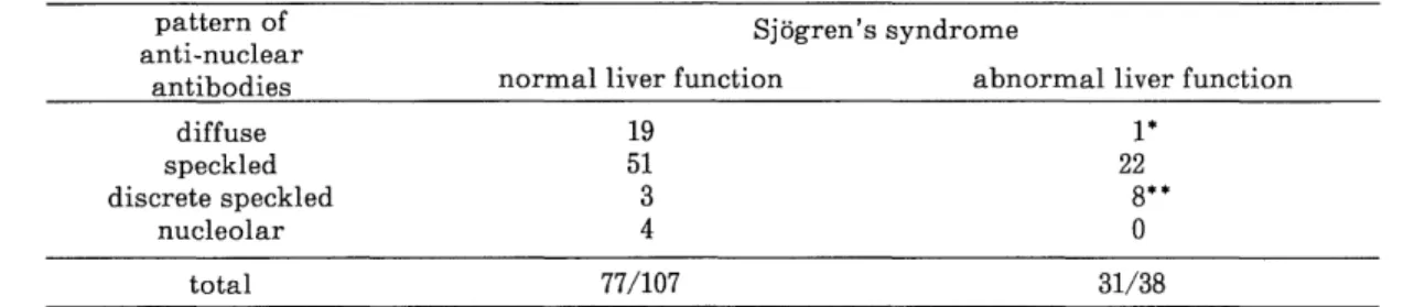
Thirty-one out of 38 sera (81.6%) from SjS patients with abnormal liver enzymes produced positive nuclear fluorescence and 2 sera (5.2%) positive cytoplasmic fluorescence (Table 4). Of the 31 ANA-positive sera with abnormal liver function tests, one (3.2%) showed a diffuse pattern, 22 (71.0%) showed a speckled pattern, and 8 (25.8%) showed a discrete speckled pattern, no patient showed a nucleolar pattern. Seventy-seven out of 107 sera (72.0%) from SjS patients with normal liver function
Table 4. Pattern of anti-nuclear antibodies and abnormal liver function in patients with Sjogren's syndrome
pattern of Sjogren's syndrome
anti-nuclear
antibodies normal liver function abnormal liver function
diffuse 19 1*
speckled 51 22
discrete speckled 3 8**
nucleolar 4 0
total 77/107 31/38
*p<0 .02 against the patients with normal liver function, **p<0.001 against the patients with normal liver function
Table 5. Relation between the presence of anti-centromere antibody and liver function tests found in patients with Sjogren's syndrome
cases complications speckled pattern titer of discrete (ELISA) ACA M2 -Ab AMA HBsAg anti-HCV Ab abnormal function tests liver diagnosis of the liver disease
1 N. T. - 320X 237 <10 - - - normal
2 H. N. - 2560X 203 <10 - - - normal
3 T. T. CREST 640X 273 <10 - - - normal
4 H. T. - 640x 222 <10 - - + not determined
5 U. R. - 640X 257 <10 - - + not determined
6 K. H. SLE 2560 x 267 99 - - + autoimmune hepatitis
7 M. K. - 160X 139 <10 - - + PBC
8 K. I. RA 160X 180 <10 + - + chronic hepatitis B
9 S. Y. CREST 2560x 284 200 - - + PBC
10 H. Y. CREST 2560X 237 <10 - - + autoimmune cholangitis
11 M. H. CREST 640X 69 <10 - - + autoimmune hepatitis
ACA : anti-nuclear antibody, AMA : anti-mitochondrial antibody
Table 6. Prevalence of antibodies to HCV and HTLV-1, and HBs-antigen in patients with Sjogren's syndrome
anti-HCV patients positive for anti-HTLV-1
positive patients HBs antigen positive patients
Age B. D.8 SjS B. D. SjS B. D SjS
positive/total (%) positive/total (%) positive/total (%) positive/total (%) positive/total (%) positive/total (% )
-19 6/3395 (0.18) 0/0 (-) 71/8031 (0.88) 0/0 (-) 8/2147 (0.37) 0/0 (- )
20-29 21/3685 (0.54) 0/2 (0) 25/2003 (1.25) 0/2 (0) 13/1588 (0.82) 1/4 (25.0)
30-39 22/1960 (1.12) 0/8 (0) 14/739 (1.89) 0/7 (0) 17/873 (1.94) 2/10 (20)
40-49 21/1642 (1.28) 0/8 (0) 7/728 (0.96) 1/10 (10.0) 29/944 (3.07) 5/14 (35.7)
* * *
50-64 44/1240 (3.55) 4/43 (9.3) 10/392 (2.55) 3/30 (10.0) 38/926 (4.10) 9/37 (24.3)
65- - 1/13 (7.7) - 0/12 (0) - 5/20 (25.0)
total 114/12102 (0.94) 5/74 (6.8) 127/11893 (1.07) 4/61 (6.6) 105/6478 (1.62) 22/85 (26.2) a B. D.: Blood Donors in Nagasaki city *P<0.001
tests were positive for ANA. Of these, 19 sera (24.7%) showed a diffuse pattern, 51 sera (66.2%) a speckled pattern, 3 sera (3.9%) a discrete speckled pattern , and 4 sera (5.2%) a nucleolar pattern. The prevalence of ANA in SjS patients with abnormal liver function tests was similar to that in patients with normal liver function tests (p = 0.24). However, discrete speckled pattern was seen more frequently in abnormal liver function tests group compared with those of normal liver function tests (p<0.001). In contrast, diffuse pattern was seen less commonly in SjS patients with abnormal liver function tests than those with normal liver function tests (p
<0.02).
All 11 sera exhibiting discrete speckled pattern contained significant amounts of anti-centromere antibody (ACA) measured by ELISA method. Table 5 shows the clinical features of SjS patients carrying these antibodies.
Abnormal liver function tests were detected in 8 of 11 sera (72.7%) with ACA. Three of the eight patients were primary SjS, and the five patients were secondary SjS complicated with SLE, RA and CREST syndrome. PBC was found in 2 patients, autoimmune hepatitis in 2 patients and autoimmune cholangitis in a patient. Case 8, who was positive for HBs-Ag, was diagnosed as chronic hepatitis B.
The etiology of abnormal liver function test in 2 other patients was not determined.
Prevalence of antibodies to HCV, HTLV-I and HBs- antigen.
The incidences of anti-HCV antibody-positive sera, HBs-Ag-positive sera and anti-HTLV-I antibody-positive sera in SjS patients were 6.8% (5 out of 74 patients), 6.6%
(4 out of 61 patients) and 26.2% (22 out of 85 patients), respectively (Table 6). The rates for the same antibodies
and the antigen in the control group were 0.9%, 1.1% and 1.6%, respectively. The incidence of positive anti-HCV antibody in the age of 50 to 64 in Sjogren's syndome was significantly higher than that of the blood donors (P<0.001). In the age of 40 to 49, and 50 to 64, the insidence of positive HBs antigen in Sj ogren's syndome was siginficantly higher (P<0.001). And in the age of 20 to 29, 30 to 39, 40 to 49, and 50 to 54, the incidence of positive HTLV-1 antibody was significantly higher than that of the blood donors (P<0.001, Table 6). To examine the relation- ship between liver function and 2 antibodies or antigen, we investigated their prevalence in SjS patients with abnor- mal liver function tests. The percentage of positive sera from these patients were 6.3% (2 out of 32 patients) for anti-HCV antibody, 7.1% (2 out of 28 patients) for HBs-antigen, and 27.3% (9 out of 33 patients) for anti- HTLV-I antibody (data not shown). There was no signifi- cant difference in the incidence of these antibodies and antigen between patients with and without abnormal liver function tests.
Discussion
Our results demonstrated the presence of abnormal liver function tests in 38 out of 145 patients with SjS (26.2 %).
The incidence of abnormal liver functions in our popula- tion samples was higher than that reported in previous studies-"').As far as we investigated the patients using abdominal ultrasound, none of the SjS patients who showed abnormal liver function tests were diagnosed as having fatty liver or alcoholic liver disease. However, it is possible that the abnormal liver function tests in some of the patients may be caused by fatty liver change.
Liver biopsy was performed in seven patients. Four patients in this group were identified to have autoimmune chronic hepatitis (type 1) according to the classification of autoimmune hepatitis"). Three other patients showed histological features consistent with PBC, including marked cellular infiltration of the portal areas and damage of the bile duct. Mild forms of interlobular inflammation and piecemeal necrosis were observed in these specimens. One of these patients (case 7 in Table 3) did not have serum anti-mitochondrial antibodies but have ACA, and responded well to treatment with prednisolone.
A subgroup of patients with autoimmune chronic active hepatitis has been recently identified as autoimmune cholangitis. This form of hepatitis is characterized by positive serum ANA but negative anti-mitochondrial antibodies, although serum biochemical values in these patients indicate the presence of cholestasis, and liver biopsy shows features of inflammatory bile duct da- mage'-'). These patients respond well to prednisolone22~
Also in common with PBC, patients with autoimmune cholangitis are at risk of acquiring other autoimmune disorders. It has also been reported that primary autoimmune cholangitis is complicated with polyarthral- gia, sicca syndrome, Raynaud's phenomenon and hy- pothyroidism",'). In the present study, one patient with autoimmune cholangitis was complicated with Sjogren's and CREST syndromes, and high titers of ACA were present in the serum.
The frequency of serum ANA in SjS patients detected in the present study using immunofluorescent techniques was comparable with that reported in previous studies"). The
observed pattern of ANA was diverse and included speck- led, diffuse, discrete speckled, nucleolar and cytoplasmic staining. Increased incidence of discrete speckled staining was found in the present study in the sera of SjS patients, particularly in sera of patients with abnormal liver
function tests. In addition, the presence of ACA was verified in sera with discrete speckled pattern, using the enzyme-linked immunosorbent assay (ELISA). Thus, all 11 sera contained significant amounts of these antibodies.
ACA has been identified in the sera of patients with the CREST variant of scleroderma2"). Patients with CREST positive for ACA are considered as a subset of patients
who do not usually progress to diffuse scleroderma or manifest major organ involvement'-'). Recent reports, however, indicate that patients with SLE, diffuse scleroderma, idiopathic Raynaud's phenomenon, drug- induced SLEn) and primary SjS" have positive titers for ACA. Our laboratory tests identified 2 SjS patients with ACA who had SLE or RA, respectively. A high incidence of Raynaud's phenomenon and limited features of CREST syndrome were identified, together with positive ACA, as described previously'-').
ACA detected by indirect immunofluorescence on HEp-2 cells are found in 10%') or 24%35) of patients with PBC. In the present study, the following diagnosis was made in 8 ACA positive patients with abnormal liver function tests ; PBC (2 patients), autoimmune hepatitis (2 patients), autoimmune cholangitis (one patient), and HBs-Ag positive chronic hepatitis B (one patient). A definite diagnosis could not be made in other 2 patients. In serum ACA positive SjS patients, the number of patients with abnormal liver function tests was significantly increased compared with that in serum ACA negative SjS patients (P<0.001), even when the patients with HBs-antigen positive chronic hepatitis were excluded from the statisti- cal analysis. This indicated that SjS patients with serum ACA positive have susceptibility to be complicated with abnormal liver function tests. In contrast, serum diffuse pattern ANA positive patients were at lower risk to show abnormal liver function tests. Relationship of the presence of ACA with liver function test abnormalities may result from the complicating disorder such as CREST syndrome.
However, even when the patients with CREST syndrome were excluded from ACA positive group, the number of patients with abnormal liver function tests were increased (5 patients) than that of patients with normal function (2 patients).
We also investigated the relationship between abnormal liver function tests and virus infections, including hepati- tis B virus, hepatitis C virus and HTLV-I. The incidence of these infections were higher than those of blood donors, consistent with those of previous studies demonstrating the presence of a significantly high HTLV-I seroprevalence rate among patients with primary SjS compared with blood donors'). Extra-hepatic immunological abnormali- ties have been shown to occur frequently in patients with chronic hepatitis C virus infection"'). These include the presence of cryoglobulin, rheumatoid factor and various autoantibodies. Salivary gland lesions, characterized by lymphocytic capillaritis, are observed in about half of the patients and sometimes associated with lymphocytic sialoadenitis resembling to that of the SjS13'. The seropre- valence rate of anti-HCV antibody in SjS patients was higher than that in blood donors, whereas there was no significant difference in the incidence of abnormal liver function tests between SjS patients seropositive for HCV and those seronegative for the same antibody. From the
above results, it is unlikely that HCV infection contributed to the high frequency of abnormal liver function tests found in SjS patients. Whether infection with HCV or HBV could be associated with the pathogenesis of SjS remains to be determined.
In summary, we have identified a high prevalence of autoimmune liver diseases in patients with SjS. The liver disease were diverse, and composed of autoimmune hepati- tis, autoimmune cholangitis and PBC. It is suggested that ACA in SjS patients may be involved in the pathogenesis of autoimmune liver diseases.
Acknowledgments
The authors thank Miss Aiko Funakoshi and Mr.
Hiroaki Matsuo in the Department of Laboratory Medicine, Nagasaki University School of Medicine for their assistance in clinical laboratory tests.
References
1) Talal, N., Moutsopoulos, H. M., Kassan, S., eds. Sjogren's syndrome : clinical and immunologic aspects. Heiderberg, Springer Veslag. 1987.
2) Fox, R.I., Kang, H-IIA.: Sjogren's syndrome. In Textbook of Rheumatology. Kelley, W. N., Harris, Jr. E. D., Ruddy, S., and Sledge,
C. B., eds. pp931-940. W. B. Saunders Co. Phildelphia, London,
Toronto, Montreal, Sydney, Tokyo, 1993.
3) Whaley, K., Goudie, R. B., Williamson, J., Nuki, G., Dick, W. C., Buchanan, W. W.: Liver disease in Sjogren's syndrome and rheumatoid
arhtirits. Lancet, 1 : 861,1970.
4) Webb, J., Whaley, K., MacSween, R. N. M., Nuki, G., Dick, W. C., Buchanan, W. W.: Liver disease in rheumatoid arhtritis and Sjogren's
syndrome : prospective study using biochemical and serological markers
of hepatic dysfunction. Ann. Rheum. Dis., 34: 70,1975.
5) Mariette, X., Gozlan, J., Clerc, D., Bisson, M., Morinet, F.: Detection of Epstein-Barr virus DNA by in situ hybridization and polymerase
chain reaction in salivary gland biopsy specimens from patients with
Sjogren's syndrome. Am. J. Med., 90: 286, 1991.
6) Couderc, L.J., D'Agay, M.F., Danon, F., Harzic, M., Brocheriou, C., Clauvel, J. P.: Sicca complex and infection with human immuno-
deficiency virus. Arch. Intern. Med., 147: 898, 1987.
7) Green, J. E., Hinrichs, S. H., Vogel, J., Jay, G.: Exocrinopathy
resembling Sjogren's syndrome in HTLV-I tax transgenic mice. Nature, 341 : 72, 1989.
8) Eguchi, K., Matsuoka, N., Ida, H., Nakashima, M., Sakai, M., Sakito, S., Kawakami, A., Terada, K., Shimada, H., Kawabe, Y., Fukuda, T.,
Sawada, T., Nagataki S.: Primary Sjogren's syndrome with antibodies
to HTLV-I : clinical and laboratory features. Ann. Rheum. Dis., 51:
769, 1992.
9) Terada, K., Katamine, S., Eguchi, K., Moriuchi, R., Kita, M., Shimada, H.,Yamashita, I., Iwata, K., Tsuji, Y., Nagataki, S.,
Miyamoto, T.: Prevalence of serum and salivary antibodies to HTLV-I
in Sjogren's syndrome. Lancet, 344: 1116, 1994.
10) Haddad, J., Deny, P., Munz-Gotheil, C., Ambrosini, J-C., Trinchet, J-C., Paterson, D., Mal, F., Callard, P., Beaugrand, M.: Lymphocytic
sialadenitis of Sjogren's syndrome associated with chronic hepatitis C virus liver disease. Lancet, 339: 321, 1992.
11) Almasio, P., Provenzano, G., Scimemi, M., Gascio, G., Graxi, A., Pagliaro, L.: Hepatitis C virus and Sjogren's syndrome. Lancet, 339:
989, 1992.
12) Pawlotsky, J. M., Ben Yahia, M., Andre, C., Voisin, M. C., Intrator, L., Roudot-Thoraval, F., Deforges, L., Duvoux, C., Zafrani, E. S.,
Duval, J.: Immunological disease in C virus chronic active hepatitis : a
prospective case-control study. Hepatology, 19: 841,1994 .
13) Pawlotsky, J. M., Roudot-Thoraval, F., Simmonds, P., Mellor, J., Ben Yahia, M., Andr6, C., Voisin, M. C., Intrator, L., Zafrani, E. S., Duval,
J., Dhumeau X.: Extrahepatic immunologic manifestations in chronic
hepatitis C and hepatitis C virus serotypes. Ann. Intern. Med., 122:
169, 1995.
14) Vitali, C., Bombardieri, S., Moutsopoulos, H. M., Balestrieri, G., Bencivelli, W., Bernstein, R. M., Bjerrum, K. B., Braga, S., Coll, J., de
Vita, S.: Preliminary criteria for the classification of Sjogren's syn- drome : results of a prospective concerted action supported by
European Community. Arthritis Rheum., 36: 340, 1993.
15) Arnett, F. C., Edworthy, S. M., Bloch, D. A., McShane, D. J., Fries, J.
F., Cooper, N. S., Healey, L. A., Kaplan, S. R., Liang, M. H., Luthra,
H. S.: The 1987 revised ARA criteria for classification of rheumatoid
arthritis. Arthritis Rheum., 31: 315, 1988.
16) Tan, E. M., Cohen, A. S., Fries, J. F., Masi, A. T., McShane, D. J., Rothfield, N. F., Schaller, J.G., Talal, N., Winchester, R.J.: The 1982
revised criteria for the classification of systemic lupus erythematosus.
Arthritis Rheum., 25: 1271, 1982.
17) Bennet, R. M.: Mixed connective tissue disease and other overlap syndromes. In Textbook of Rheumatology. Kerry, W. N., Harris, E.
D., Ruddy, S. and Sledge, C. B., eds. ppl057-1076. W.B. Saunders Co.
Phildelphia, London, Toronto, Montreal, Sydney, Tokyo, 1993.
18) Masi, A. T., Rodnan, G. P., Medsger, T. A., Altman, R. D., D'Angelo, W. A., Fries, J. F., LeRoy, E. C., Kirsner, A. B., MacKenzie, A. H.,
McShane, D. J., Myrs, A. R., Sharp, G. C.: Preliminary criteria for the
clasification of systemic sclerosis. Arthritis Rheum., 25; 581, 1980.
19) Bohan, A., Peter, J. B., Bowmann, R. L., Pearson, C. M.: A computer assisted annalsis of 153 patients with polymyositis and dermato-
myositis. Medicine, 56: 255, 1977.
20) Wechsler, F. B., Davatchi, I. F., Mizushima, Y., Hamza, M., Dilsen, N., Kansu, E., Yazici, H., Barnes, C. G., Chamberlain, M. A., Janes, D. G.,
Lehreri, T., O'Duffy, J. D., Rigby, A. S., Gregary, J., Silman, A. J.:
International Study Group for Behcet's disease. Criteria for diagnosis
of Behcet's disease. Lancet, 335 : 1078, 1990.
21) Krawitt, E. L., Vermont, B.: Autoimmune hepatitis : classification,
heterogeneity, and treatment. Am. J. Med., 96 (suppl 1A).: 23S, 1994.
22) Ben-Ari, Z., Dhillon, A. P., Sherlock, S.: Autoimmune cholangiopathy : part of the spectrum of autoimmune chronic active hepatitis.
Hepatology, 18: 10, 1993.
23) Taylor, S. L., Dean, P. J., Riely, C. A.: Primary autoimmune
cholangitis. An alternative to antimitochondrial antibody-negative
primary biliary cirrhosis. Am. J. Surg. Pathol., 18: 91, 1994.
24) Michieletti, P., Wanless, I. R., Katz, A., Scheur, P. J., Yeaman, S. J., Bessendine, M. F., Palmer, J. M., Heathcote, E. J.: Antimitochondrial
antibody negative primary biliary cirrhosis : a distinct syndrome of autoimmune cholangitis. Gut, 35: 260, 1994.
25) Tan, E. M., Rodnan, G. P., Garcia. I., Moroi, Y., Frizler, M. J., Peevles, C.: Diversity of antinuclear antibodies in progressive systemic
sclerosis, anti-centromere antibody and its relationship to CREST
syndrome. Arthritis Rheum., 23: 617, 1980.
26) Fritzler, M. J., Kinsella, T. D.: The CREST syndrome : a distinct serologic entity with anticentromere antibodies. Am. J. Med., 69: 520,
1980.
27) McCarty, G. A., Rice, J. R., Bembe, M. L., Barada, Jr. F. A.:
Anticentromere antibody : Clinical correlations and association with
favorable prognosis in patients with scleroderma variants. Arthritis
Rheum., 26: 1, 1983.
28) Tramposch, H. D., Smith, C. D., Senecal, J-L., Rothfield, N.: A long-term longitudinal study of anticentromere antibodies. Arthritis
Rheum., 27: 121, 1984.
29) Powell, F. C., Winkelmann, R. K., Venencie-Lemarchard, F., Spurbeck, J. L., Schroeter, A. R.: The anticentromere antibody : disease specificity
and clinical significance.Mayo Clin. Proc., 59: 700, 1984.
30) Catoggio, L. J., Bernstein, R. M., Black, C. M., Hughes, G. R. V., Maddison, P. J.: Serological markers in progressive systemic sclerosis
clinical correlations. Ann. Rheum. Dis., 42: 23, 1983.
31) Steen, V. D., Zeigler, G. L., Rodnan, G. P., Medsger, Jr. T. A.: Clinical and laboratory associations of anticentromere antibody in patients
with progressive systemic sclerosis. Arthritis Rheum., 27: 125, 1984.
32) Wade, J. P., Sack, B., Schur, P. H.: Anticentromere antibodies-clinical
correlates. J. Rheumatol., 15: 1759, 1988.
33) Vlachoyiannopoulos, P. G., Drosos, A. A., Wiik, A., Moutsopoulos, H.
M.: Patients with anticentromere antibodies. clinical features, diagno-
sis and evolution. British J. Rheumatol., 32: 297, 1993.
34) Hansen, B. U., Eriksson, S., Lindgrem, S.: High prevalence of autoimmune liver disease in patients with multiple nuclear dot,
anti-centromere, and mitotic spindle antibodies. Scand. J.
Gastroenterol., 26: 707, 1991.
35) Chou, M-J., Lee, S-L., Chen, T-Y., Tsay, G. J.: Specificity of antinu- clear antibodies in primary biliary cirrhosis. Ann. Rheum. Dis., 54: 148,
1995.