原 著
急性期脳梗塞の洞調律患者における左房径拡大と
発作性心房細動の検討
藤井 修一
1)2)*芝
謙作
2)井口 保之
2)坂井健一郎
2)木村 和美
2) 要旨:急性期脳梗塞患者における発作性心房細動(pAF)と左房(LA)径の関連について検討した.対象は発症 24 時間以内の脳梗塞で,入院中に経胸壁心臓超音波検査で LA 径を計測しえた 292 症例.LA 径は持続性心房細動 (cAF)群 77 例,発作性心房細動(pAF)群 32 例,非 AF 群 183 例の順に大きかった(中央値 4.7 vs 4.1 vs 3.5cm, p<0.001).pAF 群と非 AF 群を識別する至適 LA 径は 3.8cm(感度 68.8%,特異度 73.8%)であった.多変量解析 では NIHSS スコア≧8 と LA 径≧3.8cm,僧帽弁疾患が発作性心房細動の独立した関連因子であった.急性期脳梗 塞の洞調律患者において LA 径の拡大した症例は,pAF の存在を考慮する必要がある. (臨床神経,49:629―633, 2009) Key words:発作性心房細動,左房径,急性期脳梗塞 はじめに 心房細動による脳塞栓症は脳梗塞全体の 20∼25% を占め るとされる1).心房細動(atrial fibrillation:AF)は発作性, 持続性にかかわらず脳塞栓症を発症する危険性が高い2).抗凝 固療法をおこなうことによって脳塞栓症の予防をすることが できるため,心房細動を診断する意義は大きい3).ゆえに急性 期脳梗塞の洞調律患者において発作性心房細動(paroxysmal atrial fibrillation:pAF)を検出する意義は重要である. 近年,心房細動の関連因子として,年齢,男性,肥満,糖尿 病,高血圧,虚血性心疾患,心不全,僧帽弁疾患,左房拡大な どが挙げられている4)∼9).急性期脳梗塞患者において心房細 動と左房径の関連を検討した報告はない. 今回急性期脳梗塞患者において,左房径と発作性心房細動 の関連について検討をおこなった. 対象と方法 2006 年 6 月から 2008 年 4 月までに川崎医科大学脳卒中医 学教室に入院した発症 24 時間以内の急性期脳梗塞で,入院中 に経胸壁心臓超音波検査で左房径を計測しえた患者について 後ろ向きに検討した.全例入院時に 12 誘導心電図を施行し た.入院時の心電図で心房細動(atrial fibrillation:AF)をみ とめる,あるいは持続性 AF の既往があるばあいは chronic AF(cAF)群,無いばあいは洞調律(normal sinus rhythm:NSR)群とした.さらに,NSR 群において,入院後モニター 心電図あるいは 24 時間ホルター心電図で新たに発作性 AF が検出されたばあい,または発作性 AF の既往があるばあい は paroxysmal AF(pAF)群,検出されなかったばあいは非 AF 群とした.全例 CT あるいは MRI を施行し,脳卒中専門 医が脳梗塞と診断した.年齢,性別,body mass index(BMI), 血管系危険因子(高血圧,糖尿病,高コレステロール血症,喫 煙),心筋梗塞の既往,脳梗塞の既往の有無,入院時 National institutes of Health Stroke Scale(NIHSS)スコア10),左房径,
左室駆出率(ejection fraction:EF),僧帽弁疾患,肥大型心 筋症,高感度 C-reactive protein(CRP),D-dimer 値について 検討した. 血管系危険因子は以下の通り評価した.高血圧(降圧薬の服 用,脳梗塞発症前もしくは 2 週間後に収縮期血圧 140mmHg 以上,拡張期血圧 90mmHg 以上と定義する),糖尿病(経口 血糖降下薬の服用やインスリンの使用,空腹時血糖 126mg!dl 以上,glycosylated hemoglobin 6.4% 以上と定義する),高コ レステロール血症(高コレステロール血症治療薬の服用,総コ レステロール 220mg!dl 以上と定義する),喫煙(3 カ月以内の 喫煙歴と定義する).経胸壁心臓超音波検査は SONOS 7500 (Philips)をもちい計測した.アメリカ心エコー図学会の推奨 にしたがい,左房径は傍胸骨長軸像で収縮末期に前後径が最 大となるところで leading-edge-to-leading-edge により計測 した11).また駆出率は心尖部四腔断面像で Method of disks をもちいて計測した.Ejection fraction(EF)の低下は 55% 以下と定義した.僧帽弁疾患はカラードプラ法にて僧帽弁逆 * Corresponding author: 川崎医科大学脳卒中医学教室〔〒701―0192 岡山県倉敷市松島 577〕 1) 鏡野町国民健康保険病院内科 2) 川崎医科大学脳卒中医学教室 (受付日:2009 年 7 月 24 日)
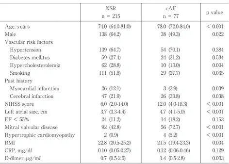
Table 1 Baseline Characteristicsofpatientswith NSR and cAF group. p value cAF n= 77 NSR n= 215 < 0.001 (72.0-84.0) 78.0 (64.0-81.0) 74.0 Age,years 0.022 (49.3) 38 (64.2) 138 Male Vascularrisk factors 0.384 (70.1) 54 (64.7) 139 Hypertension 0.534 (31.2) 24 (27.4) 59 Diabetesmellitus 0.004 (13.0) 10 (28.8) 62 Hypercholesterolemia
0.035 (37.7) 29 (51.6) 111 Smoking Pasthistory 0.039 (3.9) 3 (12.1) 26 Myocardialinfarction 0.038 (33.8) 26 (21.9) 47 Cerebralinfarction < 0.001 (4.0-18.3) 12.0 (2.0-14.0) 6.0 NIHSS score < 0.001 (4.1-5.0) 4.7 (3.3-4.4) 3.7 Leftatrialsize,cm 0.153 (18.2) 14 (11.2) 24 EF< 55% < 0.001 (72.7) 56 (42.8) 92 Mitralvalvulardisease
< 0.001 (5.2)
4 (0.9)
2 Hypertrophiccardiomyopathy
0.004 (19.4-23.3) 21.5 (20.5-25.2) 22.8 BMI 0.129 (0.06-0.46) 0.12 (0.05-0.27) 0.10 CRP,mg/dl 0.003 (0.5-2.8) 1.4 (0.5-2.0) 0.7 D-dimer,μg/ml
Data were median (interquartile range)ornumber(%).NIHSS;NationalInstitutesofHealth Stroke Scale,EF;Ejection Fraction,BMI;Body massindex,CRP;C-reactive protein
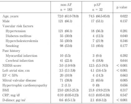
流症と狭窄症,機械弁のいずれかをみとめたものとした.肥大 型心筋症は心筋症のガイドライン12)に準じて診断した. 始めに,NSR 群と cAF 群の 2 群間の臨床背景と左房径に ついて比較検討した.さらに NSR 群を非 AF 群と pAF 群に わけて臨床背景と左房径について比較した.2 群間の比較は chi-square 検定と Mann-Whitney U 検定をもち い て 解 析 し た.非 AF 群と pAF 群を識別する各連続変数の至適 cut-off 値を receiver operating characteristics(ROC)曲線をもちい て求めた.単変量解析で p<0.1 の因子,従来指摘されている AF の関連因子4)∼9),ROC 曲線で解析した各連続変数の至適 値を独立因子として,多変量解析をおこなった.p<0.05 を有 意 差 あ り と 判 断 し た.統 計 処 理 ソ フ ト は Stat View 5.0 と SPSS(version 11)をもちいた. 結 果 急性期脳梗塞 292 例が登録された.平均年齢は 72.1±12.2 歳,男性は 193 人(66%),入院時 NIHSS スコアは 8.5±7.8, 発症から来院までの時間は 7.6±6.6 時間であった.NSR 群は 215 例(73.6%),AF 群 は 77 例(26.4%)で あ っ た.NSR 群に対し 24 時間ホルター心電図を 209 例(97.2%)に施行し た(平均 1.1 回). NSR 群と cAF 群の背景因子の比較について Table 1 に示 す.年齢(中央値(interquartile range;IQR)74.0(64.0∼81.0) vs. 78.0(72.0∼84.0)歳,p<0.001),脳 梗 塞 の 既 往(21.9% vs. 33.8%,p=0.038),入院時 NIHSS スコア(6.0(2.0∼14.0) vs. 12.0(4.0∼18.3),p<0.001),左 房 径(3.7(3.3∼4.4)vs. 4.7(4.1∼5.0)cm,p<0.001),D-dimer(0.7(0.5∼2.0)vs. 1.4(0.5∼2.8)µg!ml,p=0.003),僧帽弁疾患(42.8% vs. 72.7%, p<0.001),肥大型心筋症(0.9% vs. 5.2%,p<0.001)は cAF 群に有意に高かった.男性(64.2% vs. 49.3%,p=0.022),高 コレステロール血症(28.8% vs. 13.0%,p=0.004),喫煙(51.6% vs. 37.7%,p=0.035),心筋梗塞の既往(12.1% vs. 3.9%,p= 0.039),BMI(22.8(20.5∼25.2)vs. 21.5(19.4∼23.3),p=0.004) は NSR 群において有意に高かった.その他 2 群間で差はみら れなかった. NSR 群を非 AF 群と pAF 群に分けて,背景因子を検討し た(Table 2).年齢(72.0(61.0∼78.0)vs. 74.5(66.5∼85.0)歳, p=0.022),入院時 NIHSS スコア(3.0(1.0∼9.0)vs. 12.5(5.5∼ 19.5),p<0.001),左房径(3.5(3.1∼3.8)vs. 4.1(3.6∼4.5)cm p<0.001),僧帽弁疾患(38.8% vs. 65.6%,p=0.005),D-dimer (0.6(0.5∼1.5)vs. 2.1(0.8∼3.2)µg!ml,p<0.001)は pAF 群で有意に高値であった.一方で,糖尿病(30.0% vs. 12.5%, p=0.04)は非 AF 群に多かった.男性(66.1% vs. 53.1%,p= 0.157),高血圧(66.1% vs. 56.3%,p=0.281),高コレステロー ル血症(30.0% vs. 21.9%,p=0.346),喫煙(53.6% vs. 40.6%, p=0.177),心筋梗塞の既往(5.5% vs. 9.4%,p=0.392),脳梗 塞 の 既 往(22.4% vs. 18.8%,p=0.644),EF<55%(10.9% vs. 14.3%,p=0.663),BMI(23.0 vs. 21.8,p=0.317),CRP (0.10(0.05∼0.23)vs. 0.13(0.05∼0.36)mg!dl,p=0.547), 肥大型心筋症(1.1% vs. 0%,p=0.656)は有意差をみとめな かった.左房径は.cPA 群,pAF 群,非 AF 群の順に大きかっ た(中央値 4.7 vs. 4.1 vs. 3.5cm,p<0.001)(Fig. 1). 非 AF 群と pAF 群を区別する至適年齢,NIHSS スコア,左 房径,D-dimer について ROC 曲線をもちいて解析した.結果, 至適値は年齢 73.5 歳(感度 59.4%,特異度 56.3%),NIHSS スコア 7.5(68.8%,69.9%),左房径 3.8cm(68.8%,73.8%), D-dimer 0.95µg!ml(62.5%,62.8%)であった.
Table 2 Baseline CharacteristicsofPatientswith non-AF and pAF group. p value pAF n= 32 non-AF n= 183 0.022 (66.5-85.0) 74.5 (61.0-78.0) 72.0 Age,years 0.157 (53.1) 17 (66.1) 121 Male Vascularrisk factors 0.281 (56.3) 18 (66.1) 121 Hypertension 0.040 (12.5) 4 (30.0) 55 Diabetesmellitus 0.346 (21.9) 7 (30.0) 55 Hypercholesterolemia
0.177 (40.6) 13 (53.6) 98 Smoking Pasthistory 0.392 (9.4) 3 (5.5) 10 Myocardialinfarction 0.644 (18.8) 6 (22.4) 41 Cerebralinfarction < 0.001 (5.5-19.5) 12.5 (1.0-9.0) 3.0 NIHSS score < 0.001 (3.6-4.5) 4.1 (3.1-3.8) 3.5 Leftatrialsize,cm 0.663 (14.3) 4 (10.9) 20 EF< 55% 0.005 (65.6) 21 (38.8) 71 Mitralvalvulardisease
0.656 (0)
0 (1.1)
2 Hypertrophiccardiomyopathy
0.317 (19.9-23.9) 21.8 (20.5-25.3) 23.0 BMI 0.547 (0.05-0.36) 0.13 (0.05-0.23) 0.10 CRP,mg/dl < 0.001 (0.8-3.2) 2.1 (0.5-1.5) 0.6 D-dimer,μg/ml
Data were median (interquartile range)ornumber(%).
NIHSS;NationalInstitutesofHealth Stroke Scale,EF;Ejection Fraction, BM;Body massindex,CRP;C-reactive protein
Table 3 Multivariate logisticregression analysisforpr ob-ability ofpAF. p value 95%CI OR 0.404 0.222-1.833 0.638 Age ≧ 74 years 0.622 0.306-2.033 0.788 Male 0.197 0.180-1.424 0.507 Hypertension 0.078 0.086-1.141 0.312 Diabetesmellitus 0.698 0.221-9.555 1.452 Previousmyocardialinfarction
0.002 1.701-11.378 4.399 NIHSS score ≧ 8 < 0.001 3.238-24.268 8.882 Leftatrialsize ≧ 3.8cm 0.003 1.720-12.720 4.677 Mitralvalvulardisease
0.071 0.921-7.271
2.587 D-dimer≧ 1.0 μg/ml
NIHSS; National Institutes of Health Stroke Scale, EF; Ejection Fraction Left atr ial s ize med ian ( interquart ile range) 6 5 4 3 2 1 0 cm Non-AF cAF *p<0.001 pAF * * *
Fig. 1 Comparison of left atrial size among the groups. The median ofLA size ofthe cAF group wasthe highest, followed by the pAF group and the non-AF group.
多変量解析の結果,左房径≧3.8cm(オッズ比(OR)8.882, 95% 信 頼 区 間(CI)3.238∼24.268,p<0.001),NIHSS ス コ ア≧8(OR 4.399,95%CI 1.701∼11.378,p=0.002),僧帽弁疾患 (OR 4.677,95%CI 1.720∼12.720,p=0.003)が pAF の独立し
た関連因子であった(Table 3). 考 察 本研究は,急性期脳梗塞患者における心房細動と左房径の 関連について検討した.結果,①左房径は cAF 群,pAF 群, 非 AF 群の順に大きい,② NSR 群において 3.8cm 以上の左房 径拡大は pAF の独立した関連因子であることがわかった. 心房細動の発生・維持には電気的リモデリングと構造的リ モデリングの両者が起因していると考えられている13).心房 細動が数時間持続すると,イオンチャンネルを構成する蛋白 に変化が生じてくる.不応期が短縮し,伝導が遅延してくると リエントリーが形成されやすくなる.このように,心房筋に電 気生理学的変化が生じることを電気的リモデリングと呼ぶ. さらに数週間心房細動が持続すると,これらの電気生理学的 変化に加えて心房の拡張や繊維化などの構造学的変化も加わ り,最終的に慢性心房細動に移行すると考えられている.これ らの心房筋の解剖学的変化を構造的リモデリングと呼ぶ.心 房細動の持続時間が長いほどリモデリングがおこり,左房径 が拡大すると考えられる.Sanfilippo ら14)は,器質的心疾患を 除外した症例で AF と左房拡大の関連について報告してい る.本研究では,急性期脳梗塞患者において非 AF 群とくらべ て pAF 群で有意に左房拡大をみとめた.それゆえに,入院時 に洞調律であっても左房拡大をみとめれば,pAF が存在する 可能性が高い. 急性期脳梗塞患者において左房拡大と AF の関連を示した
報告はわれわれが検索しえたかぎりではない.左房径と AF の関連について,Psaty ら5)は community-based cohort 研究
で左房拡大が AF の独立した関連因子であると報告してい る.Di Tullio ら15)は,AF 患者において左房拡大は脳梗塞のリ
スクになると述べている.また,Corbalán ら16)は pAF 患者に おいて左房拡大は血栓塞栓症の独立した関連因子であると報 告している.それゆえに,左房拡大をみとめる症例では pAF が存在する可能性があるのみならず,塞栓症を発症するリス クも考えられる. 急性期脳梗塞患者において,pAF を検出するのに現在もっ とももちいられている検査法は 24 時間 Holter ECG であり, 新規 AF は約 5% 検出されると報告されている17).より長い 期間の心電図モニターの研究では,更に AF が 7.7%(平均測 定時間 70.1 時間)18),5.7%(159 時間)19)検出されると報告さ れ,24 時間 Holter ECG のみでは見逃す症例があると指摘さ れている.ただし長期間心電図モニターを全例おこなうこと は困難であり,pAF の可能性が高い症例に対しておこなうの が望ましい.本研究では,3.8cm 以上の左房拡大を有する患者 では,pAF が 30% と高率に検出された.それゆえに,経胸壁 心臓超音波検査で 3.8cm 以上の左房拡大をみとめたばあい は,24 時間 Holter ECG をくりかえしおこなう,あるいは長期 間の心電図モニターをおこなうべきである. 本研究の limitation は,後ろ向き研究であり当科に入院し た急性期脳梗塞患者全例に経胸壁心臓超音波検査を施行して いるわけではなく,経胸壁心臓超音波検査をおこなった患者 の選択にバイアスがかかった可能性がある.また経胸壁心臓 超音波検査を施行した期間が一定でない.今後入院直後に経 胸壁心臓超音波検査をおこない,左房径と pAF の関連につ いて前向きに検討する必要性がある.またホルター心電図を 平均 1.1 回おこなっているが,それでも pAF を見逃している 可能性が否定できない. 結 語 入院時 NSR の急性期脳梗塞患者において,3.8cm 以上の左 房拡大は pAF の独立した関連因子であった.左房拡大をみ とめるばあい,ホルター心電図をくりかえしおこなう,あるい は長期間モニター心電図を施行し,pAF を検索すべきであ る. 文 献
1)Hart RG: Atrial fibrillation and stroke prevention. N Engl J Med 2003; 349: 1015―1016
2)Inoue H, Atarashi H: Risk factors for thromboembolism in patients with paroxysmal atrial fibrillation. Am J Cardiol 2000; 86: 852―855
3)Hart RG, Pearce LA, Koudstaal PJ: Transient ischemic at-tacks in patients with atrial fibrillation. Implications for secondary prevention : the European Atrial Fibrillation Trial and Stroke Prevention in Atrial Fibrillation III trial.
Stroke 2004; 35: 948―951
4)Benjamin EJ, Levy D, Vaziri SM, et al: Independent risk factors for atrial fibrillation in a population-based cohort. The Framingham Heart Study. JAMA 1994; 271: 840― 844
5)Psaty BM, Manolio TA, Kuller LH, et al: Incidence of and risk factors for atrial fibrillation in older adults. Circula-tion 1997; 96: 2455―2461
6)Kannel WB, Wolf PA, Benjamin EJ, et al: Prevalence, inci-dence, prognosis, and predisposing conditions for atrial fibrillation : population-based estimates. Am J Cardiol 1998; 82: 2N―9N
7)Wang TJ, Parise H, Levy D, et al: Obesity and the risk of new-onset atrial fibrillation. JAMA 2004; 292: 2471―2477 8)Krahn AD, Manfreda J, Tate RB, et al: The natural
his-tory of atrial fibrillation: incidence, risk factors, and prog-nosis in the Manitoba Follow-Up Study. Am J Med 1995; 98: 476―484
9)Frost L, Hune LJ, Vestergaard P: Overweight and obe-sity as risk factors for atrial fibrillation or flutter: the Dan-ish Diet, Cancer, and Health Study. Am J Med 2005; 118: 489―495
10)Lyden P, Brott T, Tilley B, et al: Improved reliability of the NIH Stroke Scale using video training. NINDS TPA Stroke Study Group. Stroke 1994; 25: 2220―2226 11)Lang RM, Bierig M, Devereux RB, et al:
Recommenda-tions for chamber quantification : a report from the American Society of Echocardiography s Guidelines and Standards Committee and the Chamber Quantification Writing Group, developed in conjunction with the Euro-pean Association of Echocardiography, a branch of the European Society of Cardiology. J Am Soc Echocardiogr 2005; 18: 1440―1463
12)穂積健之,吉川純一:肥大型心筋症.心筋症 診断の手引 きとその解説,厚生労働省難治性疾患克服研究事業特発 性心筋症調査研究班,北畠 顕,友池仁暢 編,かりん舎, 北海道,2005,pp 37―50
13)Wijffels MC, Kirchhof CJ, Dorland R, et al: Atrial fibrilla-tion begets atrial fibrillafibrilla-tion. A study in awake chroni-cally instrumented goats. Circulation 1995; 92: 1954―1968 14)Sanfilippo AJ, Abascal VM, Sheehan M, et al: Atrial en-largement as a consequence of atrial fibrillation. A pro-spective echocardiographic study. Circulation 1990 ; 82 : 792―797
15)Di Tullio MR, Sacco RL, Sciacca RR, et al: Left atrial size and the risk of ischemic stroke in an ethnically mixed population. Stroke 1999; 30: 2019―2024
16)Corbalán R, Arriagada D, Braun S, et al: Risk factors for systemic embolism in patients with paroxysmal atrial
fib-rillation. Am Heart J 1992; 124: 149―153
17)Liao J, Khalid Z, Scallan C, et al: Noninvasive cardiac monitoring for detecting paroxysmal atrial fibrillation or flutter after acute ischemic stroke. A systematic review. Stroke 2007; 38: 2935―2940
18)Barthélémy JC, Féasson-Gérard S, Garnier P, et al: Auto-matic cardiac event recorders reveal paroxysmal atrial
fibrillation after unexplained strokes or transient ischemic attacks. Ann Noninvasive Electrocardiol 2003; 8: 194―199
19)Schuchert A, Behrens G, Meinertz T: Impact of long-term ECG recording on the detection of paroxysmal atrial fib-rillation in patients after an acute ischemic stroke. Pacing Clin Electrophysiol 1999; 22: 1082―1084
Abstract
Relationship between left atrial size and paroxysmal atrial fibrillation in acute ischemic stroke Shuichi Fujii, M.D.1)2)
, Kensaku Shibazaki, M.D.2)
, Yasuyuki Iguchi, M.D.2)
, Kenichiro Sakai, M.D.2)
and Kazumi Kimura, M.D.2) 1)
Department of Internal Mecicine, Kagamino Hospital
2)
Department of Stroke Center, Kawasaki Medical School
We investigated the relationship between parosysmal atrial fibrillation (pAF) and left atrial (LA) size in pa-tients with acute ischemic stroke. Between June 2006 and April 2008, we retrospectively enrolled 292 papa-tients with acute ischemic stroke within 24 hours of onset, who measured LA size by transthoracic echocardiography. The patients were classified according to the presence or absence of chronic AF on admission (cAF and normal si-nus rhythm (NSR) group). The NSR group was subdivided based on the pAF (pAF and non-AF group) . We com-pared LA size among each groups. Furthermore in the NSR group, the factors associated with pAF were investi-gated by multivariate logistic regression analysis.
Among the enrolled patients, cAF (cAF group) had 77 (26.4%), pAF (pAF group) had 32 (11.0%) and non-AF group was 183 (62.7%). The median of LA size of the cAF was highest (4.7 cm), followed by the pAF group (4.1 cm) and the non-AF group (3.5 cm) (p<0.001). Median age (72.0 for the non-AF group vs. 74.5 years for the pAF group, p<0.001), NIHSS score on admission (3.0 vs. 12.5, p<0.001), D-dimer (0.6 vs. 2.1µg!ml, p=0.003), LA size (3.5 vs. 4.1 cm, p<0.001) were higher in the pAF group than in the non-AF group. The optimal cut-off value, sensitivity and specificity of LA size to distinguish pAF from non-AF were 3.8 cm, 68.6% and 73.8%, respectively. Multivariate lo-gistic regression analysis demonstrated that a NIHSS score of"8 (odds ratio [OR], 4.399; 95% confidence interval [CI], 1.701 to 11.378, p=0.002), LA size of"3.8 cm (OR, 8.882; 95% CI, 3.238 to 24.268, p<0.001) and mitral valvular disease (OR, 4.677; 95% CI, 1.720 to 12.720, p=0.003) were independent factors associated with pAF. We should consider the presence of pAF when LA size is over 3.8 cm in acute ischemic stroke patients with sinus rhythm.
(Clin Neurol, 49: 629―633, 2009)