Warming Effect on Miriplatin.Lipiodol Suspension as a Chemotherapeutic Agent for Transarterial Chemoembolization for Hepatocellular Carcinoma: Preliminary Clinical Experience
Shinichi Kora ・ Hiroshi Urakawa ・ Toshimichi Mitsufuji ・ Akinobu Osame ・ Hideyuki Higashihara ・ Kengo Yoshimitsu
Abstract
Purpose: To retrospectively elucidate the preliminary clinical impact of warmed miriplatin/lipiodol suspension (MPT-LPD) when used as a chemotherapeutic agent of transarterial chemoembolization (TACE) for hepatocellular carcinoma (HCC).
Materials and Methods: Between June and Dec. 2010, TACE was performed using MPT/LPD at room temperature (RT group), and after Jan. 2011, TACE using MPT- LPD warmed upto 40℃ was performed (W group). The intraarterial appearance of MPT-LPD immediately after injection through microcatheters at the second-order branches was compared between the two groups using a 5-point grading system. Local therapeutic effects of HCCs as assessed on follow-up computed tomography (CT) obtained 1-3 months after TACE were compared between the groups with a 4-point grading system (TE1-TE4). After April 2011, angiography-assisted CT was routinely performed at TACE, and HCCs that revealed apparent corona enhancement (CE) were retrospectively selected. The degree of concordance between CE and MPT-LPD accumulation as assessed by CT immediately after TACE was assessed using 3-point grading.
Results: MPT-LPD therapy resulted in a smooth and continuous appearances in the W groups (grades 1, 2, 3, 4, and 5=1, 2, 11, 18, and 4) compared to the RT group (4, 0,
1, 2, and 0). The W group (TE1, TE2, TE3, and TE4 were 1, 9, 11, and 12) revealed better local therapeutic effects than the RT group (6, 3, 9, and 0) (p < 0.05). CE was found in 29 HCC nodules, and concordance between CE and MPT-LPD accumulation was observed in 66% (grade 1, 2, and 3 were, respectively, 2, 8, and 19).
Conclusion: Warmed MPT-LPD flowed more smoothly within vascular lumen, passed through tumor sinusoid of HCC, and had better local therapeutic effects at short-term observation than MPT-LPD at room temperature.
Introduction
Transarterial chemoembolization (TACE) is one of the accepted therapeutic choice for hepatocellular carcinomas (HCC), usually indicated for relatively large (> 3cm) or multiple (> 3) lesions [1,2]. For TACE, usage of emulsion of chemotherapeutic agents, such as epirubicin or cisplatinum, or iodized oil (lipiodol, LPD; Guerbet, Villepinte, France) is recommended to enhance anti-cancer effect [1, 2]. To maximize the therapeutic effect of TACE, anti-cancer agents with higher solubility and stability in LPD, also for gradual release within tumors, are considered ideal.
Miriplatin (MPT) is theoretically a good candidate for this kind of agent [3, 4].
However, in actual clinical practice, many interventional radiologists have pointed out that injection of MPT-LPD suspension requires unusually high pressure, particularly through a microcatheter system, compared to LPD emulsion with conventional chemotherapeutic agents. This is due to the high viscosity of MPT- LPD suspension.
To overcome this problem, we proposed a warming MPT-LPD method and reported its basic in vitro data [5], in which simple warming of MPT-LPD to 40℃ significantly reduced its viscosity and thus the injection pressure through microcatheters. Since then, after obtaining institutional review board (IRB) approval, we applied this
warming method to clinical cases, expecting MPT-LPD would flow more smoothly and with less resistance within microcatheters and huma arteries. The high viscosity of MPT-LPD means that it may choke the vessels before reaching HCC lesion, which would greatly hamper its antitumor effects, although a lesser antitumor effect of MPT-LPD has recently been reported compared to conventional TACE uses epirubicine, when used at room temperature [6].
According to previous investigators [7, 8], anticancer drug emulsions and LPD pass through the tumor sinusoids within HCC and reach the peritumoral liver parenchyma via venous drainage routes, which enhances the antitumor effect of subsegmental TACE by obstructing both the arterial and portal sides of the tumor vascular system.
On the basis of these concepts, if we could demonstrate that warmed MPT-LPD can reach the venous drainage areas around the tumor, namely the areas of corona enhancement (CE) [9], it would indirectly suggest that MPT-LPD can pass through tumor sinusoids and hence may have sufficient antitumor effect.
Our current study first addresses the appearances of MPT-LPD within the arteries immediately after its injection in patients who underwent TACE with MPT-LPD at room temperature (RT group) and with warmed MPT-LPD (W group). We expected that the change in viscosity of MPT-LPD would result in a morphological change.
Second, the degree of MPT-LPD accumulation was compared on the follow-up computed tomography (CT) obtained 1-3 months after TACE between RT and W groups. Finally, for HCC nodules that exhibited apparent CE on the second phase of CT during hepatic arteriography (CTHA), the degree of concordance between MPT- LPD distribution and CE was assessed by CT obtained immediately after TACE in W group.
Materials and Methods
Before the IRB approval of clinical use of warmed MPT-LPD, 15 patients had been treated with MPT-TACE at room temperature (RT group) between June and December 2010. After we received IRB approval, 37 patients were treated with TACE that used MPT-LPD warmed to 40℃ (W group) between January and October 2011. From these patients, further selection was applied for each assessment.
Assessment of the intraarterial appearance of MPT-LPD
To assess the intraarterial appearance of MPT-LPD, the whole process of MPT-LPD injection was routinely recorded via digital subtraction angiography, at three frames per second, between September 2010 and March 2011 (Multistar TOP; Siemens, Erlangen, Germany). Two experienced abdominal and interventional radiologists blinded to the patients’ information retrospectively reviewed the digital subtraction angiographic images with MPT-LPD injection, and treated branches of similar caliber, namely second-order branches, were selected for review. The appearance of MPT- LPD was qualitatively graded into five categories by consensus (Fig. 1). The grades were compared between the two groups by Mann-Whitney test.
Short term local therapeutic effect assessed on follow-up CT
For the patients in both the RT and W groups, follow-up CT examinations after
TACE were evaluated. Among these patients, those with nodular HCCs with a total sum of diameters less than 6cm were retrospectively selected to exclude patients treated with an insufficient amount of MPT-LPD, because the maximum amount of MPT-LPD currently approved in Japan is 6ml, and previous report has suggested that 1 ml of emulsion of chemotherapeutic agent and lipiodol correspond to 1cm diameter of HCC to be treated by TACE [10].
TACE was performed according to the usual method as reported, as previously reported [11]. A 3F coaxial microcatheter was advanced to second-order or more peripheral branches of the hepatic artery supplying the tumors, where MPT-LPD was injected, followed by injection of 1 mm gelatin sponge particle (GelPart ;Nippon Kayaku, Tokyo, Japan), finally obtaining complete stoppage of the arterial flow.
MPT-LPD injection was performed under fluoroscopic guidance. At the end of the procedure, angiography was repeated to confirm complete disappearance of tumor stain.
Follow-up CT was typically obtained 1-3 months after TACE, depending on the condition of the patient, referring to the blood test data, including tumor markersα- fetoprotein and protein induced by vitamin K absence/antagonist-II. Parameters for CT scanning are as follows: equipment, 64-row multidetector CT (Aquilion 64;
Toshiba Medical systems, Ootahara, Japan), 0.5mm collimation, 2 mm reconstruction, pitch 53, 120 kVp; Auto-mAs: quadruple scanning consisting of precontrast, arterial dominant (40 s delay), portal venous (70 s delay), and equilibrium phases (240 s delay). A total of 600 mg I/kg was delivered intraveneously in 30 s with 370 mg I/mL contrast medium (Iopamiron 370; BayerHealthCare, Osaka, Japan) or 350 mg I/mL contrast medium (Omnipaque 350, Daiichi-Sankyo Co., Tokyo , Japan; or
Iomeron 350, Eisai Co., Ltd., Tokyo, Japan). To compare the local therapeutic effect of TACE between the RT and W groups, two experienced abdominal and interventional radiologists blinded to the patients’ information reviewed and
evaluated these images in consensus. The degree of LPD accumulation was assessed and subclassified into four grades (TE1 through TE4) according to the General Rules the Clinical and Pathological Study of Primary Liver Cancer [12] on follow-up CT.
Definitions of the four grades are as follows; TE1, tumor enlargement more than 25 %;
TE2, tumor response between TE1 and TE3; TE3, tumor necrosis or dense LPD accumulation of more than 50 %; and TE4, difuse accumulation of LPD associated with tumor size reduction, suggesting100% tumor necrosis.
The severity of postembolization syndrome was compared between the groups using the maximum level of C-reactive protein after TACE and duration of hospitalization, in days, as arbitrary indices.
Evaluation of LPD Distribution Immediately after TACE in correlation to Areas of CE
In April 2011, a combined CT and angiography system, the so-called IVR-CT (Artis Zee Ceiling and Somatom Emotion 16; Siemens, Erlangen, Germany) was installed in our institute. This system consisted of flat-panel angiography equipment and a 16-row multidetector CT, and it replaced the old angiography system (Multistar T.O.P.;
Siemens). With this IVR-CT apparatus, CT during arterial portography (CTAP) and two-phase CTHA were routinely obtained before TACE, and plain CT was obtained at the end of TACE to confirm the distribution of LPD. Parameters of CTAP and CTHA
were as follows; 0.5 mm collimation, 1.5 mm reconstruction, pitch 0.8, 120kVp, and Auto-mAs. A total of 100 mg I/mL contrast medium with a total volume of 120 mL was injected into the superior mesenteric artery at a rate of 6 mL/s, and CTAP was obtained with a 30 sec delay time. For CTHA, the same contrast medium as that for CTAP was injected into proper or common hepatic artery at a rate of 2-3 mL/s, and the first phase was obtained with a 5 s delay time. The total volume of contrast medium was defined so that the injection lasts until the first phase scanning was completed.
The second phase was started in 10 s after the completion of the first-phase scan.
CE was defined as areas surrounding HCC with stronger enhancement than nontumorous liver parenchyma, with the tumor per se revealing less enhancement corresponding to washout, as observed on the second phase of CTHA [7, 13].
For the patients in the W group who underwent TACE with a sufficient amount of warmed MPT-LPD in the IVR-CT system, HCCs with apparent CE, as observed on the second phase of CTHA, were retrospectively selected by two radiologists in consensus who were blinded to the patients’ information. The areas of CE and of LPD accumulation as observed by distribution of LPD were compared, and their degree of concordance was subclassified into three grades (Fig. 2). Grade 1 indicated no accumulation of MPT-LPD in CE, grade 2 partial coverage of CE by MPT-LPD, and, grade 3 complete and sufficient coverage of CE by MPT-LPD. This
assessment was carried out not only on axial dimension, but also three-dimensionally, utilizing the multiplanar reconstruction function equipped with our picture archiving and communication system (Rapid Eye, Toshiba Medical System, Ootahara, Japan).
Finally, the degree of LPD-CE concordance (grades 1-3) and that of short-term treatment effects (TE1-TE4) were also correlated.
Results
Assessment of the intraarterial appearance of MPT/LPD
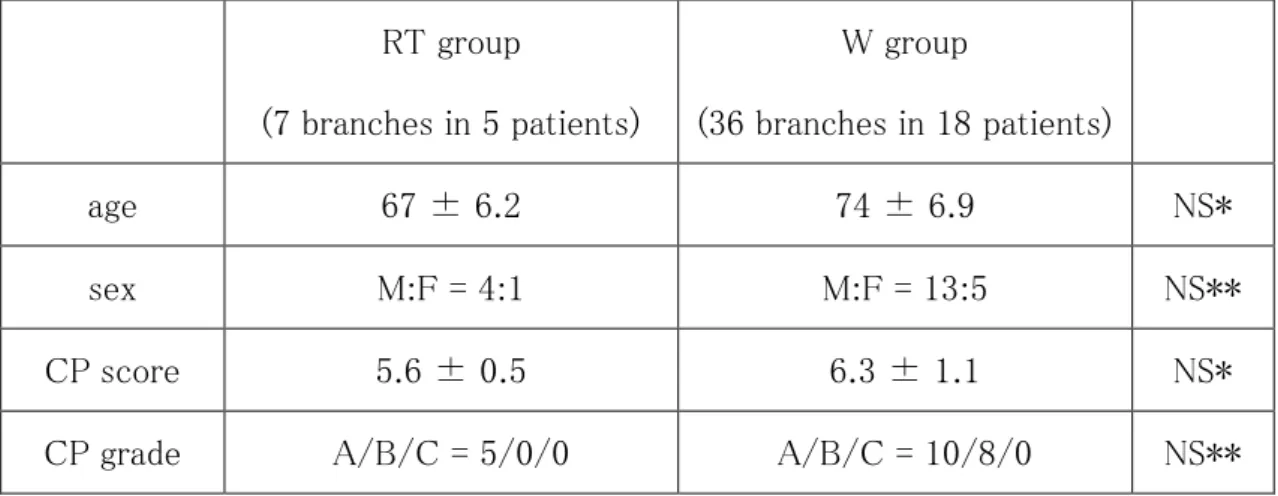
There were seven branches in five patients in RT group and 36 branches in 18 patients in W group for this evaluation. Demographic data of the two groups are listed in Table 1. There was no difference between the groups in age, sex, largest tumor size, and level of liver dysfunction.
The samples of morphological grades 1, 2, 3, 4, and 5 were, respectively 4, 0, 1, 2, and 0 for RT group, and 1, 2, 11, 18, and 4 for W group, revealing a statistically significant difference between the groups, with W group demonstrating higher grades than the RT group (p = 0.015 < 0.05, Mann-Whitney U test). In other words, warmed MPT-LPD tended to result in smooth, continuous features, whereas MPT- LPD at room temperature exhibited a beaded or globular appearance.
Representative cases are shown in Fig. 3.
Short term local therapeutic effect assessed on follow-up CT
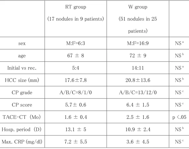
The demographic data of the two groups that underwent follow up CT are listed in Table 2. There were 17 HCC nodules in 9 patients in the RT group, and 51 HCC nodules in 25 patients in the W group that met the inclusion criteria for this
evaluation. There was no difference between the groups in age, sex, the largest tumor size, and level of liver dysfunction. No significant difference was observed in the period from TACE to discharge, or in the maximum C-reactive protein level after
TACE. The period between TACE and follow-up CT was slightly shorter in RT group (Table 2). The TE1, TE2, TE3, and TE4 degrees of LPF accumulation were respectively, 6, 3, 8, and 0 for the RT group, and 15, 5, 10, and 21 for the W group, which was a statistically significant difference (p<0.05, Mann-Whitney U test).
That is , the W group exhibited significantly better short-term therapeutic effect than the RT group. Representative cases are shown in Figs. 4.
Evaluation of LPD Distribution Immediately after TACE in Correlation to CE
There were 29 HCCs in 16 patients that demonstrated apparent CE on the second phase of CTHA and that were treated with sufficient amount of warmed MPT-LPD.
There were 13 men and three women aged 70 ± 10 years. The size of the tumors was 15 ± 9.2 mm. Child-Pugh grades were A, B, and C scores were , respectively, 8, 8, and 0 (6.5 ± 1.4).
As for the 29 lesions, the concordance of LPD accumulation and CE were 2, 8, and 19 for grades 1, 2, and 3, respectively. Namely, in 66 % (19 of 29) of HCC nodules demonstrating apparent CE, MPT-LPD completely covered CE of the nodules.
Finally, the degree of LPD-CE concordance (grades 1-3) and that of short term treatment effects (TE1-TE4) were significantly correlated (ρ = 0.434, p = 0.022, Spearman’s rank correlation) (Table 3). In other words, the more complete coverage of CE by MPT-LPD, the better the short-term therapeutic effect.
Representative cases are shown in Fig. 4.
.
Discussion
In our previous report [5], we demonstrated that warming MPT-LPD to 40 ℃ reduces its viscosity to almost one half of that at room temperature (25℃), and it also reduces the pressure required to inject the substance through microcatheters.
In the current study, we first confirmed that warmed MPT-LPD exhibited a more smooth and continuous appearance immediately after injection into arteries compared to MPT-LPD at room temperature. This indicates warmed MPT-LPD may behave , for example, more like liquid contrast medium, which smoothly and continuously distributes within the vascular lumen. The viscosity of iodine contrast medium (350-370 mg I/mL) at 37 ℃ have been reported to be approximately 10 mPa/S [14]. Another possible mechanism for these findings is the difference in the
injection rate. Because we did not use a consistent injection rate during MPT-LPD injection, but rather paid attention to not causing regurgitation under fluoroscopy guidance, it was possible that less viscosity in W groups resulted in more rapid injection, and thus a continuous appearance.
Our data also suggested that more than 60% of warmed MPT-LPD may pass through the tumor sinusoids of HCC and reach around the tumor at the areas of CE.
According to Terayama et al. [7], emulsion of epirubicine and LPD reached the areas of CE via tumor sinusoids in 64% of the HCC nodules demonstrating apparent CE;
they considered that this may represent the mechanism of embolization of both the arterial and portal sides of the tumor, leading to an excellent antitumor effect. Our data with warmed MPT-LPD are at least comparable to theirs, although the
backgrounds of the treated lesions are not identical. We also confirmed that the more complete the coverage of CE by MPT-LPD, the better short-term therapeutic
effect.
Thus, we might reasonably infer from our results that warmed MPT-LPD with reduced viscosity flowed smoothly within the vascular lumen, passing through the tumor sinusoids to the portal venous sides of the tumor, resulting in better short- term therapeutic effect than that at room temperature.
Nevertheless, our results were not satisfactory: although warming MPT-LPD greatly improved its short-term therapeutic effect, there are still several examples of poor response (TE1) in patients treated with warmed MPT-LPD. This may at least partly be attributable to the cooling effect of warmed MPT-LPD during the procedure. To solve this problem, some devices that will keep the injection syringes warm while under sterile condition may need to be developed. However, considering that seven of 18 nodules (more than one third) demonstrating complete coverage of CE by MPT-LPD (grade 3) resulted in TE1, improvement in the delivery of embolic materials may also be required. Further investigation is needed to clarify these issues.
There are several limitations to this preliminary clinical study, including its retrospective nature. We did not evaluate the concordance between MPT-LPD distribution and CE in the RT group, but only in the W group. This is because we already had some data suggesting inferiority of the RT group in terms of the local control rate. After we installed the IVR-CT apparatus, it was ethically inappropriate to perform MPT-LPD TACE at room temperature as a control. This is also a reason why we could not increase the number of control subjects (that is, RT group) in this study. Our short follow-up period is another limitation. Further, we only compared MPT-LPD delivered warmed and at room temperature; we did not compare
conventional chemotherapy agents in this study. To address these limitation, We have begun a prospective study comparing TACE performed with warmed MPT-LPD and TACE performed with epirubicine in a selected patient group in our institute.
In conclusion, MPT-LPD warmed to 40 ℃ flowed smoothly within the vascular lumen and reached the portal venous sides of the tumor in 60% of the cases, presumably passing through the tumor sinusoids. TACE with warmed MPT-LPD provided significantly better local control of HCC at short-term observation
compared to room-temperature MPT-LPD. Warming MPT-LPD may prove to be a simple and promising method that contributes to the therapeutic effect of MPT-LPD in HCC.
References
1. Kokudo N, Makuuchi M. Evidence-based clinical practice guidelines for hepatocellular carcinoma in Japan: the J-HCC guidelines. J Gastroenterol.
2009;44 Suppl 19:119-21.
2. Arii S, Sata M, Sakamoto M, Shimada M,et al. Management of hepatocellular carcinoma: Report of Consensus Meeting in the 45th Annual Meeting of the Japan Society of Hepatology (2009). Hepatol Res. 2010 Jul;40(7):667-85.
3. Hanada M, Baba A, Tsutsumishita Y, et al. Intra-hepatic arterial administration with miriplatin suspended in an oily lymphographic agent inhibits the growth of tumors implanted in rat livers by inducing platinum-DNA adducts to form and massive apoptosis, Cancer Chemother. Phamacol. 2009; 64 : 473-483 ,
4. Hanada M Baba A, Tsutsumishita Y, et al. Intra-hepatic arterial administration with miriplatin suspended in an oily lymphographic agent inhibits the growth of human hepatoma cells orthotopically implanted in nude rats , Cancer Sci.
2009 ;100: 189-194.,
5. Kora S, Urakawa H, Mitsufuji T, et al. Warming effect on miriplatin-Lipiodol suspension for potential use as a chemotherapeutic agent for transarterial chemoembolization of hepatocellular carcinoma: in vitro study. submitted 6. Miyayama S, Yamashiro M, Shibata Y, et al.Comparison of local control effects of
superselective transcatheter arterial chemoembolization using epirubicin plus mitomycin C and miriplatin for hepatocellular carcinoma. Jpn J Radiol. 2012 Apr;30(3):263-70.
7. Tearayama N, Matsui O, Gabata T, et al. Accumulation of Iodized Oil Within the Non-Neoplastic Liver Adjacent to Hepatocellular Carcinoma via the Drainage Routes of the Tumor After Transcatheter Arterial Embolization. Cardiovasc Intervent Radiol. 2001; 24: 383-387.
8. Miyayama S, Matsui o, Yamamoto Y, et al. Ultraselective transcathter arterial chemoembolization with a 2-Fr tip microcatheter for small hepatocellular
carcinomas: relationship between local tumor recurrence and visualization of the portal vein with iodized oil. J Vas Interv Raiol 2007;18:365-376
9. Ueda K, Matsui O, Kawamori Y, et al. Hypervascular hepatocellular carcinoma:
evaluation of hemodynamics with dynamic CT during hepatic aeteriography.
Radiology 1998; 206:161-166
10.Matsuo N, Uchida H, Sakaguchi H, et al. Optimal lipiodol volume in transcatheter arterial chemoembolotherapy for hepatocellular carcinoma: study based on
lipiodol accumulation patterns and histopathologic findings. Semin Oncol.
1997;24:S6-61 - S6-70.
11.Higashihara H, Okazaki M. Transcatheter arterial embolization of hepatocellular carcinoma : a Japanese experience. Hepato-Gastroenterology , 2002;49: 72-78 , 12.Liver Cancer Study Group of Japan. General Rules for the Clinical and
Pathological Study of Primary Liver Cancer, 3rd English ed., Kanehara Co., Ltd, Tokyo
13. Pugacheva O, Matsui O, Kozaka K, et al. Detection of small hypervascular hepatocellular carcinomas by EASL criteria: comparison with double-phase CT during hepatic arteriography. Eur J Radiol. 2011 Dec;80(3):e201-6.
14.Galloti A, Uggeri F, Favilla A, Cabrini M, de Haen C. The chemistry of iomeprol and physic-chemical properties of its aqueous solutions and pharmaceutical formulations. Eur J Radiol. 1994 18 suppl l;Sl-S12
Figure legends
Fig. 1 Grading system for the assessment of the intraarterial appearance of MPD-LPD suspension, ranging from the discontinuous, beaded appearance of grade 1 to the smooth, continuous appearance of grade 5
Grade 1 Grade 2 Grade 3
Grade 4 Grade 5
Fig. 2 Grading system for the assessment of the concordance between CE and LPD accumulation, with no, partial, and complete coverage of CE by LPD, corresponding to grades 1, 2, and 3, respectively. T represents the main body of the tumor. The yellow zone around T represents the area of CE; the shaded circle in the lower row represents LPD accumulation
Grade 1 Grade 2 Grade 3
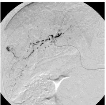
Fig. 3 Intraarterial appearance of MPD-LPD suspension immediately after injection A RT group (67-year-old man). Injection from the posterior branch. Note globular or beaded appearance, which was categorized as grade 2
A
T T T
B W group (70-year-old man). Injection from the anterior branch. Note smooth and continuous appearance as compared to Fig. 3a, which was categorized as grade 4 B
Fig. 4 Short-term therapeutic effect in 69-year-old man in the W group.
A Arterial phase CT 1 month before treatment shows 3 cm enhancing nodule with necrotic component (arrow).
B Second phase of CT during CTHA. Thin CE is seen surrounding the tumor (arrowhead). Necrotic component remains low dense (arrow).
C Plain CT immediately after treatment. Sufficient LPD covers the whole CE, suggesting grade 3 LPD accumulation. No LPD accumulation is seen at the necrotic part (arrow)
D Arterial phase CT 2 months after the treatment. Note complete accumulation of LPD throughout the nodule and the decrease in size of the lesion. Necrotic component remains low dense (arrow); TE4 was assigned
A
B
C
D
Table 1 Demographic data of the two groups that underwent transarterial
chemoembolization with MPD-LPD at room temperature (RT group) and with warmed MPD-LPD (W group) to assess the intraarterial appearance within the vascular lumen
RT group
(7 branches in 5 patients)
W group
(36 branches in 18 patients)
age 67 ± 6.2 74 ± 6.9 NS*
sex M:F = 4:1 M:F = 13:5 NS**
CP score 5.6 ± 0.5 6.3 ± 1.1 NS*
CP grade A/B/C = 5/0/0 A/B/C = 10/8/0 NS**
RT: transarterial chemoembolization using miriplatin at room temperature W: transarterial chemoembolization using warmed miriplatin
CP: Child-Pugh
*unpaired t-test
** Fisher’s exact probability test
Table 2 Demographic data of RT and W groups for short-term local therapeutic effect compared by follow-up computed tomography
RT group
(17 nodules in 9 patients)
W group (51 nodules in 25
patients)
sex M:F=6:3 M:F=16:9 NS a
age 67 ± 8 72 ± 9 NS b
Initial vs rec. 5:4 14:11 NS a
HCC size (mm) 17.6±7.8 20.8±13.6 NS b
CP grade A/B/C=8/1/0 A/B/C=13/12/0 NS c
CP score 5.7± 0.6 6.4 ± 1.5 NS c
TACE-CT (Mo) 1.6 ± 0.4 2.5 ± 1.6 p <.05 Hosp. period (D) 13.1 ± 5 10.9 ± 2.4 NS b
Max. CRP (mg/dl) 7.2 ± 5.5 3.6 ± 4.5 NS c
MPT-LPD miriplatin-lipiodol suspension, HCC hepatocellular carcinoma, CRP: C-reactive protein, TACE-CT: interval between TACE and follow-up computed tomography,
CRP maximum serum C-reactive protein level after TACE
a Chi-square test b unpaired t test c unpaired t test, marginal
Table 3 Correlation between the lipiodol-corona enhancement concordance grades and the short-term therapeutic effects in 29 nodules
TE1 TE2 TE3 TE4
Grade 1 2 0 0 0
Grade 2 3 1 1 4
Grade 3 7 0 1 10
TE1 tumor enlargement more than 25%;
TE2 tumor response between TE1 and TE3;
TE3 tumor necrosis or dense lipiodol (PLD) accumulation of more than 50%;
TE4 difuse accumulation of LPD associated with tumor size reduction, suggesting 100% tumor necrosis
ρ = 0.434, p = 0.022, Spearman’s rank correlation