九州大学学術情報リポジトリ
Kyushu University Institutional Repository
前腕における末梢磁気刺激対電気刺激後の運動皮質 興奮性の調整
佐藤, 綾
https://doi.org/10.15017/1831395
出版情報:Kyushu University, 2017, 博士(システム生命科学), 課程博士 バージョン:
権利関係:
Modulation of motor cortical excitability after peripheral magnetic versus
electrical stimulation in the forearm
2016
Aya Sato
- 3 -
Abstract
This study found that inhibition of motor cortical excitability was induced by peripheral magnetic and electrical stimulation, and investigated the possibility that peripheral stimulation has similar effects to transcranial magnetic stimulation (TMS) on motor cortical excitability. In recent years, techniques combining movement and TMS have been used to aid the recovery of motor function after stroke. The presence of abnormal transcallosal inhibition of intact motor cortex following stroke may inhibit the activity of lesioned motor cortex in the contralateral hemisphere, which has been suggested to disrupt the recovery of motor function. In addition, several studies have suggested that the inhibition of intact motor cortex might be effective for the recovery of motor function. However, TMS is not an appropriate treatment for all patients, because of potential adverse events caused by the strong magnetic fields involved.
Therefore, the current study focused on the effects of peripheral stimulation on cortical excitability in motor cortex.
Previous studies have reported that the peripheral afferents resulting from voluntary movement, passive movement, electrical stimulation, and magnetic stimulation affect cortical excitability in the primary motor cortex.Ridding et al. reported that facilitation of cortical excitability was induced by 10 Hz electrical mixed nerve stimulation for 2 h.
Moreover, in another study, 20 Hz peripheral magnetic stimulation was applied transcutaneously to the area of muscle supplying the terminal nerve branches of the finger and hand extensor muscles using a circular coil.Similarly, previous studies have examined the use of peripheral electrical and magnetic stimulation to aid the recovery of motor function by facilitating cortical excitability of the motor cortex. Other reports have confirmed the facilitation of cortical excitability of the primary motor cortex via high-frequency and long-duration stimulation. However, few studies have examined whether magnetic and electrical stimulation can induce inhibition of motor cortical excitability under certain conditions.
- 4 -
TMS induces different effects on cortical excitability with different magnetic stimulation parameters, such as stimulation frequency and intensity. For example, low-frequency repetitive TMS (rTMS) of 1 Hz has been reported to decrease motor cortical excitability, and many previous studies have used TMS of supra-threshold intensity. In addition, muscle twitch caused by TMS is thought to be involved in the inhibition of motor cortical excitability.In the current study, peripheral magnetic and electrical stimulation were administered over the forearm to induce muscle twitch similar to that induced by TMS, to investigate the inhibition of motor cortical excitability caused by peripheral stimulation. The effect of peripheral stimulation was evaluated by comparing the motor evoked potential (MEP) amplitude evoked by TMS applied to the primary motor cortex before and after peripheral stimulation. Next, the stimulation frequency (above 1 Hz) and stimulation site (the contralateral or ipsilateral forearm for the target motor cortex) were altered to investigate the effects (facilitation or inhibition) of motor cortical excitability induced by peripheral stimulation. In addition, the MEP amplitude evoked by magnetic and electrical stimulation was compared to investigate the differences in motor cortical excitability induced by each stimulation type.
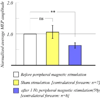
The results revealed that 1 Hz magnetic and electrical stimulation of the contralateral forearm caused a decrease in MEP amplitude in response to TMS applied to the left motor cortex after stimulation, compared with before stimulation. These results were similar to the effects of 1 Hz supra-threshold rTMS, suggesting that inhibition of motor cortical excitability may have been induced by 1 Hz magnetic and electrical stimulation.
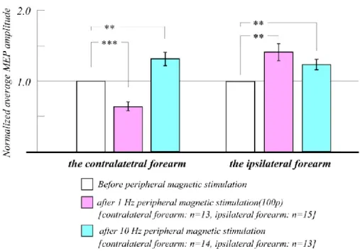
Moreover, changing the stimulation frequency and stimulation site of magnetic and electrical stimulation over the forearm increased MEP amplitude after stimulation under some conditions. Taken together, these findings confirmed that both magnetic and electrical stimulation induced similar modulation of MEP amplitude, suggesting that magnetic and electrical stimulation exert similar effects on motor cortical excitability.
The current findings suggest that 1 Hz magnetic and electrical stimulation over the contralateral forearm evoked inhibition of motor cortical excitability that was similar to
- 5 -
that induced by supra-threshold TMS of 1 Hz. In addition, modulation of motor cortical excitability was evoked by changing the stimulation frequency and stimulation site of magnetic and electrical stimulation. Importantly, these effects occurred both for magnetic stimulation and electrical stimulation.
- 6 -
Contents
Abstract 3
Contents 6
Chapter 1
General Introduction 8
1-1 Transcranial magnetic stimulation (TMS) 9
1-1-1 History and characteristic of TMS
1-1-2 Principle and structure of TMS 10
1-1-3 Nerve conduction in TMS 13
1-1-4 The alteration of cortical excitability with TMS 14
1-2 Motor evoked potentials (MEPs) 19
1-3 Influence of peripheral stimulation on cerebral cortex 22
1-4 The purpose of this study 29
Chapter 2
The modulation of motor cortical excitability by supra-threshold rTMS 32
2-1 Introduction 33
2-2 Experimental methods 35
2-3 Results 38
2-4 Discussion 41
2-5 Conclusion 44
- 7 -
Chapter 3
Alteration of motor cortical excitability by peripheral magnetic stimulation 46
3-1 Introduction 47
3-2 Experimental methods 50
3-3 Results
3-3-1 Change of stimulation site of peripheral magnetic stimulation 56 3-3-2 Effect of different stimulation frequencies of peripheral magnetic stimulation 62 3-3-3 Changes in MEP amplitude decrease after peripheral magnetic stimulation 64
3-4 Discussion 65
3-5 Conclusion 74
Chapter 4
Alteration of motor cortical excitability with peripheral electrical stimulation 75
4-1 Introduction 76
4-2 Experimental methods 79
4-3 Results 83
4-4 Discussion
Influence of peripheral electrical stimulation on left motor cortical excitability 86
Comparison of peripheral magnetic and electrical stimulation 91
4-5 Conclusion 96
Conclusion 98
Acknowledgement 100
References 101
Chapter 1
General introduction
- 9 -
1-1 Transcranial magnetic stimulation (TMS)
1-1-1 History and characteristics of TMS
Investigation of brain function has traditionally required invasive surgery to open the skull, limiting the scope of feasible research, particularly in healthy subjects. In 1980, Merton et al. reported a noninvasive method for stimulating the brain directly using high-voltage electrical stimulation applied through electrodes on the scalp (Merton et al., 1980).This was called transcranial electrical stimulation (TES). In the TES paradigm, the delivery of high-voltage electric shocks to the primary motor area was reported to induce muscle responses. Electric stimulation was applied not only to the brain, but also to the spinal cord (Merton et al. 1982). TES operates via stimulation of the cortex by an electric current flowing between two electrodes positioned on the scalp. The electric current flows radially from an anode to a cathode, and the partial current penetrates the scalp and activates neurons.However, because the brain is electrically protected by the high impedance of the skull, scalp and hair, electrical stimulation can cause pain and discomfort to participants. In addition, localization of electrical stimulation to a target area of the cortex is difficult.Transcranial magnetic stimulation (TMS) was developed as a method for direct brain stimulation in 1985 by Barker and colleagues, to resolve several of the limitations associated with TES (Barker et al., 1985a). TMS has been applied in various fields, and has a range of benefits: 1) magnetic stimulation is a noninvasive and relatively painless method, 2) because stimulation is not attenuated by the high impedance of the skull and scalp, the brain can be stimulated directly by a magnetic stimulation coil positioned on the scalp, 3) the magnetic stimulation coil can be positioned on the skull to alter cortical excitability in a specific brain region (Barker et al., 1985b, Hallett, 2000).
- 10 -
1-1-2 Principle and structure of TMS
In magnetic stimulation, a high-current pulse is produced by a magnetic wire coil.
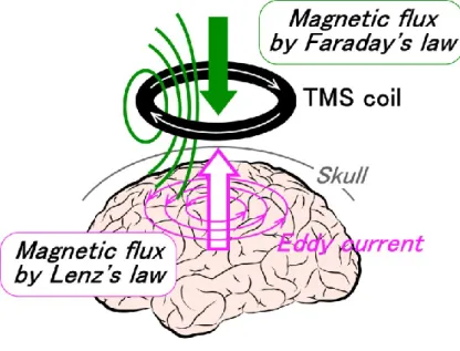
Magnetic fields are produced with lines of flux passing perpendicularly to the plane of the coil when placed on the scalp in a tangential position. In TMS, magnetic fields can reach up to approximately 2 T, and typically last for around 100 µs. Moreover, these magnetic fields generate an electric current that flows in the opposite direction to the electric current in the magnetic coil, called the eddy current (Figure 1-1). The eddy current flows in loops that are parallel to the plane of the magnetic coil. The generation of the eddy current is explained by Faraday's law of electromagnetic induction, as well as Lenz's law. The strongest eddy current occurs close to the circumference of the magnetic coil itself. The eddy current becomes weaker near the center of the magnetic coil, and there is no current at the center.Although it has been established that neuronal networks are activated by induced eddy currents, the precise extent of neuronal activation is currently unclear. However, it has been found that neuronal activation varies with the intensity of magnetic stimulation, and that cortical excitability can be modulated by eddy currents (Maeda et al., 2000, Touge et al., 2001, Ridding and Rothwell 2007, Hallett, 2007).
Figure 1-1 Direction of current flow in a magnetic coil and the induced current in the brain
- 11 -
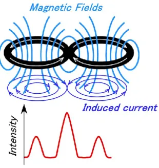
TMS involves several functional limitations. Because the magnetic field falls off rapidly with distance from the magnetic coil surface, direct stimulation is limited to the outer parts of the cerebral cortex located near the skull (Hallett, 2000, Hallett, 2007, Ridding et al., 2007). As magnetic stimulation techniques have developed, magnetic coils of various shapes have been introduced. A round coil has been used since the development of TMS, inducing a powerful magnetic field that enables extensive stimulation. However, round coils do not produce a focal site of stimulation. In contrast, figure-eight-shaped coils enable focal stimulation with a focal resolution of 5 mm, enabling stimulation of a much smaller cortical area of 1–2 cm2. Figure-eight-shaped coils are constructed so that two round coils are attached at an angle (under 180 degrees), with an area of overlap. The overlapping part of the coil increases the magnetic field intensity, enabling figure-eight-shaped coils to produce more effective and focal magnetic stimulation (Figure 1-2) (Ueno et al., 1988, Ueno et al., 1990, Thielscher et al. 2004, Ravazzani et al., 1996). Moreover, figure-eight-shaped coils with an angle of approximately 90–100 degrees exhibit increased power at the intersection, and can stimulate deeper regions of cortex.
Figure 1-2 Magnetic coil shape and resultant stimulus intensity in the brain
- 12 -
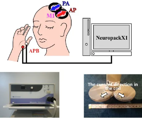
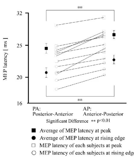
The development of figure-eight-shaped flat coils has enabled TMS stimulation with directionality. The descending volley elicited by TMS with a figure-eight-shaped flat coil depends on the direction of the induced current. For example, when the coil is placed on a hand-related region of the motor area, the thresholds for a monophasic stimulator are lowest when the eddy current of a magnetic coil flows in the posterior-anterior (PA) direction, perpendicular to the central sulcus (Sakai et al., 1997, Mills et al., 1992). In addition, responses with the shortest latency occur when the eddy current flows in a latero-medial direction (Werhahn et al., 1994, Di lazzaro, 2003). Thus, the effect of TMS varies with the direction of the induced current, and depends on the pulse waveform output by the stimulator (Brasil et al., 1992), which can be biphasic or monophasic.For a monophasic pulse, the direction of induced current is always in the postero-anterior or antero-posterior direction. This method has been used in a number of previous studies examining the motor threshold (Mills et al., 1997, Rossini et al., 1992, Triggs et al., 1994). In addition, monophasic pulses in the induced current passing through the motor cortex in the postero-anterior direction are more effective than those traveling in the antero-posterior direction (Kammer et al., 2001). Alternatively, biphasic pulses can be applied with repetitive transcranial magnetic stimulation (rTMS). rTMS was introduced by Pascual-Leone et al. in 1987, and has been suggested to have value as a treatment for the pathological physiology of depression (Pascual-Leone et al., 1996).High-frequency rTMS has since been used for treatment of a range of psychiatric conditions, in addition to depression. (Pascual-Leone et al., 1996a, George et al., 1997).
- 13 -
1-1-3 Nerve conduction in TMS
Many of the basic principles of TES also apply to TMS (Di Lazzaro et al., 2004). The effects of TMS applied over the primary motor area appear to be similar to those of TES.
Both TMS and TES have been shown to excite corticospinal neurons at short latencies, consistent with direct activation of neurons in the cortex, and at longer latencies, as measured by direct recording from the surface of the spinal cord (Boyd et al., 1986, Burke et al., 1993). However, the two methods vary in terms of the latency of responses to stimulation.
In the motor cortex, it has been reported that TMS tends to recruit I-waves at threshold, whereas TES tends to recruit D-waves (Thompson et al., 1991, Burke et al., 1993).In electrical stimulation of the brain, the vertical component of current flow has been found to favor direct excitation of pyramidal tract neurons (Day et al. 1989). Thus, the production of early D-wave activity by TES appears to reflect direct activation of descending axons in the corticospinal tract (Patton et al. 1954). With higher intensities of stimulation, D-wave activity is followed by I-wave activity, with a periodicity of approximately 1.5 ms (Patton et al. 1954, Day et al. 1989). In contrast, magnetic stimulation appears to induce eddy currents under the magnetic coil, predominantly in the horizontal plane, stimulating horizontally-oriented neurons within the cortex, including interneurons, pyramidal tract axon collaterals and afferent axons from cortical and subcortical sites.
TMS over the motor cortex tends to activate I-waves at lower intensities than D-wave activity (Thompson et al., 1991, Berardelli et al., 1990). In addition, the latency of I-wave activity is delayed compared with D-wave activity. I-wave latencies are commonly categorized by numbers corresponding to the duration of the latency, in order: I1, I2, and I3 (Kernell and Chien-Ping, 1967). The interval of each descending volley is 1.5 ms, corresponding to the latency of an individual synapse (Kernell and Chien-Ping, 1967). The D-wave produced by a TMS pulse is recruited at intensities much greater than threshold. TMS over the motor cortex typically evokes I-wave activity, and the resulting effect of descending volley stimulation can be observed in
- 14 -
motor-evoked potential (MEP) recordings. The MEP latency induced by TMS is typically around 1.5 ms longer than that evoked by TES (Berardelli et al., 1990, Edgley et al., 1990).The MEP latency of TMS shortens and becomes equal to that induced by TES, because stronger TMS evokes D-waves (Kaneko et al., 1996, Di lazzaro, 2003).
Recruitment of various components of the corticofugal discharge by TMS depends on the intensity of stimulation, the shape of the coil, the pulse configuration, and the relative threshold of each volley to the direction of the induced current flow in the cortex (Hallett, 2007, Rossini et al. 2015, Groppa et al., 2012).
1-1-4The alteration of cortical excitability with TMS
Several studies have reported that low-frequency magnetic stimulation of 1 Hz decreases motor cortex excitability, whereas high-frequency magnetic stimulation of above 5 Hz increases motor cortex excitability (Wassermann et al., 1998, Pascual-Leone A et al. 1994, Chen et al., 1997). Pascual-Leone et al. reported that the characteristics of MEPs depend on the intensity and frequency of repetitive stimulation (Pascual-Leone et al., 1994). In one study, stimulus frequencies of 1, 3, 5, 10, 20 Hz were tested, and rTMS began at the threshold intensity (100% threshold) and progressively increased in steps of 10% of threshold up to the maximal intensity of the stimulator’s output (Pascual-Leone et al., 1994). The results showed that rTMS of 20 pulses at 5 Hz at 150% threshold evoked larger MEPs in subjects at rest, and the MEP amplitude induced by rTMS of 20 pulses at 10 Hz gradually increased for all participants at an intensity of 110%–130% of motor threshold. With rTMS trains of 1 Hz stimulation, no consistent alteration of MEP amplitude was observed. Adjustment of the amplitude of MEPs produced in the target muscle by magnetic stimulation showed effects that depended on rTMS frequency and intensity, including inhibition and facilitation of cortical excitability (Pascual-Leone et al. 1994).
Berardelli et al. examined the neurophysiological mechanisms underlying the facilitation of MEPs after serial magnetic stimulation. They applied a single TMS pulse
- 15 -
to the motor cortex at 200–900 ms after a train of 20 pulses of 5 Hz rTMS with a 1-min interval between trains. The results revealed that applying 5 Hz rTMS to the motor cortex at an intensity of 120% of motor threshold produced a transient increase in cortical excitability. Moreover, their findings suggested that the after-train facilitation of MEPs was cortical in origin, showing that MEPs induced by rTMS (which tends to activate the same neurons via corticocortical synapses) were facilitated, whereas MEPs induced by transcranial electrical stimulation (TES) (which tends to activate corticospinal neurons directly) were not. In addition, although the afferent input from the twitches evoked by each type of TMS might have been expected to increase spinal cord excitability, H-reflexes after the train were suppressed rather than enhanced.
Therefore, the results suggested that facilitation of cortical excitability may occur in the cortex, but not in the spinal cord. The facilitation of cortical excitability may induce activity in the stimulated cortical region by increasing the excitability of pyramidal cells and their excitatory inputs. Thus, the excitatory reduction of cortical inhibition mechanisms may play a role in the facilitatory effects of magnetic stimulation (Berardelli et al., 1998).
In contrast, low-frequency TMS has been found to cause inhibition of cortical excitability. Chan et al. applied 0.1 Hz rTMS at an intensity of 105% of the motor threshold to the left motor cortex for 1 h (a total of 360 pulses). The average MEP amplitude induced by each group of 30 pulse stimuli (5 min) was normalized using the average MEP amplitude of the first 60 pulse stimuli (10 min), which was defined as the baseline. The results demonstrated that the MEP amplitude of the right abductor pollicis brevis (APB) muscle was not significantly reduced or increased by 1 h of rTMS.
Moreover, 90 pulses of 0.1 Hz rTMS (15 min) were applied before and after an intervention stimulus of 0.9 Hz rTMS at an intensity of 115% of the motor threshold for 810 magnetic stimulation pulses (15 min). The results indicated that MEP amplitude in the APB was reduced following rTMS compared with before rTMS, and this effect continued for 15 min. These findings suggest that low-frequency rTMS may decrease the efficacy of cortico-cortical synapses or the excitability of postsynaptic corticospinal
- 16 -
neurons, possibly reflecting changes in extrinsic input to the motor cortex. In addition, the results suggested that the observed decrease in motor cortical excitability may be mediated by long-term depression (LTD), because the stimulus parameters used in the study (1 Hz, 900 pulses) resembled the stimulation conditions used for inducing LTD in CA1 cells of hippocampal slices (Chen et al., 1997, Dudek et al., 1992, Mulkey et al., 1992). In another study, Maeda et al. applied 240 pulses of 1, 10 and 20 Hz rTMS at an intensity of 105%, on two different days. The results indicated a significant decrease in MEP amplitude following 1 Hz rTMS, and a significant increase of MEP amplitude following 20 Hz rTMS, on both day 1 and day 2. This effect was greater on day 2. The researchers suggested that this effect may have been related to LTD- and long-term potentiation (LTP)-like effects (Maeda et al., 2000).
Moreover, decreased cortical excitability is associated not only with stimulation frequency but also stimulation intensity (sub- or supra-motor threshold). Romero et al.
studied the effects of sub-threshold 1 Hz rTMS on cortical excitability. They applied 1 Hz rTMS at sub-threshold intensity (90% at the resting motor threshold, RMT) for 10 min, and the position of TMS was determined as the scalp position from which TMS evoked MEPs of greatest amplitude in the first dorsal interosseous (FDI) muscle. The effect was evaluated by measuring MEP amplitude induced by intracortical facilitation and inhibition.The results revealed that a significant decrease of mean MEP amplitude was induced by intracortical facilitation, while intracortical inhibition was not affected.
In addition, this report suggested that the effect of sub-threshold stimulation with 1 Hz rTMS was intracortical, and that the effect of an rTMS train may preferentially result in decreased intracortical facilitation via an inhibitory mechanism (Romero et al., 2002).
Several possible mechanisms underlying the decrease of cortical excitability with supra-threshold 1 Hz rTMS have been proposed. Fitzgerald et al. applied 1 Hz rTMS at an intensity of 85% (sub-threshold) or 115% (supra-threshold) at the RMT or the active motor threshold (AMT) for 15 min.The results showed a significant decrease in MEP amplitude following 1 Hz rTMS only with stimulation of 115% of the AMT. However, no changes were observed in cortical inhibition, cortical facilitation or the cortical silent
- 17 -
period (CSP). In terms of decreased cortical excitability with supra-threshold 1 Hz rTMS, this report suggested that supra-threshold rTMS may change the magnitude of MEPs via a spread of stimulation to adjacent premotor areas rather than the primary motor cortex itself. This suggestion is supported by a previous study demonstrating that stimulation of the dorsal pre-motor cortex at an intensity of 90% of the AMT decreased MEP amplitude, while stimulation of the same intensity was insufficient to produce changes when applied directly to the motor cortex (Gerschlager et al. 2001).
In one study, Lang et al. applied 1 Hz rTMS at the sub-threshold (90% of RMT) and supra-threshold level (115% of RMT) to the left motor cortex for 15 min. To assess changes of corticospinal and intracortical excitability in response to rTMS, MEPs induced by TMS were recorded from the contralateral FDI muscle, before and after rTMS. Inter-stimulus intervals of 2 ms and 12 ms were used to investigate short-latency intracortical inhibition (SICI) and intracortical facilitation (ICF), and the CSP was measured to assess the duration. A 1 Hz repetitive electrical nerve stimulation (rENS) was applied to the right ulnar nerve at the wrist as a control experiment to test corticospinal excitability by supra-threshold 1 Hz rTMS for 15 min. The results revealed that a decrease in MEP amplitude was induced by 1 Hz rTMS supra-threshold stimulation and 1 Hz rENS, but was not observed with 1 Hz rTMS sub-threshold stimulation. Regarding the effects on intracortical excitability, prolongation of the CSP and decreased ICF were confirmed, but no alteration of SICI was observed. Importantly, the results confirmed that corticospinal excitability and intracortical inhibition were induced by 1 Hz supra-threshold rTMS. Moreover, this report indicated that supra-threshold low-frequency rTMS was superior to sub-threshold low-frequency rTMS for facilitating inhibitory circuits in the stimulated primary motor cortex. As a potential mechanism of the increased efficacy of supra-threshold 1 Hz rTMS, these findings suggest that the stronger after-effect may be due to reafferent feedback activation of primary motor cortex caused by rTMS-evoked muscle twitches. Moreover, this hypothesis is also in accord with evidence from functional neuroimaging studies.
Bestmann et al. applied rTMS at supra-threshold (110% of RMT) or sub-threshold
- 18 -
intensity (90% of AMT) to primary motor cortex, functional magnetic resonance imaging (fMRI) results confirmed that supra-threshold rTMS caused a consistent increase in regional synaptic activity, while sub-threshold rTMS elicited no fMRI-detectable activity in the stimulated region. Moreover, it was suggested that reafferent feedback from evoked muscle twitches may represent the dominant input to the motor system via motor cortex during supra-threshold rTMS (Baudewig et al., 2001, Bestmann et al., 2004).
These findings suggest that the modulation of cortical excitability may depend on stimulation parameters, such as frequency, intensity, duration and inter-train interval of magnetic stimulation. Based on these characteristics of TMS, the modulation of cortical excitability by high- and low-frequency rTMS has enabled detailed investigation of motor and cognitive function (Torii et al., 2012, Maeda et al., 2000, Touge et al., 2001, Hallett, 2007, Birgit et al., 2010, Hartwig et al., 2000). Moreover, TMS has been utilized as a valuable tool when combined with various diagnostic instruments, including electroencephalography (EEG), single-photon emission computed tomography (SPECT), fMRI, and positron emission tomography (PET) (Kähkönen et al., 2005, George et al., 1999, Nahas et al., 2001, Paus et al., 1997). However, for therapeutic applications, TMS is not appropriate for all patients, exclusion criteria include the presence of metallic hardware, implanted brain electrodes, pregnancy, and heart disease (Rossi et al. 2009).
- 19 -
1-2 Motor evoked potentials (MEPs)
Methods using MEPs involve stimulating motor cortex while recording the surface electromyogram from a target muscle. When TMS or TES is applied to the motor cortex at appropriate stimulation intensity, MEPs can be recorded in the contralateral extremity muscles (Ridding et al., 2007, Hasey, 2001, Mano et al., 2003, Mano et al., 1993a, Pascual-Leone et al., 1998). Barker et al. reported that TMS can be used to activate the corticospinal tract (Barker et al., 1985a). Activation by TMS was found to cause contraction of the contralateral muscle of the stimulation point on the cortex, examined by measuring the latency and amplitude of evoked potentials in electromyography (EMG) recordings from the muscles. MEPs were constructed from the EMG record, providing valuable information about the physiology of the corticospinal tract (Petersen et al., 2003). The mechanism underlying the production of MEPs can be described as follows. Excitatory postsynaptic potentials (EPSPs) are produced when stimulation reaches the anterior horn of spinal cord motor neurons.In the case of weak stimulation, EPSPs do not fire in response to the arrival of a single spinal cord volley.By increasing stimulation intensity, several subsequent I-waves (I2 or I3) produce temporal summation in EPSPs. EPSP firing then occurs when spinal cord volleys arrive at the membrane potential, which is necessary for depolarization. Therefore, the timing of firing is delayed compared with the arrival of the first spinal cord volley (Edgley et al., 1997, Kernell et al., 1967). Because EPSPs induced by TMS are initially small, they do not arrive at the same time as motoneuronal firing. In contrast, in D-wave activity induced by TES, EPSPs exhibit greater magnitude than those induced by TMS, because the conduction velocity is faster.Therefore, D-wave activity fires easily at the time of arrival (Edgley et al., 1997). EPSPs produce depolarization, and excitation occurs when the membrane potential reaches a threshold.The excitation transmits along a peripheral axon as a nerve impulse, causing muscle fibers to contract via the myoneural junction.
As a result, an EMG response is produced in the contralateral muscle to the cortex
- 20 -
receiving magnetic stimulation.
MEPs provide information about transmission in the corticospinal pathways and spinal motoneurons. The sum of these synaptic events and postsynaptic excitability determines the activation of corticospinal cells. In addition, indirect pathways through spinal interneurons may also contribute to the activation of spinal motoneurons. MEP amplitude is determined by the excitability of spinal motoneurons, their intrinsic membrane properties and the distribution of the synaptic activation that crosses the motoneuronal pool (Petersen et al., 2003). MEP amplitude is also affected by synapses of corticospinal cells onto spinal motoneurons because of changes in transmitter release (Gandevia et al., 1999). Therefore, MEP amplitude can be used to evaluate the excitability of the corticospinal tract, which is a cerebrocortical output pathway (Petersen et al., 2003). MEP amplitude is also related to EEG changes, and is affected by factors that affect corticospinal excitability (Rossini et al., 1991, Pascual-Leone et al., 1998). For example, MEPs depend on the intensity and frequency of repetitive magnetic stimulation (Pascual-Leone et al., 1994).MEPs recorded during volitional contraction of the target muscle are reported to be increased compared with a resting period, and this effect was found to depend on muscle contraction (Hess et al., 1986, Rothwell et al.
1991, Taylor et al. 1997, Di Lazzaro et al. 1999a, Martin et al. 2006). Another study found that mental imagery of contraction of the target muscle produced facilitation of MEPs similar to that produced by volitional contraction (Rossini et al., 1991). This facilitation was not found to occur in MEPs evoked by TES (Pascual-Leone et al., 1998).
Because the response evoked by TES is not typically affected by the excitability of corticospinal cells, the difference in this effect provides insight into the intracortical mechanisms of TMS (Pascual-Leone et al., 1998, Petersen et al., 2003). Moreover, the modulation of MEP amplitude evoked by rTMS may be associated with the cortex (Berardelli et al., 1998). Taken together, these findings suggest that MEPs evoked by TMS can provide an appropriate index of excitatory cerebral changes. In terms of the motor threshold, MEP amplitude can be used as a valuable measure for determining the appropriate stimulation intensity of TMS and TES. The motor threshold is defined as
- 21 -
the lowest stimulation intensity at which particular MEP amplitude can be induced. This measure can be divided into the AMT and the RMT. Many studies have used MEP amplitude to evaluate the cortical effects of stimulation over peripheral nerves or muscles, while MEP latency provides information about the conduction time from the site of stimulation in the cortex (Petersen et al., 2003).
- 22 -
1-3 Influence of peripheral stimulation on cerebral cortex
Peripheral stimulation is commonly used for the recovery of motor function in the human brain and typically consists of motor tasks, electrical stimulation, and magnetic stimulation. A number of studies have used MEP amplitude to evaluate the cortical effects of stimulation over peripheral nerves or muscles (Cramer et al., 1999, Reid et al., 2014, Kaelin-Lang et al., 2002, Chen et al., 1999, Struppler et al., 2007). As described in the previous section, MEP amplitude can be used to evaluate the excitability of the corticospinal tract. Thus, it is possible that peripheral stimulation-related changes in MEP amplitude reflect modulation at the level of the motor cortex. Alternatively, the effect may be related to spinal motoneuronal changes. The F-wave, M-wave and H-reflex have been used to investigate the effects of peripheral stimulation on spinal motoneurons. F-waves reflect the excitability of only a sub-portion of the spinal motoneuron pool, providing an indicator of excitability changes in spinal motoneurons (Mercuri et al., 1996). In contrast, M-waves reflect excitability changes occurring at the neuromuscular junction and muscle, and the H-reflex is considered an index of spinal motoneuron excitability (Mercuri et al., 1996).
In a previous study of peripheral electrical stimulation, Ridding et al. investigated whether prolonged, repetitive mixed nerve stimulation of the ulnar nerve leads to a change in cortical excitability in primary motor cortex. They delivered electrical stimulation to the ulnar nerve on the wrist via surface electrodes in trains of 1 ms duration square-wave pulses with a frequency of 10 Hz, for 2 h. MEPs generated in the hand muscles induced by focal TMS were recorded before and after a period of prolonged repetitive electrical nerve stimulation of the ulnar nerve at the wrist. TMS was applied at 120% of RMT at the optimal scalp site for evoking responses in the right FDI. To confirm the excitability of spinal motoneurons after 2 h of ulnar nerve stimulation, F-waves were recorded from the left FDI muscle, evoked by supra-threshold electrical stimulation of the ulnar nerve at the wrist. The result indicated that MEPs evoked in the FDI were increased after prolonged repetitive electrical nerve
- 23 -
stimulation to the ulnar nerve. In addition, this report found that F-wave activity in the hand muscles of the FDI were not altered after prolonged repetitive electrical nerve stimulation. Thus, this report suggested that MEP changes were not caused by stimulus-induced increases in the excitability of spinal motoneurons (Ridding et al., 2000).
Kaelin-Lang et al. investigated the mechanisms underlying the increase in cortico-motoneuronal excitability to stimulated body parts following somatosensory stimulation. Using similar protocols to those described in a previous study by Ridding et al., they applied electrical stimulation to the ulnar nerve at the wrist with a stimulation frequency of 10 Hz, duration of 1 ms, and stimulation time of 2 h. The stimulation intensity was 50–100 V. MEP amplitude and M-responses from FDI, the APB and abductor digiti minimi (ADM) were recorded before and after a 2-h period of ulnar nerve electrical stimulation at the wrist. Moreover, to differentiate excitability changes at cortical and subcortical sites, they recorded supramaximal peripheral M-responses and MEPs to brainstem electrical stimulation (BES). The results revealed an increase of MEP amplitude induced by TMS at an intensity of 140% of RMT in the ADM.
Moreover, a 2-h period of ulnar nerve electrical stimulation that caused increases of TMS-evoked MEP amplitudes did not affect amplitudes or areas of the MEPs following BES, or maximal peripheral M responses. These results suggested the lack of significant excitability changes in the muscle, neuromuscular junction and spinal cord. Thus, this report suggested that the site of the interaction of somatosensory stimulation is the motor cortex itself, which receives somatotopically organized projections from the primary somatosensory cortex. It is also possible that other structures of the motor system that receive somatotopically organized somatosensory inputs are involved in this sensorimotor interaction (Kaelin-Lang et al., 2002).
Tinazzi et al. investigated alteration of cortical excitability using a higher stimulation frequency than that used in Kaelin-Lang et al.’s study described above. An electrical stimulation frequency of 150 Hz was applied to the flexor carpi radialis (FCR) muscle via a pair of self-adhesive electrodes in trains of 100 μs duration with an asymmetrical
- 24 -
rectangular biphasic waveform for 30 min. Stimulation intensity was approximately 1.5 mA below the motor threshold, and did not induce muscle twitch or pain.MEPs were recorded simultaneously from the FCR and extensor carpi radialis (ECR) muscle belly induced by TMS at 130% of RMT, before and after electrical stimulation. To investigate whether the observed excitability was caused in cortical or spinal sites, they recorded MEPs to TMS and H-waves, as well as maximal peripheral M responses, from forearm flexor muscles before and after 30 min of electrical stimulation. Moreover, they investigated the effects of cutaneous input on cortical excitability of forearm flexor muscles by stimulating the lateral forearm cutaneous nerve. The results indicated that the MEP evoked by TMS was decreased after electrical stimulation over the FCR. In contrast, it was increased after electrical stimulation over the ECR. Electrical stimulation at a stimulation frequency of 150 Hz did not modify the amplitude of maximal peripheral M responses or of H-waves, suggesting the lack of significant changes at the muscle, neuromuscular junction or spinal cord. Based on these results, the authors suggested that the observed inhibition and facilitation of MEPs occurred within the motor cortex.Moreover, stimulation of the cutaneous afferents of the forearm cutaneous nerve that innervates the skin close to the FCR muscle did not significantly inhibit MEPs measured from the forearm flexor muscles. In addition, the results suggested that cutaneous input did not significantly contribute to motor cortical inhibition, and muscle afferents were likely to be mainly responsible for the observed modulation of motor cortical excitability (Tinazzi et al. 2005).
Because magnetic stimulation can be used non-invasively in humans, it is appropriate not only for brain stimulation, but also afferent stimulation. Magnetic stimulation applied to the area of muscle that supplies terminal branches has been proposed as an alternative method of transcutaneous electrical stimulation, known as peripheral magnetic stimulation. Muscle contractions caused by peripheral magnetic stimulation are elicited by depolarization of the terminal motor branches, which mostly activates proprioceptive afferents. The primary effects of peripheral magnetic stimulation at the central level are caused by this proprioceptive inflow to the central nervous system. In
- 25 -
addition, peripheral magnetic stimulation avoids activation of cutaneous receptors as well as the activation of mechanoreceptor afferents from the skin and fiber groups III and IV, because the biologically effective electrical field of peripheral magnetic stimulation is considerably smaller than that of transcutaneous electrical stimulation.In addition, because the magnetic field depends upon the ion environment, it can penetrate deeper areas of muscle, whereas the current caused by an electrical field will take the path of lowest resistance, thus being relatively spatially limited to areas near the surface (Struppler et al., 2004, Struppler et al., 2007).
Moreover, Stefan et al. demonstrated the importance of the conjoint activity of somatosensory afferents and intrinsic cortical motor circuits using paired stimulation that synchronized peripheral nerve electrical stimulation and TMS. Using this method, MEPs were increased when the somatosensory input of electrical stimulations was synchronous at the level of the motor cortex (Stefan et al., 2000).
Struppler et al. used PET to investigate the central reorganization mechanisms involved in the improvement of repetitive finger movements in patients under treatment with repetitive peripheral magnetic stimulation. Between PET scanning sessions, repetitive peripheral magnetic stimulation was transcutaneously applied to the area of muscle supplying the terminal nerve branches of the finger and hand extensor muscles, using a circular TMS coil. Repetitive peripheral magnetic stimulation was applied at a stimulation frequency of 20 Hz, with a total of 5000 stimuli and average amplitude of 1.2 T. After every 30 impulses, a break of 4 s was provided, to induce repetitive contractions and relaxations simultaneously with the sensation of movement and vibration. To investigate the conditioning effect of the repetitive peripheral magnetic stimulation, a simple motor task (index finger extension) was used. A simple motor task was executed before and 45 min after the repetitive peripheral magnetic stimulation.
Improvement of repetitive finger movements was evaluated using the displacement amplitude and displacement velocity of finger extension, and the motor activity due to convulsions was recorded using EMG of the index finger flexors and extensors. The results revealed an improvement of displacement amplitude and displacement velocity
- 26 -
in the flexors and extensors after repetitive peripheral magnetic stimulation. In addition, motor activity due to convulsions was significantly decreased. PET results after repetitive peripheral magnetic stimulation indicated significant increases of regional cerebral blood flow (rCBF) in the contralateral premotor cortex and posterior parietal cortex, compared with before repetitive peripheral magnetic stimulation. In addition, the results suggested that patients’ improvement in the motor task was related to an increase of neural activation. Based on these results, the authors proposed a possible mechanism to explain the effects of repetitive peripheral magnetic stimulation, as follows. First, repetitive peripheral magnetic stimulation causes controlled muscle contractions, generating proprioceptive inflow to the central nervous system. Two pathways may be involved, activation of mechanoreceptors of the stimulated muscles during the induced contraction (indirect), and via the direct activation of the underlying sensorimotor afferents. The afferent inflow to the central nervous system projects via the fast conducting myelinated nerve fibers primarily to the systems related to the movement of the finger and hand. In addition, the impulses reach the primary and secondary somatosensory cortex via the thalamus, and are processed further in the premotor and parietal areas. Thus, simultaneously to the induced movement, the proprioceptive inflow leads to concomitant perception of movement and vibration.These findings suggest that increased activation of the parieto-premotor network following repetitive peripheral magnetic stimulation reflects a significant conditioning effect of peripheral magnetic stimulation at the cortical level (Struppler et al., 2007).
Behrens et al. applied repetitive peripheral magnetic stimulation over the soleus muscle belly in a healthy subject, and compared spinal excitability with a sham stimulation condition. Repetitive peripheral magnetic stimulation was applied at a frequency of 15 Hz, with 20 trains of 100 stimuli (a total of 2000 stimuli) and 40% of stimulator output. The maximal H-reflex and maximal M response evoked by electrical stimulation of the posterior tibial nerve were used for evaluating spinal excitability of repetitive peripheral magnetic stimulation. Spinal excitability was measured before and 2 min after repetitive peripheral magnetic stimulation or sham stimulation. While the
- 27 -
results revealed no effect on H-reflex amplitudes, a significant decrease of maximal M-responses was found following repetitive peripheral magnetic stimulation. Thus, the authors concluded that repetitive peripheral magnetic stimulation did not influence spinal excitability (Behrenset al., 2011). In addition, peripheral magnetic stimulation has been used in a range of clinical research, including the palliation of pain (Pujol et al., 1998, Lo et al., 2011, Leung et al., 2014), improvement of spasticity (Flamand et al., 2012, Krewer et al., 2014, Nielsen et al., 1996, Krause et al., 2005), and treatment after stroke (Struppler et al., 2003, Struppler et al., 2007). In addition, peripheral magnetic stimulation has been used for measuring muscle function (respiratory and skeletal) (Man et al., 2004). Taken together, the findings of previous studies suggest that changes induced by electrical stimulation and peripheral magnetic stimulation affect cortical activity.
Moreover, changes of MEP amplitude related to motor tasks at the cortical level have been found in peripheral muscular movement, including the finger, hand and arm.
Tinazz et al. reported that although an increase in MEP amplitude was demonstrated during voluntary movement of the finger, the H-reflex remained unchanged (Tinazzi et al., 1998). Thus, the authors suggested that the change of bilateral MEP amplitude occurs at the cortical level. In addition, comparison of the effects of TMS and TES suggests the cortical neuronal networks targeting pyramidal neurons may be the sites at which most of the facilitation of motor cortical excitability induced by TMS take place.
Stedman et al. applied transcutaneous electrical stimulation to the spinal cord to investigate spinal cord excitability (Stedman et al., 1998). During the volitional contraction of the dominant FDI, MEP amplitude induced by TMS indicated a significant increase, while no significant difference was found with transdermal electrical stimulation of the spinal cord. Hence, the authors concluded that the facilitation of cortical excitability occurred at the cortical level. Several brain imaging studies have used fMRI (Cramer et al., 1999, Kim et al., 1993, Rao et al., 1993) and PET (Shibasaki et al., 1993) to examine the activation of the motor cortex in contralateral and ipsilateral finger movements. Similar results have been reported in the
- 28 -
voluntary movement of the hand or arm (Reid and Serrien, 2014, Ziemann et al., 2001, Mazzocchio et al. 1994, Kawashima et al., 1998). Taken together with the findings described above (Ridding et al., 2000, Kaelin-Lang et al., 2002, Stedman et al., 1998, Behrens et al., 2011, Struppler et al., 2007), the current findings suggest that changes induced by peripheral magnetic and electrical stimulation may influence cortical activity.
- 29 -
1-4 The purpose of this study
The cortical excitability of the motor cortex can be altered by TMS, and by peripheral stimulation. TMS has been found to affect the excitability of the cerebral cortex via facilitation and inhibition, depending on the stimulation parameters used, including the frequency, intensity, duration and inter-train interval of magnetic stimulation. Previous studies have indicated the possibility that high-frequency TMS of greater than 5 Hz increases motor cortex excitability, whereas low-frequency TMS of 1 Hz decreases motor cortex excitability (Chen et al., 1997, Romero et al., 2002, Maeda et al. 2000, Pascual-Leone et al. 1994, Berardelli et al., 1998, Wassermann et al., 1998). MEP amplitude reflects corticospinal excitability, and has been found to be affected by a number of the same factors that affect corticospinal excitability (Pascual-Leone et al.
1998). Many previous studies have used MEPs to evaluate cortical excitability induced by TMS in the primary motor cortex (Chen et al., 1997, Romero et al., 2002, Maeda et al. 2000, Pascual-Leone et al. 1994, Berardelli et al., 1998, Wassermann et al., 1998).
Berardelli et al. suggested that the facilitation of cortical excitability induced by 5 Hz rTMS applied to the primary motor cortex occurs in the cortex, but not in the spinal cord. In addition, they reported that the facilitation of cortical excitability may induce activity in the stimulated cortical region by increasing the excitability of pyramidal cells and their excitatory inputs (Berardelli et al., 1998). In contrast, Chen et al. found that MEP amplitude decreased following 0.9 Hz rTMS to the primary motor cortex, showing an inhibitory effect on cortical excitability. Based on these findings, the authors suggested that low-frequency rTMS may decrease the efficacy of corticocortical synapses or the excitability of postsynaptic corticospinal neurons (Chen et al., 1997).
Moreover, the cortical excitability induced by rTMS in the primary motor cortex may depend on the stimulation intensity as well as the stimulation frequency (sub- or supra- motor threshold) (Romero et al., 2002, Lang et al. 2006). Low-frequency repetitive TMS (rTMS) at a frequency of 1 Hz has been reported to decrease motor cortical
- 30 -
excitability, and many studies have used supra-threshold stimulation intensity (Chen et al., 1997, Maeda et al. 2000). The inhibition of motor cortical excitability is thought to play a role in muscle twitch induced by TMS. However, not all types of TMS alter cortical excitability. Chen et al. confirmed that 0.1 Hz rTMS at an intensity of 105% of the motor threshold for the primary motor cortex for 1 h did not affect cortical excitability (Chen et al., 1997).
Previous studies have suggested that the alteration of cortical excitability induced by peripheral stimulation (electrical or magnetic) occur at the cortical level rather than in the spinal cord. These findings suggest that the electrical or magnetic stimulation with a high frequency or long duration (several minutes or hours) applied over the periphery can cause facilitation of motor cortical excitability (Ridding et al., 2000, Kaelin-Lang et al., 2002, Struppler et al., 2007, Behrens et al., 2011).
As discussed above, it has been well established that electrical or magnetic stimulation with a high frequency or long duration over the periphery can facilitate cortical excitability in the primary motor cortex (Ridding et al., 2000, Kaelin-Lang et al., 2002, Stedman et al., 1998, Behrens et al., 2011, Struppler et al., 2007). However, few other studies have reported that magnetic and electrical stimulation can induce inhibition of motor cortical excitability under certain conditions. Thus, the current study focused on the inhibitory effects of induced cortical excitability in the motor cortex using supra-threshold rTMS.The current study hypothesized that if inhibition of cortical excitability is induced by muscle twitch caused by supra-threshold rTMS, inhibition of cortical excitability would be elicited by direct stimulation for the forearm. Thus, the current study investigated the possibility that peripheral stimulation has similar effects to TMS on motor cortical excitability, by examining the inhibition of motor cortical excitability induced by peripheral stimulation. To induce inhibition of motor cortical excitability, a stimulation frequency of 1 Hz was used, similar to the frequency used in previous studies of supra-threshold TMS over the forearm. Moreover, the stimulation frequency and stimulation site of peripheral stimulation were varied to investigate the underlying changes in cortical excitability in the primary motor cortex induced by
- 31 -
peripheral stimulation. In this study, magnetic stimulation and electrical stimulation were used as methods of peripheral stimulation. Both magnetic stimulation and electrical stimulation cause muscle twitches by stimulating muscle fibers. This study compared the effects of magnetic and electrical stimulation on motor cortical excitability to investigate differences in the alteration of motor cortical excitability caused by each stimulation method. The effects of peripheral stimulation were evaluated by comparing the MEP amplitude induced by TMS applied to the primary motor cortex before and after peripheral stimulation. For the measurement of MEPs, we used TMS parameters that do not affect cortical excitability, with a stimulation frequency 0.1 Hz and stimulation intensity of 105% of RMT (Chen et al., 1997).
Chapter 2
The modulation of motor cortical excitability by supra-threshold rTMS
Reference paper
Modulation of amplitude and latency of motor evoked potential by direction of
transcranial magnetic stimulation, Aya Sato, Tetsuya Torii, Masakuni Iwahashi, Yuji Itoh, and Keiji Iramina, Journal of Applied Physics 115, 17B304, 2014
- 33 -
2-1 Introduction
Transcranial electrical stimulation (TES) is a noninvasive technique for stimulating the human brain, first reported by Merton et al. about 35 years ago (Merton et al., 1980).
This technique involves direct stimulation of the brain using high-voltage electrical stimuli delivered through electrodes on the scalp. TES attracted early attention as a breakthrough technique for noninvasive stimulation of the brain and spinal cord, and was applied to the investigation of movement and brain function, as well as being used as a diagnostic tool. However, the electrical stimulation involved in TES is painful, limiting its usefulness with patients and healthy participants. Five years after the development of TES, transcranial magnetic stimulation (TMS) was introduced by Barker et al. (Barker et al., 1985a). Repetitive TMS (rTMS) in which trains of TMS stimuli are delivered, has been used by Pascual-Leone et al. for the treatment and investigation of neurological diseases of the central nervous system (Pascual-Leone et al., 1996). Because TMS stimulates the brain using eddy currents produced by a magnetic coil, it is not affected by the high impedance of the skull and scalp. Therefore, in contrast with TES, participants do not experience discomfort during stimulation. The effects of TMS are similar to TES, and both are thought to affect cortical excitability (Merton et al., 1980, Nitsche et al., 2001, Barker et al., 1985b, Hallett, 2000, Ridding et al., 2007). However, the latency of motor evoked potentials (MEPs) induced by rTMS are slightly longer compared with the latency of MEPs induced by TES. In addition, stimulating the cortex through the intact scalp by rTMS using a figure-eight-shaped coil enables more focal stimulation in comparison with TES, in which localization is relatively imprecise (Merton et al., 1980, Ueno et al., 1990). The effects of magnetic stimulation are thought to depend on a range of stimulus parameters, including frequency, intensity, and duration (Pascual-Leone et al., 1994, Touge et al., 2001, Fitzgerald et al., 2002, Torii et al., 2012). For example, high-frequency (5–25 Hz) rTMS has been found to induce an increase in motor cortical excitability during or following trains of stimulation (Pascual-Leone et al., 1994, Maeda et al., 2000, Peinemann et al.,
- 34 -
2000, Romeo et al., 2000). In contrast, decreased motor cortical excitability has been reported to be induced by low-frequency (e.g., 1 Hz) rTMS (Chen et al., 1997, Siebner et al., 1999, Maeda et al., 2000, Muellbacher et al., 2000, Touge et al., 2001). Chen et al.
reported that rTMS delivered to the primary motor cortex with an intensity of 115% of the resting motor threshold (RMT) and a frequency of 0.9 Hz decreased MEP amplitude for 15 min after the end of the train, while MEP amplitude was unaffected by rTMS of an intensity of 105% of the RMT and a frequency of 0.1 Hz for 1 h (Chen et al., 1997).
Moreover, with the same stimulus frequency (1 Hz), it was reported that the effect of magnetic stimulation depended on whether the stimulus intensity was supra-threshold or sub-threshold (Fitzgerald et al., 2002). Hence, the effect of magnetic stimulation does not change cortical excitability under all conditions, and appears to depend on stimulation parameters. In addition, the effects of magnetic stimulation on cortical excitability may vary in a complex way depending on the particular combination of stimulation parameters.
Various combinations of stimulation parameters have been utilized in previous studies and treatment methods. However, some reports have investigated the effects of changing both the stimulus intensity and the direction of the eddy current. The current study focused on inhibition of motor cortical excitability, and investigated changes in cortical excitability in the motor cortex induced by alteration of the magnetic coil direction and rTMS with higher stimulation intensity than that used in previous studies.
MEPs during rTMS were measured, and confirmed temporal changes of MEP amplitude.
Moreover, changes in MEP latency induced by the direction of the stimulation coil were investigated. Thus, the current study investigated alterations of motor cortical excitability induced by magnetic stimulation of low frequency and supra-threshold intensity.