Regulation of functional KCNQ1OT1
lncRNA by β-catenin
Naohiro sunamura
1, takahito ohira
1, Miki Kataoka
1, Daigo Inaoka
1, Hideyuki tanabe
2,
Yuji Nakayama
3, Mitsuo oshimura
4& Hiroyuki Kugoh
1,4Long noncoding RNAs (lncRNAs) have been implicated in many biological processes through epigenetic mechanisms. We previously reported that KCNQ1OT1, an imprinted antisense lncRNA in the human KCNQ1 locus on chromosome 11p15.5, is involved in cis-limited silencing within an imprinted KCNQ1 cluster. Furthermore, aberration of KCNQ1OT1 transcription was observed with a high frequency in colorectal cancers. However, the molecular mechanism of the transcriptional regulation and the functional role of KCNQ1OT1 in colorectal cancer remain unclear. Here, we show that the KCNQ1OT1 transcriptional level was signiicantly increased in human colorectal cancer cells in which β-catenin was excessively accumulated in the nucleus. Additionally, overexpression of β-catenin resulted in an increase in KCNQ1OT1 lncRNA-coated territory. on the other hand, knockdown of β-catenin resulted in signiicant decrease of KCNQ1OT1 lncRNA-coated territory and an increase in the mRNA expression of the SLC22A18 and PHLDA2 genes that are regulated by KCNQ1OT1. We showed that β-catenin can promote KCNQ1OT1 transcription through direct binding to the KCNQ1OT1 promoter. our evidence indicates that β-catenin signaling may contribute to development of colorectal cancer by functioning as a novel lncRNA regulatory factor via direct targeting of KCNQ1OT1.
A number of long non-coding RNAs (lncRNAs) have recently been identified through rapid advances in high-throughput analyses of transcriptomes1,2. Several of these lncRNAs are antisense lncRNAs, which are tran- scribed from the antisense strand of transcriptional units3. Additionally, evidence has been provided that anti- sense lncRNAs, such as the antisense noncoding RNA in the INK4A locus (ANRIL), the HOX transcript antisense RNA (HOTAIR) and the KCNQ1 opposite strand/antisense transcript 1 (KCNQ1OT1, also known as LIT1) can epigenetically regulate the expression of neighboring genes in cis or of distant genes in trans4–6. In contrast, alter- ation of those lncRNAs directly leads to mis-regulation of the lncRNA target genes, which ultimately resulting in the contraction of various diseases including cancers4,5,7. hese indings suggest that epigenetic regulation of gene expression by antisense lncRNA is closely associated with and contributes to many cellular functions. However, the molecular mechanism by which transcriptional regulatory factors control antisense lncRNA remains unclear. We previously identiied the antisense lncRNA KCNQ1OT1 as an imprinted gene at the KCNQ1 cluster on human chromosome 11p15.5 by using a novel in vitro system that was developed for the screening of imprinted genes using human monochromosomal hybrids6. KCNQ1OT1 is stably accumulated at its own gene region and that some imprinted genes on the KCNQ1 cluster, which lie outside of the KCNQ1OT1 lncRNA transcriptional domain throughout the cell cycle, suggesting that KCNQ1OT1 may play a signiicant role as a regulatory factor at a speciic domain such as at an imprinted cluster6,8,9. hus, these previous studies indicated that KCNQ1OT1 lncRNA may control gene expression by accumulation at a sub-chromosomal region in a way that resembles X chromosome inactivation (XCI) by XIST RNA10. In contrast, it was previously reported that colorectal tissues and cancer cell lines harbor aberrations of KCNQ1OT1 transcription and epigenetic statuses including histone modi- ications and DNA methylation at the KCNQ1 cluster9. hese evidences suggested that KCNQ1OT1 transcription may be closely related to initiation and/or progression of colorectal cancer. However, as yet little is known regard- ing the mechanism by which KCNQ1OT1 lncRNA is regulated and its functional role in cancer development.
1Department of Biomedical Science, institute of Regenerative Medicine and Biofunction, Graduate School of Medical Science, Tottori University, 86 Nishi-Cho, Yonago, Tottori 683-8503, Japan. 2Department of evolutionary Studies of Biosystems Science, School of Advanced Sciences, SOKENDAI (The Graduate University for Advanced Studies), Shonan Village, Hayama, Kanagawa 240-0193, Japan. 3Division of Functional Genomics, Research Center for Bioscience and Technology, Tottori University, 86 Nishi-Cho, Yonago, Tottori 683-8503, Japan. 4Chromosome Engineering Research Center, Tottori University, 86 Nishi-Cho, Yonago, Tottori 683-8503, Japan. Correspondence and requests for materials should be addressed to H.K. (email: kugoh@med.tottori-u.ac.jp)
Received: 12 October 2015 accepted: 11 January 2016 Published: 12 February 2016
opeN
he majority of colorectal cancers are driven by aberration of the Wnt/β -catenin signaling pathway11. In the absence of Wnt signals, β -catenin that is located in the cytoplasm is degraded by a protein complex consisting of adenomatous polyposis coil (APC), axis inhibitor (AXIN), casein kinase 1α (CK1α) and glycogen synthase kinase 3β (GSK3-β)12. he β -catenin level in the cytoplasm was shown to be increased in colorectal cancers that have an active Wnt signaling pathway, and this β -catenin eventually translocates to the nucleus, leading to the transcription of target genes such as cell proliferation-associated genes12–14. Furthermore, a lncRNA E2F4 tran- scription was directly activated by the β -catenin that accumulated in the nucleus in colorectal cancers, resulting in cancer progression15. his inding suggested that aberrations in KCNQ1OT1 lncRNA in colorectal cancers could be caused by an efect of β -catenin activity.
In the present study, we showed that KCNQ1OT1 transcription in colorectal cancer cell lines is driven by direct binding of β -catenin to its promoter region. Moreover, both KCNQ1OT1 transcription and the extent of KCNQ1OT1 lncRNA-coated territory changed remarkably depending on β -catenin activity. hese data provided additional novel evidences that the regulation of KCNQ1OT1 lncRNA by β -catenin signaling may be involved in the multiple processes of colorectal cancer development.
Results
the KCNQ1OT1 transcription level is associated with the amount of nuclear β-catenin in
colorectal cancer cell lines.
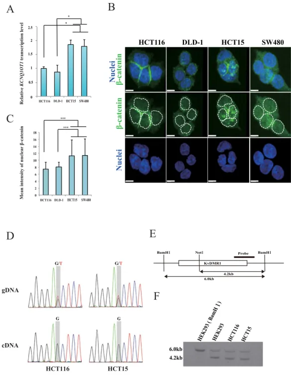
We have previously reported that aberration of KCNQ1OT1 transcription is frequently observed in colorectal cancer tissues and cell lines9. Here, we irst investigated KCNQ1OT1 expres- sion status in four colorectal cancer cell lines using quantitative reverse transcription PCR (qRT-PCR). he KCNQ1OT1 transcription level was increased 1.8-fold in HCT15 and SW480 cells compared to that in HCT116 and DLD-1 cells (Fig. 1A, *p < 0.05).An aberrant Wnt signaling pathway is implicated in the multistep processes for colorectal cancer development. In particular, nuclear accumulation of the key oncogenic factor β -catenin indicates activation of its target genes, which facilitate cancer promoting functions such as cell proliferation11,12,15. To determine if the KCNQ1OT1 lncRNA transcription status depends on the amount of β -catenin protein in the nucleus, we performed immunolu- orescence staining analysis using a β -catenin antibody and measured the relative intensities of the luorescence sig- nals of β -catenin in the nuclei of HCT116, DLD-1, HCT15 and SW480 cells (Fig. 1B,C and Supplementary Fig. S1). As shown in Fig. 1B,C, the luorescence intensity of nuclear β -catenin in HCT15 and SW480 cells was higher than that in HCT116 and DLD-1 cells (***p < 0.001). hese results indicated that KCNQ1OT1 lncRNA transcription levels could be increased by nuclear accumulation of β -catenin in colorectal cancer, suggesting that KCNQ1OT1 transcription could be regulated by β -catenin activity. Next, we performed single polymorphism nucleotide (SNP) analysis of KCNQ1OT1 transcript (bdSNP: rs231359) in order to validate the status of the allelic expression of KCNQ1OT1 in HCT116 and HCT15 cells which exhibit diferential distribution of β -catenin to cytoplasm and nuclear compartments of cells. Both the G and T alleles were detected in the genomic DNA. In contrast, only G allele was detected in cDNA samples (Fig. 1D). Furthermore, methylation-sensitive southern hybridization anal- ysis of those cells revealed that the methylated (6.0 kb) and unmethylated (4.2kb) alleles were detected at diferen- tially methylated regions (KvDMR) which play a crucial role in maintenance of the parent-of-origin-speciic gene expression pattern6 (Fig. 1E,F). In addition, two copies of KCNQ1OT1 locus are frequently observed in HCT116 and HCT15 cells by DNA luorescence in situ hybridization (DNA-FISH) analysis using a KCNQ1OT1 speciic DNA probe (Supplementary Table S1 online). hus, these results indicate that KCNQ1OT1 is monoallelically transcribed in HCT116 and HCT15 cells, suggesting that increase expression levels of KCNQ1OT1 lncRNA were attributed to an excess of β -catenin into nuclear.
overexpression of β-catenin increases KCNQ1OT1 transcription and expands lncRNA-coated
territory in colorectal cancer.
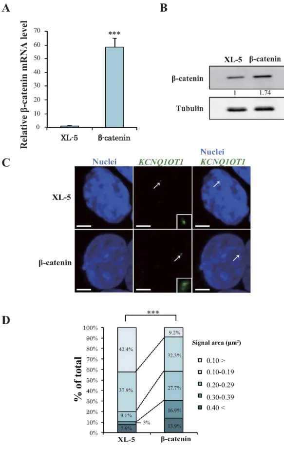
KCNQ1OT1 lncRNA has been reported to accumulate on its own gene and on targeted regulatory genes. Moreover, the genes on which it accumulates change depending on cell-type and/ or developmental stage8,16,17. To further explore the efect of β -catenin on KCNQ1OT1 transcription, we gener- ated HCT116 cells that transiently overexpressed β -catenin or the control vector (XL-5). Analysis by qRT-PCR and western blotting indicated that the transcription and protein level of β -catenin, respectively, was markedly increased in the β -catenin overexpressing clone compared with that in the control clone (Fig. 2A,B, ***p < 0.001). To investigate whether the KCNQ1OT1 lncRNA signal in the nucleus, i.e., lncRNA-coated territory, was increased by β -catenin activity, we performed RNA-luorescence in situ hybridization (RNA-FISH). We used a speciic probe to detect KCNQ1OT1 lncRNA, in β -catenin overexpressing HCT116 cells and measured the area of the lncRNA-coated territory. KCNQ1OT1 lncRNA-coated territory expanded in β -catenin overexpressing HCT116 cells compared with that in the cells transfected with control vector (Fig. 2C). Figure 2D summarizes the score of KCNQ1OT1 lncRNA-coated territory area in the β -catenin overexpressing and control cells. Compared to the control cells, the clone overexpressing β -catenin displayed a 3.0- and 5.6-fold increase in KCNQ1OT1 lncRNA-coated territory with a signal area of 0.20–0.29 and 0.30–0.39 µ m2 respectively, and a 4.6-fold decrease in territory with a signal area of < 0.10 µ m2 (Fig. 2D). To investigate whether endogenous KCNQ1OT1 transcription was also increased in β -catenin overexpressing HCT116 cells, we performed qRT-PCR analysis. As observed in Supplementary Fig. S3, overexpressing clones of β -catenin resulted in a 1.3-fold increase in KCNQ1OT1 tran- scription (*p < 0.05). hese results suggested that an increase in KCNQ1OT1 transcription through β -catenin activity eventually leads to an increase in its lncRNA-coated territory.Downregulation of β-catenin results in contraction of KCNQ1OT1 lncRNA-coated territory and
dysregulation of KCNQ1OT1-regulated genes in colorectal cancer.
To further explore the associa- tion between β -catenin activity and the area of KCNQ1OT1 lncRNA-coated territory, we performed knockdown of β -catenin transcription using short interfering RNA (siRNA) in the HCT15 cells that showed the highestFigure 1. Comparison of the levels of KCNQ1OT1 transcription and β-catenin protein in colorectal cancer cell lines. he colorectal cancer cell lines HCT116, DLD-1, HCT15 and SW480 were analyzed as follows. (A) Relative KCNQ1OT1 transcription levels were determined using qRT-PCR analysis. Transcription levels were normalized to GAPDH mRNA control in the same cell line. he transcription level in HCT116 cells was arbitrarily assigned as 1. Error bars represent means ± S.D. of three independent experiments (*p < 0.05). (B) Representative immunoluorescence staining of the expression of β -catenin (green). Nuclei (blue) were stained with DAPI. he dotted lines show individual nuclei. Scale bars represent 10 µ m. (C) Quantiication of nuclear β -catenin. he relative mean intensity of nuclear β -catenin staining of the colorectal cancer cell lines is shown. he graphs represent the average mean intensity of a single nucleus in each cell line (HCT116, n = 51; DLD-1, n = 56; HCT15, n = 53; SW480, n = 59; ***p < 0.001). Error bars represent means ± S.D. (D) Sanger sequencing chromatograms of KCNQ1OT1 genome DNA (gDNA; top) and cDNA (bottom), respectively. (E) Schematic representation of the KvDMR1 region is indicated. Black bar shows probe using methylation- sensitive southern hybridization. BamH I and Not I site show digestion site for using methylation-sensitive southern hybridization. (F) he 6.0 kb BamHI-digested fragment encompassing the KvDMR1 was digested with Not I, resulting in a 4.2 kb fragment. HEK293 cells were digested with BamH I and observed 6.0 kb digested fragment. he methylation-sensitive southern hybridization indicates statuses of methylation (6.0 kb) and unmethylation (4.2 kb) at the KvDMR1 in HEK293, HCT116 and HCT15 cells. Cropped blot images were used in this igure. Full-length blots and full-length gel image are presented in Supplementary Fig. S2.
Figure 2. Overexpression of β-catenin enhances transcription and lncRNA-coated territory of KCNQ1OT1 in HCT116 cells. HCT116 cells were transfected with a β -catenin expression vector or with the control XL-5 vector, and the cells were analyzed 24 h ater transfection as follows. (A) qRT-PCR analysis of relative β -catenin mRNA expression levels. Expression levels were normalized to GAPDH mRNA control. he expression level in HCT116 cells transfected with the XL-5 vector was arbitrarily assigned as 1. Error bars represent means ± S.D. of three independent experiments (***p < 0.001). (B) Western blotting analysis of β -catenin protein levels. Band intensities were densitometrically assayed and analyzed using FUJIFILM Multi Gauge sotware. he protein
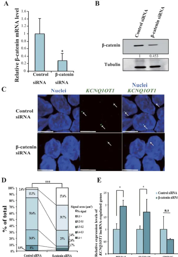
amount of nuclear β -catenin protein of the colorectal cancer cell lines tested (Fig. 1C). Knockdown of β -catenin in HCT15 cells reduced its mRNA expression to 35% of that of control cells (*p < 0.05; Fig. 3A). Additionally, the protein level of β -catenin was reduced by 50% (Fig. 3B). To evaluate the efect of decreased β -catenin expression on the extent of KCNQ1OT1 lncRNA-coated territory in HCT15 cells, we analyzed the KCNQ1OT1 lncRNA signal pattern with single-cell resolution using RNA-FISH. KCNQ1OT1 lncRNA-coated territory was contracted by downregulation of β -catenin (Fig. 3C). As summarized in Fig. 3D, compared to control cells, β -catenin siRNA transfected-cells showed a 10.6-fold increase nuclei without KCNQ1OT1 lncRNA signals and a 2.9-fold increase in KCNQ1OT1 lncRNA-coated territory area with a signal area of < 0.10 µ m2. Moreover, a 2.0- and 7.2-fold decrease in territory with a signal area of 0.10–0.19 and 0.2–0.29 µ m2, respectively. hese results suggested that a decrease in KCNQ1OT1 transcription by downregulation of β -catenin caused contraction of the KCNQ1OT1 lncRNA-coated territory in HCT15 cells.
We previously reported that KCNQ1OT1 lncRNA accumulates on its own gene region and that the solute carrier family 22 member 18 gene, SLC22A18, pleckstrin homology-like domain, family A member 2 gene, PHLDA2 and cyclin-dependent kinase inhibitor 1C gene, CDKN1C, which lie outside of the KCNQ1OT1 lncRNA transcriptional domain, is regulated by KCNQ1OT1 lncRNA spreading to other gene regions6,8. To examine whether reduction in β -catenin activity in the nucleus induces dysregulation of the SLC22A18, PHLDA2 and CDKN1C genes through contraction of KCNQ1OT1 lncRNA-coated territory, we analyzed the expression level of SLC22A18, PHLDA2 and CDKN1C mRNA in β -catenin siRNA-transfected and control siRNA-transfected HCT15 cells using qRT-PCR. he β -catenin siRNA-transfected HCT15 cells displayed a 1.9- and 1.7-fold increase in PHLDA2 and SLC22A18 mRNA levels compared with the control cells, respectively (Fig. 3E, *p < 0.05). In con- trast, no remarkable change in the CDKN1C mRNA level was observed (Fig. 3E), indicating that the remaining KCNQ1OT1 lncRNA-coated territory may function on CDKN1C adjacent to the KCNQ1OT1 transcription site. hus, these results indicated that a decrease in β -catenin activity can cause at least dysregulation of the expression of KCNQ1OT1-targeted genes through contraction of KCNQ1OT1 lncRNA-coated territory from SLC22A18 to PHLDA2 locus, suggesting that luctuation in KCNQ1OT1 lncRNA by the accumulation of nuclear β -catenin may play a role as an important step in colorectal cancer development.
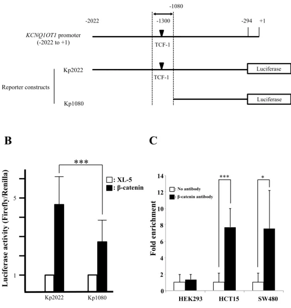
KCNQ1OT1 transcription is directly regulated by β-catenin in colorectal cancer. To investigate whether β -catenin regulates KCNQ1OT1 lncRNA transcription through association with the KCNQ1OT1 pro- moter and regulation of promoter activity, we investigated the presence of TCF consensus binding sites, which are known to be important for β -catenin regulation of other promoters, on the promoter region of KCNQ1OT1. We irst selected a TCF binding site that is predicted to be located closest to the transcription start site on KCNQ1OT1, based on the search sotware (MatInspector) for transcription factor binding sites that supports Genomatix sot- ware. Second, we constructed human KCNQ1OT1 promoter-luciferase reporter plasmids; plasmid Kp2022 con- tained a 2020-bp fragment from within the KCNQ1OT1 promoter region that included this TCF binding site, and plasmid Kp1080 contained a truncated fragment of the 5′ region of the KCNQ1OT1 promoter that did not include this TCF binding site (Fig. 4A). We then examined the efect of co-transfection of a β -catenin expressing vector or the control XL-5 vector on the transcriptional activity of these KCNQ1OT1 promoters by measurement of luciferase activity in HCT116 cells. KCNQ1OT1 promoter activity in the β -catenin/Kp1080 transfected cells, whose reporter lacked the selected TCF binding site, was only 40.6% that of the β -catenin/ Kp2022 transfected cells, whose reporter included the TCF binding site (Fig. 4B, ***p < 0.001). his result suggested that β -catenin regulated KCNQ1OT1 lncRNA transcription through an efect on the KCNQ1OT1 promoter.
To determine whether β -catenin directly binds to a TCF site in the KCNQ1OT1 promoter, we performed chromatin immunoprecipitation (ChIP) analysis. In this assay, crosslinking of β -catenin to the TCF binding site in the KCNQ1OT1 promoter in cells of the colorectal cancer cell lines HCT15 and SW480 was assayed (Fig. 4C). he result indicated that β -catenin directly binds to a TCF-1 site on the KCNQ1OT1 promoter. hus, in vivo, β -catenin can directly regulate KCNQ1OT1 transcription suggesting that the regulation of KCNQ1OT1 lncRNA by β -catenin signaling may be involved in the multiple processes of colorectal cancer development.
Discussion
We reported here that the β -catenin directly regulated KCNQ1OT1 lncRNA transcription through target- ing its promoter region. Moreover, β -catenin afected the extent of KCNQ1OT1 lncRNA-coated territory in a dose-dependent manner, resulting in at least dysregulation of KCNQ1OT1-targeted genes in colorectal cancer.
Diferentially methylated regions (DMR) associated with imprinted clusters function as important regions for maintenance of the parent-origin-speciic gene expression pattern, which is known as the imprinting con- trol region (ICR). he ICR of the KCNQ1 cluster harbors the KCNQ1OT1 promoter region7. A previous study reported that the transcription factor NF-Y directly binds to the Kcnq1 ICR, resulting in loss of Kcnq1ot1 tran- scription18. NF-Y mediated Kcnq1ot1 transcription thereby plays a crucial role in regulating the bidirectional silencing of neighboring imprinted genes18. Here, we found that β -catenin also binds to the proximal region of levels of β -catenin were normalized to the levels of tubulin. he protein level in HCT116 cells transfected with the XL-5 vector was arbitrarily assigned as 1. Cropped blot images were used in this igure. Full-length blots are presented in Supplementary Fig. S3. (C) Representative RNA-FISH analysis of KCNQ1OT1 lncRNA-coated territory (green; arrows) and the insets show enlarged images of KCNQ1OT1 lncRNA. Nuclei (blue) were stained with DAPI. Scale bars represent 3 µ m. (D) Quantiication of KCNQ1OT1 lncRNA-coated territory in RNA-FISH analysis. he 100% stacked bar charts indicate the percentage of nuclei displaying the indicated signal area of KCNQ1OT1 lncRNA-coated territory (β -catenin, n = 65; XL-5, n = 66, ***p < 0.001)
Figure 3. Downregulation of β-catenin reduced transcription, contracted lncRNA-coated territory of KCNQ1OT1, and increased in PHLDA2 and SLC22A18 expression in HCT15 cells. HCT15 cells were transfected with a β -catenin siRNA or a control siRNA, and the cells were analyzed 72 h ater transfection as follows. (A) qRT-PCR analysis of relative β -catenin mRNA expression levels. Expression levels were normalized to GAPDH mRNA control in each transfected cell type. he expression level in HCT15 cells transfected with control siRNA was arbitrarily assigned as 1. Error bars indicate the means ± S.D. of three independent experiments (*p < 0.05). (B) Western blotting analysis of the β -catenin protein level. Band intensities were densitometrically assayed and analyzed using FUJIFILM Multi Gauge sotware. he protein levels of β -catenin were normalized to the levels of tubulin. he protein level in HCT15 cells transfected with control siRNA was arbitrarily assigned as 1. Cropped blot images were used in this igure. Full-length blots are presented in Supplementary Fig. S4. (C) Representative RNA-FISH analysis of KCNQ1OT1 lncRNA-coated territory (green;
the ICR within the KCNQ1OT1 promoter, and afects the expression of KCNQ1OT1 and of KCNQ1OT1-targeted genes (Figs 2,3 and 4). In addition, an excess of β -catenin protein in cells is a key factor for the development of colorectal cancer. Indeed, the accumulation of β -catenin in the nucleus is frequently observed in colorectal cancer cell lines (Fig. 1)13. It is therefore likely that at least two distinct KCNQ1OT1 regulatory factors exist at the KCNQ1 ICR.
he lncRNA-mediated gene silencing behavior of KCNQ1OT1 lncRNA resembles that of Xist RNA in terms of chromatin association and cis-limited epigenetic silencing8. Xist RNA is essential for X chromosome inacti- vation (XCI). Xist RNA accumulates and spreads along the X chromosome, which expresses its RNA, and then recruits gene silencing complexes that include histone methyltransferases and the polycomb group proteins Eed/Ezh2/Suz12, which ultimately establishes XCI by induction of heterochromatin formation10. Furthermore, we found that genes such as Jarid1c or Utx that escape from XCI are always located at the periphery or out- side of the Xist RNA-coated territory in C2C12 normal mouse myoblast cells19. In contrast, during the stage of prenatal development, the mouse Kcnq1ot1 lncRNA-coated territory area in placental derived cells is largely expanded compared to that of embryonic cells17,20. his expansion is because the Kcnq1ot1 lncRNA-silencing target locus difers in the placenta and the embryo17,20. hus, these evidences suggest that expansion of cis-limited lncRNA-coated territory is coordinated with the silencing behavior of its coated-genes. In the present study, we demonstrated that the mRNA expression of SLC22A18 and PHLDA2, which are regulated by KCNQ1OT1 lncRNA spreading, was increased by the knockdown of β -catenin in colorectal cancer cells (Fig. 3). herefore, aberration of the extent of the KCNQ1OT1 lncRNA-coated territory that is caused by an excess of nuclear β -catenin may play a signiicant role in the process of cancer development. However, We do not rule out the possibility that aberra- tion of KCNQ1OT1 lncRNA-coated territory may signiicantly afect genes and microRNAs involved in cancer initiation or progression rather than the expression of speciic genes that located on chromosome 11, which would ultimately lead to disruption of the balance of gene expression in a whole cell.
Aberration of lncRNA transcription including that of lncRNA-p21, KCNQ1OT1, colorectal cancer associ- ated transcript 1 long isoform (CCAT1-L), HOTAIR, E2F4 antisense transcript and metastasis associated lung adenocarcinoma transcript 1 (MALAT1) has been observed in colorectal cancer that harbors alteration of Wnt/β -catenin signaling pathways9,15,21–24. In this respect it is interesting that dysregulation of the E2F4 antisense transcript and of MALAT1 transcription, which cause accumulation of β -catenin in the nucleus, strongly contrib- utes to the development of colorectal cancer15,24. We demonstrated that β -catenin directly activates KCNQ1OT1 transcription through binding to its promoter region (Figs 2,3 and 4). hese combined evidences indicated that aberrations of some lncRNAs are strongly associated with Wnt/β -catenin signaling pathways that contribute to colorectal cancer progression, and which are important during the multistep processes of neoplastic development.
We found that accumulation of nuclear β -catenin induced dysregulation of KCNQ1OT1 transcription in colorectal cancer cells (Figs 2 and 3). his phenomenon has also been observed in various other cancers including melanoma, ovarian carcinoma and gastric cancer25–27. hus, these indings strongly support the hypothesis that dysregulation of KCNQ1OT1 transcription by nuclear β -catenin may be involved in the development of various cancers.
In conclusion, our study demonstrated that excessive nuclear β -catenin causes aberration in the extent of KCNQ1OT1 lncRNA-coated territory, suggesting that a change in its lncRNA-coated territory proile may strongly contribute to the multistep processes that lead to the establishment of malignant colorectal cancer. However, the mechanism by which gene transcription is inluenced by aberration of the extent of KCNQ1OT1 lncRNA-coated territory remains to be clariied. Further studies aimed at identiication of KCNQ1OT1 lncRNA-targeted genes and the factors that maintain and regulate KCNQ1OT1 lncRNA-coated territory will be necessary in order to clar- ify the signiicance of the regulation of the extent of KCNQ1OT1 lncRNA-coated territory through an oncogenic signaling pathway such as Wnt/β -catenin in cancer development.
Materials and Methods
Cell lines and Cell culture.
HCT116, DLD-1, HCT15, SW480 and HEK293 cells were purchased from the ATCC (#CCL-247, #CCL-221, #CCL-225, #CCL-228, #CRL-1573, respectively). HCT116, DLD-1, HCT15 and HEK293 cells were cultured in Dulbecco’s modiied Eagle’s medium (DMEM; Sigma-Aldrich, St. Louis, MO, USA) supplemented with 10% fetal bovine serum (FBS; HyClone, Logan, UT, USA). SW480 cells were cultured in Leibovitz’s L-15 medium (hermo Fisher Scientiic, Gibco Cell Culture, Rockford, IL, USA) supplemented with 10% FBS.plasmid transfection and siRNA-mediated knockdown.
Cells were transfected with plasmid or siRNA using Lipofectamine 2000 (hermo Fisher Scientiic, Invitrogen). For overexpression of β -catenin, 1 × 10⁶ HCT116 cells were seeded in each well of six wells plates and were transfected 24 h ater seeding with 4 µ g of β -catenin expression vector or XL-5 vector. Both the β -catenin expression vector and XL-5 were purchased from OriGene (#SC107921, OriGene, Rockville, MD, USA). For knockdown of β -catenin, 5 × 10⁵ HCT15 cells were arrows). Nuclei (blue) were stained with DAPI. Scale bars indicate 10 µ m. (D) Quantiication of KCNQ1OT1 lncRNA-coated territory in RNA-FISH analysis. he 100% stacked bar charts indicate the percentage of nuclei displaying the indicated signal area of KCNQ1OT1 lncRNA-coated territory and no signal (β -catenin siRNA, n = 116; control siRNA, n = 113, ***p < 0.001). (E) qRT-PCR analysis of the relative mRNA expression levels of KCNQ1OT1 lncRNA-regulated genes. Expression levels were normalized to the level of GAPDH mRNA in each transfected cell type. he expression level in HCT15 cells transfected with control siRNA was arbitrarily assigned as 1. Error bars indicate the means ± S.D. of three independent experiments (*p < 0.05).-294
-2022 +1
TCF-1 -1080
Luciferase
Kp2022 Luciferase
TCF-1 KCNQ1OT1 promoter
(-2022 to +1)
Reporter constructs
-1300
5
1
Kp2022
***
Luciferase activity (Firefly/Renilla)
A
B C
Kp1080
Kp1080 : XL-5 : β-catenin
0 2 4 6 8 10 12 14
HEK293 HCT15 SW480
*** *
Fold enrichment
: No antibody : β-catenin antibody
Figure 4. β-catenin directly regulated KCNQ1OT1 promoter activity in vivo in colorectal cancer cells via binding to a TCF-1 site in the KCNQ1OT1 promoter. (A) Schematic representation of the KCNQ1OT1 promoter region and the reporter constructs used for the luciferase assay. he region of the KCNQ1OT1 promoter shown is from the transcription start site (TSS, + 1) to − 2022. he predicted β -catenin binding site (TCF-1, black inverted triangle) is located − 1300 up-stream from the TSS. Arrows indicate the PCR ampliication region − 1211 to − 1351 for the ChIP assay. he two luciferase reporter vectors used in the luciferase assay are indicated. One reporter contains the KCNQ1OT1 promoter region from − 294 to − 2022 (Kp2022). he other reporter contains a truncated promoter in which the TCF-1 region was deleted (Kp1080). (B) hese reporter or control vectors were co-transfected with into HCT116 cells. 24 h later, luciferase activity was measured. Renilla luciferase values were normalized to total protein concentration. Luciferase activity in HCT116 cells transfected with Kp2022 and the XL-5 vector or Kp1080 and the XL-5 were arbitrarily set at 1, respectively. Data are presented as the means ± S.D. of six independent experiments (***p < 0.001). (C) he ChIP assay of β -catenin binding to the TCF site in the KCNQ1OT1 promoter in the human embryonic kidney (HEK293) cell line and the colorectal cancer cell lines HCT15 and SW480. he assay was performed with or without β -catenin antibody. DNA was recovered from immunoprecipitated and nonimmunoprecipitated (input) chromatin and analyzed by qRT-PCR. he fold enrichment of target sequence in precipitated DNA compared with input DNA was calculated by comparison of the threshold cycle value of the sample of precipitated DNA with the standard curve generated from those of input DNA. Data are presented as the means ± S.D. of three independent experiments (***p < 0.001 and *p < 0.05).
seeded in each well of 6 plates and were transfected 72 h ater seeding with 100 pmol of β -catenin siRNA (siRNA ID 42816, hermo Fisher Scientiic, Ambion) or control (#SN-1003, Bioneer, Seoul, Korea).
Luciferase assay.
he Luciferase assay was performed as described previously28. In brief, a segment of the human KCNQ1OT1 promoter region containing the predicted binding site for TCF-1 based on the Transcription Element Search System (http://www.cbil.upenn.edu/tess), Genomatix GEMS Lancher (http://www.genom- atix.de) and Genetyx Ver.10 sotware (Genetyx, Tokyo, Japan) was PCR-ampliied from cDNA and inserted into a BglII/Acc65I-digested luciferase reporter vector pGL3-basic (Promega, Madison, WI, USA). he PCR primer sets were designed to amplify the upstream KCNQ1OT1 promoter region from -2022 to -294. he PCR primer sets used were: forward primer: 5′ -GGGGTACCCCAGGTGACAAGGTGCAGGCGC and reverse primer: 5′ -ACAGAGTTCCTCGTTGGGAGCTTGAAGATCTTC. For the truncated luciferase reporter con- struct without the TCF-1 site (Kp1080), deletion of the upstream KCNQ1OT1 promoter region − 2022 from− 1080 was performed using PCR-based site-directed mutagenesis (Toyobo, Osaka, Japan). Kp1080 was PCR-ampliied from the Kp2022 luciferase reporter construct according to the manufacturer’s protocol. he PCR primer sets used were: forward primer: 5′ -CGGAGGTGGGAATCCCCGTTG, and reverse primer: 5′ -GGGGTACCTATCGATAGAGAAATG.
Methylation-sensitive southern hybridization.
Methylation-sensitive southern hybridization was per- formed using DIG High Prime DNA Labeling and Detection Kit (Roche Applied Science, Penzberg, Germany) according to the manufacturer’s instructions. In brief, 20 µ g of genome DNA from normal and colorectal cancer cell lines were digested with BamHI and NotI overnight, resolved by gel electrophoresis, and transferred to High bond N + (GE Healthcare, Piscataway, NJ, USA). he PCR products were labeled by Digoxigenin-11-dUTP and used as probe. he PCR primer sets used were: forward primer: 5′ - TCTCTCTGGGAGGGTTTGAA and reverse primer: 5′ - TTACTTCGCCCCCTAATTCCT. Immunoreactive bands were analyzed using a Luminescent Image Analyzer LAS-4000 (Fujiilm, Tokyo, Japan).Western blotting.
Western blotting was performed as described previously28. he membranes were blot- ted with a rabbit monoclonal antibody against the human β -catenin antigen (1:2000, #8480, Cell Signaling Technology, Tokyo, Japan), or with a rabbit polyclonal antibody against the human α /β -tubulin antigen (1:2000,#2148, Cell Signaling Technology) and the appropriate standard peroxidase-labeled anti-rabbit IgG secondary antibody was used according to the manufacturer’s instructions (GE Healthcare). Immunoreactive bands were analyzed using a Luminescent Image Analyzer LAS-4000 (Fujiilm) and β -catenin protein levels were quantiied using Multi Gauge V3.0 sotware (Fujiilm).
sNp analysis.
Search of allelic speciic SNP on KCNQ1OT1 locus was used dbSNP website (http://www.ncbi. nlm.nih.gov/SNP/) and PCR products spanning SNP from colorectal cancer cell lines were sequenced (Euroins Genomics, Tokyo, Japan). he PCR primer sets were: forward primer: 5′ -GGGTAGGCTGGTCACGTTTA and reverse primer: 5′ -AGTCCCCTGTAGATTCTGGG. he sequence data was analyzed using Finch TV (http:// www.geospiza.com).ChIp assay.
ChIP assay was performed as described previously9. A mouse monoclonal antibody against human β -catenin (#610153, BD Japan, Tokyo, Japan) was used for the ChIP assay. Precipitated DNA was ampliied using an Applied Biosystems StepOne thermal cycler system and a SYBR green PCR kit (hermo Fisher Scientiic, Applied Biosystems) and the following TCF-1 site-speciic detection primers: forward; 5′ - GGTTCTGAGTCCGCGCTATT, and reverse; 5′ - GGATTCCCACCTCCGATCCT.Immunofluorescence staining.
Cells (2.4 × 10⁴) were seeded on 24 × 60 mm micro cover glass and were incubated at 37 °C for 24 h. he cells were then ixed with 4% paraformaldehyde at room temperature for 10 min. Ater two cold PBS washes, the cells were treated 0.5% saponin (Sigma-Aldrich) and 0.5% Triton X-100 (Sigma-Aldrich) at room temperature for 20 min and were then kept in 20% glycerol (Wako, Osaka, Japan) / PBS for 2 h. Subsequently, the cells were passed six times through liquid nitrogen. Ater two PBS washes, the cells were blocked with 5% bovine serum albumin (Sigma-Aldrich) in PBS (5% BSA/PBS). Rabbit monoclonal antibody against the human β -catenin antigen diluted to 1:500 in 3% BSA/PBS was applied to the cells. he cells were washed twice in PBS with 0.05% Tween-20 (PBST; Sigma-Aldrich), followed by addition of the sec- ondary antibody Alexa Fluor 488 goat-anti-rabbit IgG (hermo Fisher Scientiic, Invitrogen) diluted to 1:600 in 3% BSA/PBS. Ater a PBST wash, the cells were stained with DAPI (Vector Laboratories, Burlingame, CA). Immunoluorescence staining was observed by using the confocal microscope LSM780 (Carl Zeiss, Oberkochen, Germany). Intensity was measured using the ZEN 2010 sotware (Carl Zeiss). Note that all Immunoluorescence staining experiments were performed in parallel and, for detection, exposure and interval time was kept the same in several experiments.RNA-FIsH.
RNA-FISH analysis was performed as previously described29. Note that micro cover glass seeded cells were not heat-denatured in order to avoid hybridization of probes to genomic DNA. Probes were labe- led with DNP-11-dUTP (Perkin Elmer Japan, Kanagawa, Japan) using a Nick translation Mix (Roche Applied Science). Anti-DNP-rabbit IgG (Sigma-Aldrich) and Alexa Fluor 488 goat-anti-rabbit IgG (Thermo Fisher Scientiic, Invitrogen) were diluted to 1:500 and used to detect DNP-labeled probes. he U90095 P1-derived arti- icial chromosome (PAC) genomic probe was used for RNA-FISH. he U90095 PAC was obtained from BACPAC Resource Center (BPRC) at Children’s Hospital Oakland Research Institute (CHORI). Signals of the KCNQ1OT1transcript were detected using the confocal microscope LSM780 (Carl Zeiss). Images were minimally enhanced for brightness and contrast to resemble that which was seen by eye through the microscope. In setting the confo- cal microscope, the objective lens used was the Plan-Apochromat 63x/1.40 Oil DIC M27, and the lasers used were 405 nm (DAPI) and 488 nm (Alexa 488). he image sizes were: x, 512; y, 512; z, 13, and 12-bit. Area was measured using the ZEN 2010 sotware (Carl Zeiss). Note that all RNA-FISH experiments were performed in parallel and, for detection, exposure and interval time was kept the same in several experiments.
DNA-FIsH.
DNA-FISH analysis was performed as previously described30. Metaphase images were captured digitally with a cooled CCD camera equipped with an ISIS (Carl Zeiss) and then copy number of KCNQ1OT1 locus were counted.qRt-pCR.
RNA isolation and reverse transcriptase (RT)-PCR were performed as described p re v i o u s l y2 8. KC N Q 1 OT 1 t r a n s c r i p t i o n , a n d β - c at e n i n a n d S LC 2 2 A 1 8 m R NA e x p re s - sion were detected using qRT-PCR. KCNQ1OT1 transcription, and β -catenin and SLC22A18 mRNA expression, were analyzed using the following specific primers. KCNQ1OT1: forward, 5′ -CT T TGCAGCAACCTCCT TGT; reverse, 5′ -TGGGGTGAGGGATCTGAA. β -catenin: for- ward, 5′ -TCTGATAAAGGCTACTGTTGGATTGA; reverse, 5′ -TCACGCAAAGGTGCATGATT; SLC22A18: forward, 5′ -CATCTTGCTTACCTACGTGCTG; reverse, 5′ -CCCAGTTTCCGAGACAGGTA. PHLDA2: forward, 5′ - TCCAGCTATGGAAGAAGAAGC ; reverse, 5′ - GTGGTGACGATGGTGAAGTACA. CDKN1C: forward, 5′ - CTCCGCAGCATCCACGAT; reverse, 5′ - GGTGCGCACTAGTACTGGGA. cDNA was ampliied using an Applied Biosystems StepOne thermal cycler system and a SYBR green PCR kit (hermo Fisher Scientiic, Applied Biosystems). he mRNA level was normalized to human GAPDH mRNA (PCR primers: for- ward, 5′ -AGCCACATCGCTCAGACAC; reverse, 5′ -GCCCAATACGACCAAATCC).statistical Analysis.
Data from more than two separate experiments are presented as means ± S.D. Signiicance was established at P-values less than 0.05 using an unpaired Two-tailed Student’s t test. In RNA-FISH analyses, we used the Chi-squared test to determine whether there was any signiicant diference in the distribu- tion of area ratio between control cells and β -catenin expressing plasmid or siRNA transfected-cells.References
1. homas, D. J. et al. he ENCODE Project at UC Santa Cruz. Nucleic Acids Res 35, D663–7 (2007).
2. Banfai, B. et al. Long noncoding RNAs are rarely translated in two human cell lines. Genome Res 22, 1646–57 (2012). 3. Pelechano, V. & Steinmetz, L. M. Gene regulation by antisense transcription. Nat Rev Genet 14, 880–93 (2013).
4. Kotake, Y. et al. Long non-coding RNA ANRIL is required for the PRC2 recruitment to and silencing of p15(INK4B) tumor suppressor gene. Oncogene 30, 1956–62 (2011).
5. Bhan, A. & Mandal, S. S. LncRNA HOTAIR: A master regulator of chromatin dynamics and cancer. Biochim Biophys Acta 1856, 151–64 (2015).
6. Mitsuya, K. et al. LIT1, an imprinted antisense RNA in the human KvLQT1 locus identiied by screening for diferentially expressed transcripts using monochromosomal hybrids. Hum Mol Genet 8, 1209–17 (1999).
7. Horike, S. et al. Targeted disruption of the human LIT1 locus deines a putative imprinting control element playing an essential role in Beckwith-Wiedemann syndrome. Hum Mol Genet 9, 2075–83 (2000).
8. Murakami, K., Oshimura, M. & Kugoh, H. Suggestive evidence for chromosomal localization of non-coding RNA from imprinted LIT1. J Hum Genet 52, 926–33 (2007).
9. Nakano, S. et al. Expression proile of LIT1/KCNQ1OT1 and epigenetic status at the KvDMR1 in colorectal cancers. Cancer Sci 97, 1147–54 (2006).
10. Chaligne, R. & Heard, E. X-chromosome inactivation in development and cancer. FEBS Lett 588, 2514–22 (2014).
11. Cancer Genome Atlas, N. Comprehensive molecular characterization of human colon and rectal cancer. Nature 487, 330–7 (2012). 12. Sebio, A., Kahn, M. & Lenz, H. J. he potential of targeting Wnt/beta-catenin in colon cancer. Expert Opin her Targets 18, 611–5
(2014).
13. Rosin-Arbesfeld, R., Clife, A., Brabletz, T. & Bienz, M. Nuclear export of the APC tumour suppressor controls beta-catenin function in transcription. EMBO J 22, 1101–13 (2003).
14. Han, J. et al. Nuclear expression of beta-catenin promotes RB stability and resistance to TNF-induced apoptosis in colon cancer cells. Mol Cancer Res 11, 207–18 (2013).
15. Yochum, G. S., Cleland, R., McWeeney, S. & Goodman, R. H. An antisense transcript induced by Wnt/beta-catenin signaling decreases E2F4. J Biol Chem 282, 871–8 (2007).
16. hakur, N., Kanduri, M., Holmgren, C., Mukhopadhyay, R. & Kanduri, C. Bidirectional silencing and DNA methylation-sensitive methylation-spreading properties of the Kcnq1 imprinting control region map to the same regions. J Biol Chem 278, 9514–9 (2003). 17. Redrup, L. et al. he long noncoding RNA Kcnq1ot1 organises a lineage-speciic nuclear domain for epigenetic gene silencing.
Development 136, 525–30 (2009).
18. Pandey, R. R. et al. NF-Y regulates the antisense promoter, bidirectional silencing, and diferential epigenetic marks of the Kcnq1 imprinting control region. J Biol Chem 279, 52685–93 (2004).
19. Murakami, K. et al. Identiication of the chromatin regions coated by non-coding Xist RNA. Cytogenet Genome Res 125, 19–25 (2009).
20. Kanduri, C. Kcnq1ot1: a chromatin regulatory RNA. Semin Cell Dev Biol 22, 343–50 (2011).
21. Zhai, H. et al. Clinical signiicance of long intergenic noncoding RNA-p21 in colorectal cancer. Clin Colorectal Cancer 12, 261–6 (2013).
22. Xiang, J. F. et al. Human colorectal cancer-speciic CCAT1-L lncRNA regulates long-range chromatin interactions at the MYC locus. Cell Res 24, 513–31 (2014).
23. Svoboda, M. et al. HOTAIR long non-coding RNA is a negative prognostic factor not only in primary tumors, but also in the blood of colorectal cancer patients. Carcinogenesis 35, 1510–5 (2014).
24. Ji, Q. et al. Resveratrol inhibits invasion and metastasis of colorectal cancer cells via MALAT1 mediated Wnt/beta-catenin signal pathway. PLoS One 8, e78700 (2013).
25. Clements, W. M. et al. beta-Catenin mutation is a frequent cause of Wnt pathway activation in gastric cancer. Cancer Res 62, 3503–6 (2002).
26. Rimm, D. L., Caca, K., Hu, G., Harrison, F. B. & Fearon, E. R. Frequent nuclear/cytoplasmic localization of beta-catenin without exon 3 mutations in malignant melanoma. Am J Pathol 154, 325–9 (1999).
27. Saegusa, M. & Okayasu, I. Frequent nuclear beta-catenin accumulation and associated mutations in endometrioid-type endometrial and ovarian carcinomas with squamous diferentiation. J Pathol 194, 59–67 (2001).
28. Ohira, T. et al. miR-19b regulates hTERT mRNA expression through targeting PITX1 mRNA in melanoma cells. Sci Rep 5, 8201 (2015).
29. Habermann, F. A. et al. Arrangements of macro- and microchromosomes in chicken cells. Chromosome Res 9, 569–84 (2001). 30. Tomizuka, K. et al. Functional expression and germline transmission of a human chromosome fragment in chimaeric mice. Nat
Genet 16, 133–43 (1997).
Acknowledgements
his work was supported in part by grants from the Sanyo Broadcasting Foundation of Japan.
Author Contributions
H.K. and N.S. designed the experiments and analyzed the data. N.S., T.O., M.K., D.I., Y.N. and H.T. performed the experiments and contributed to discussion. H.K. and N.S. wrote the manuscript. H.T., Y.N. and M.O. contributed to data analysis and discussion. H.K. conceived and managed the project. All authors revised and edited the manuscript.
Additional Information
Supplementary information accompanies this paper at http://www.nature.com/srep Competing inancial interests: he authors declare no competing inancial interests.
How to cite this article: Sunamura, N. et al. Regulation of functional KCNQ1OT1 lncRNA by β-catenin. Sci. Rep. 6, 20690; doi: 10.1038/srep20690 (2016).
his work is licensed under a Creative Commons Attribution 4.0 International License. he images or other third party material in this article are included in the article’s Creative Commons license, unless indicated otherwise in the credit line; if the material is not included under the Creative Commons license, users will need to obtain permission from the license holder to reproduce the material. To view a copy of this license, visit http://creativecommons.org/licenses/by/4.0/