INVITED PAPER
Special Section on Electronic DisplaysSynthesis and Photoluminescence Properties of HEu 1 − x Gd x (MoO 4 ) 2 Nanophosphor
Mizuki WATANABE†a), Kazuyoshi UEMATSU††, Sun Woog KIM†, Kenji TODA†b), andMineo SATO††,Nonmembers
SUMMARY New HEu1−xGdx(MoO4)2 nanophosphors were synthe- sized by a simple one-step ion-exchange method. These nanophosphors have rod-like particle morphology with 0.5–15µm in length and outer di- ameters in the range of 50–500 nm. By optimization of the composition, the highest emission intensity was obtained for the samples withx=0.50 for both KEu1−xGdx(MoO4)2and HEu1−xGdx(MoO4)2.
key words: Nanophosphor, soft chemical process, rare earth molybdate, layered structure.
1. Introduction
Thin films with dispersed nanophosphor have been inves- tigated for their use in displays, LEDs, and solar cells be- cause of their excellent optoelectronic properties and low light-scattering intensity [1]–[5]. In particular, transparent displays with thin films containing dispersed nanophosphor attracted much attention in the context of next-generation displays [6]. Such transparent displays have been demon- strated mainly in the field of organic light-emitting de- vices [7]. In addition, stable low-scattering suspensions of inorganic phosphors in organic solvents have to be devel- oped to improve the durability of flexible and transparent displays [8], [9]. However, almost all inorganic phosphors with high luminescence efficiency contain micron-sized par- ticles, which have strong scattering characteristics. The desired insignificant scattering can be obtained for parti- cles smaller than 50 nm [10]. Therefore, nano-sized inor- ganic phosphors are required for the fabrication of flexible and transparent displays. However, such nanophosphors ag- glomerate easily in organic solvents. Therefore, the fabrica- tion of stable suspensions of nanophosphors is required.
Several wet chemical methods have been reported for the preparation of nanophosphors, such as the hydrother- mal method [11], polymerized complex method [12], sol- gel method [13], and others. These methods offer several advantages, such as homogeneity, phase purity, and narrow size distribution. However, these methods require a special
Manuscript received March 5, 2014.
Manuscript revised June 15, 2014.
†The authors are with Graduate School of Science and Tech- nology, Niigata University, 8050 Ikarashi 2-nocho, Niigata 950- 2181, Japan.
††The authors are with Department of Chemistry and Chemical Engineering, Niigata University, 8050 Ikarashi 2-nocho, Niigata 950-2181, Japan.
a) E-mail: [email protected] b) E-mail: [email protected]
DOI: 10.1587/transele.E97.C.1063
reactor and special precursor materials to achieve complete dissolution in solvent solutions. In addition, methods for the synthesis of clear suspensions of nanophosphors have not been well established until now [14].
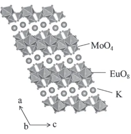
In this paper, we propose a new simple synthesis method via ion-exchange for preparing nanophosphors with high luminescence efficiency. We selected a triclinic al- kali rare earth molybdate, KEu(MoO4)2 as the precur- sor material to synthesize emissive nanophosphors. Tri- clinic KEu(MoO4)2 is a well-known phosphor material and presents excellent red emission due to the 4f−4f transition of Eu3+[15]. Furthermore, triclinic KEu(MoO4)2has a lay- ered structure, which is shown in Fig. 1, using the VESTA program [16]. In the crystal structure of KEu(MoO4)2, the polyhedral EuO8 layers are situated between two tetra- hedral MoO4 layers along the c-axis with the exchange- able potassium cations located between these (Eu(MoO4)2)n
layers. Therefore, the K+ ions in KEu(MoO4)2 are ex- pected to be exchanged by other ions via ion-exchange methods with corresponding easy control of the particle size and morphology. However, KEu(MoO4)2is easily dis- solved in concentrated acidic solutions; therefore, there have been no reports on the successful ion-exchange of molyb- date and the related synthesis of nanophosphors. On the other hand, we have successfully synthesized HEu(MoO4)2
nanophosphor with rod-like particle morphology by sim- ple ion-exchange methods in dilute acid, and the solution with the dispersed HEu(MoO4)2nanophosphor showed high transparency and strong red-emission due to the f–f transi- tion of Eu3+[17]. In order to further enhance the emission
Fig. 1 Crystal structure of KEu(MoO4)2.
Copyright c⃝2014 The Institute of Electronics, Information and Communication Engineers
efficiency of HEu(MoO4)2nanophosphor, part of the Eu3+ ions in the HEu(MoO4)2lattice were substituted by smaller Gd3+ ions, and the particle morphology and luminescence properties of HEu1−xGdx(MoO4)2were investigated.
2. Experimental
KEu1−xGdx(MoO4)2 (0.00≤x≤1.00) were synthesized by a conventional solid-state reaction method. K2CO3 (pu- rity 99.95%; Kanto Chemical Co. Inc.), Gd2O3 (pu- rity 99.99%; Shinetsu Chemical Co. Inc.), Eu2O3 (purity 99.99%; Shinetsu Chemical Co. Inc.), and MoO3 (purity 99.99%; Kojundo Chemical Co. Inc.) were mixed using a mortar and pestle with acetone; the mixture was then cal- cined at 700◦C for 6 h in air. HEu1−xGdx(MoO4)2samples were obtained by the H+exchange of KEu1−xGdx(MoO4)2
in HNO3solution (0.01 M, 100 mL) at room temperature for 7 days. After stirring, the solutions were isolated by suc- tion filtration using a membrane filter (ADVANTEC MFS, INC., mixed cellulose ester, pore size: 0.45µm, diameter:
47 mm). The samples were washed with deionized water for 12 h and then dried at 50◦C for 24 h. The obtained HEu1−xGdx(MoO4)2 powder samples were redispersed in deionized water (pH=7).
The obtained samples were characterized by powder X- ray diffraction (XRD, MX-Labo; Mac Science Ltd.) to iden- tify the crystal structure and the sample composition was determined by X-ray fluorescence analysis (XRF, SII, SEA 1200 VX). The sample morphology was characterized by scanning electron microscopy (SEM, Hitachi, S-4300SD).
The emission (PL) and excitation (PLE) spectra were mea- sured at room temperature with a spectrofluorometer (Jasco, FP-6500/6600); emission spectra were obtained for exci- tation at 309 nm, and excitation spectra were obtained for emission at 614 nm.
3. Results and Discussion
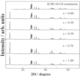
Figure 2 shows the XRD patterns of the precursor KEu1−xGdx(MoO4)2(0.00≤x≤1.00) phosphors. The XRD patterns of all samples were in good agreement with single- phase triclinic alkali rare earth molybdate.
Figure 3 shows excitation and emission spectra of the precursor KEu1−xGdx(MoO4)2(x=0.00 and 0.50) phos- phors. The excitation spectra of all samples consisted of a strong broad band in the range from 220 to 350 nm, cor- responding to the charge-transfer (CT) transition of O2−– Mo6+. Some strong narrow peaks are observed between 360 and 500 nm and are attributed to the 4f–4f transitions of the Eu3+ ion. On the other hand, the CT band of Eu3+–O2−is not clearly observed in the excitation spectra, possibly due to the overlap of the CT band with that of the molybdate group [15], [17]–[19]. In the emission spectra, all peaks corresponded to the Eu3+ 4f–4f transition. The emission peak intensity corresponding to the5D0–7F2 electric dipole transition at 614 nm is higher than that of the5D0–7F1mag- netic dipole transition at 592 nm, suggesting that the Eu3+
Fig. 2 XRD patterns of KEux−1Gdx(MoO4)2(0.00≤x≤1.00) synthesized by the solid-state reaction method at 700◦C for 6 h in air.
Fig. 3 Excitation (broken line) and emission (solid line) spectra of KEu1−xGdx(MoO4)2(x=0.00 ((a), gray line) andx=0.50 ((b), black line)) phosphors.
ions occupy sites in the KEu(MoO4)2 lattice without in- version symmetry. The ratio I614 nm(5D0–7F2)/I592 nm(5D0–
7F1) of KEu(MoO4)2 (12.5) decreased upon Gd3+ doping and was 10.8 in case of KEu0.50Gd0.50(MoO4)2. This re- sult indicates that the symmetry of the Eu3+ site in the KEu1−xGdx(MoO4)2lattice was higher than that of the sam- ple without the Gd3+ doping. It is well known that the
5D0–7F2 electric dipole transition of Eu3+ is sensitively affected by the change of the site symmetry in the host lattice. The peak intensity corresponding to the 5D0–
7F1 transition is relatively higher than that of the 5D0–
7F2 transition when Eu3+ is located at a site having high symmetry (inversion symmetry site) in the host lattice, such as in case of Ba2GdNbO5:Eu3+, NaLuO2:Eu3+, and InBO3:Eu3+phosphors [20]. Concerning the presently stud- ied KEu1−xGdx(MoO4)2, since the ionic radius of Gd3+ (0.1193 nm for 8-fold coordination [21]) is smaller than that of Eu3+(0.1206 nm for 8-fold coordination [21]), a lattice distortion is induced by doping of Gd3+at the Eu3+site of KEu(MoO4)2, which leads to a change of the local environ-
Fig. 4 Dependence of the emission intensity on the Gd3+content in KEu1−xGdx(MoO4)2(0.00≤x≤1.00) phosphors.
ment and symmetry of the Eu3+ site in the crystal lattice.
On the contrary, the emission intensity of KEu(MoO4)2was effectively enhanced by doping of Gd3+ into the host lat- tice. This can be explained by the suppression of the con- centration quenching on decreasing Eu3+ concentration in KEu1−xGdx(MoO4)2.
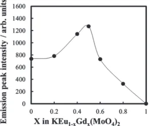
Figure 4 shows the compositional dependence of the emission intensity excited at 309 nm of KEu1−xGdx(MoO4)2
(0.00≤x≤1.00). The emission intensity initially increases with increasing amount of Gd3+ and reaches a maxi- mum at x=0.50. However, the emission intensity de- creases on further increasing Gd3+content beyond the op- timum concentration, which is attributed to the decrease of the Eu3+ concentration. To synthesize nanophosphors, the obtained KEu1−xGdx(MoO4)2 phosphors were stirred in HNO3 solution at room temperature. However, the KEu1−xGdx(MoO4)2powders were completely dissolved in highly concentrated HNO3 solution above 0.05 M during the stirring process. In contrast, for the samples stirred in 0.01 M aqueous HNO3 solution for 7 days, the K+ions of the KEu1−xGdx(MoO4)2 phosphors were successfully ex- changed by H+ without particle dissolution. The compo- sition of KEu1−xGdx(MoO4)2before and after H+exchange was analyzed by XRF. The amount of K+ions in the phos- phors after H+exchange was reduced to about 30% com- pared with the precursor materials KEu1−xGdx(MoO4)2.
To compare the photoluminescence property of K0.3H0.7Eu1−xGdx(MoO4)2 (H+-form) with that of KEu1−x
Gdx(MoO4)2(K+-form), the H+-form powders were washed with deionized water for 12 h and then dried at 50◦C for 24 h. Figure 5 shows excitation and emission spectra of dried K+-form and H+-form powders. The behavior ob- served in the excitation and emission spectra of all H+-form powders was similar. The highest emission intensity was obtained for the H+-form withx=0.50.
The peak wavelength of the CT bands of these phos- phors depends on the excitation energy for electron transfer from O2−to Mo6+. Since the electronegativity of H+(2.1)
Fig. 5 Excitation (broken line) and emission (solid line) spectra of dried (a) KEu0.50Gd0.50(MoO4)2 (K+-form) and (b) K0.3H0.7Eu0.50
Gd0.50(MoO4)2(H+-form) powders prepared by stirring in 0.01 M HNO3
solution for 7 days.
Fig. 6 Dependence of the emission intensity on the Gd3+content of dried K0.3H0.7Eu1−xGdx(MoO4)2(0.00≤x≤1.00) phosphor powders prepared by stirring in 0.01 M HNO3solution for 7 days.
is larger than that of K+(0.8), the electronic attractive force between O2−and Mo6+is increased by H+exchange which, in turn, is causative for the increase of the excitation energy for the electron transfer from O2−to Mo6+. As a result, the excitation absorption band corresponding to the CT transi- tion of O2−–Mo6+is shifted toward shorter wavelength by H+exchange. In addition, the peak intensity of the CT tran- sition of O2−–Mo6+ decreased in comparison with that of the K+-form. This is probably because H2O persisted in the interlayers as a result of using dilute acid.
Figure 6 shows the compositional dependence of the luminescence intensity excited at 309 nm of K0.3H0.7Eu1−x
Gdx(MoO4)2(0.00≤x≤1.00). Similar to the results obtained for the K+-form samples that are shown in Fig. 4, the emis- sion intensity was effectively enhanced by doping of Gd3+ into the K0.3H0.7Eu(MoO4)2lattice and the highest emission intensity was obtained for K0.3H0.7Eu0.50Gd0.50(MoO4)2.
Figure 7 shows SEM images of dried K+-form and H+-
Fig. 7 SEM images of dried (a) KEu0.50Gd0.50(MoO4)2(K+-form) and (b) K0.3H0.7Eu0.50Gd0.50(MoO4)2 (H+-form) powders and (c) H+-form particles redispersed in deionized water (pH=7) and corresponding photo- graph of the clear colloidal solution (the concentration of H+-form powder was 1.0×10⁻³ mol/dm3).
form powders. The K+-form powder has a granular parti- cle morphology with average particle size of 10 µm. In contrast, although a small amount of granular particles re- mained, the dried H+-form powders have rod-like particle morphology with 115µm in length and outer diameters in the range of 50–500 nm. These results indicate that the par- ticle size and morphology of KEu1−xGdx(MoO4)2were suc- cessfully changed by H+exchange at the K+sites. Although the mechanism of the particle-morphology change remains unexplained in detail, it is possible to consider exfoliation of the Eu1−xGdx(MoO4)nlayers by the substitution of K+by H+ions, which may has contributed to the change of the par- ticle morphology. In addition, the rod-like particles formed larger aggregates, like fascicles of fibers. The dispersion of nanophosphors in solution is of high significance for their use in transparent displays. Figure 7(c) shows a SEM image of H+-form powder redispersed in deionized water (pH=7).
A corresponding photograph of the suspension after 1 h is also shown in Fig. 7. The concentration of the H+-form powders were adjusted to 1.0×10⁻³mol/dm3. The Tyndall effect was confirmed by the scattering of a laser beam in the colloidal nanophosphor solution, suggesting that the H+- form nanophosphors were fully suspended in deionized wa- ter without precipitation. From Fig. 7(b) and (c), the ag- gregation of H+-form nanophosphor particles was signifi- cantly reduced by the redispersion in deionized water and, as a result, the particles formed small units. These results
suggest that K0.3H0.7Eu0.50Gd0.50(MoO4)2 might be used as a nanophosphor for transparent displays.
4. Conclusion
HEu1−xGdx(MoO4)2 nanophosphors were synthesized by one-step ion exchange achieved by stirring KEu1−xGdx
(MoO4)2 phosphors in 0.01 M aqueous HNO3 solu- tion at room temperature for 7 days. The obtained K0.3H0.7Eu1−xGdx(MoO4)2 nanophosphors have rod-like particle morphology with 0.5–15µm in length and outer di- ameters in the range of 50–500 nm. The emission inten- sity of these phosphors was effectively enhanced by Gd3+ doping and the highest emission intensity was obtained for the samples with x=0.50, both for KEu1−xGdx(MoO4)2
and K0.3H0.7Eu1−xGdx(MoO4)2phosphors. The aggregated dried nanophosphor powders could be redispersed in deion- ized water. Thus, it is expected that these phosphors can be applied as a transparent display material.
References
[1] M. V. Shestakov, V. K. Tikhomirov, D. Kirilenko, A. S. Kuznetsov, L. F. Chibotaru, A. N. Baranov, G. Van Tendeloo, and V. V.
Moshchalkov, “Quantum cutting in Li (770 nm) and Yb (1000 nm) co-dopant emission bands by energy transfer from the ZnO nano- crystalline host,” Opt. Express, vol.19, no.17, pp.15955–15964, Aug. 2011.
[2] W.-T. Hsu, W.-H. Wu, and C.-H. Lu, “Synthesis and luminescent properties of nano-sized Y3Al5O12:Eu3+ phosphors,” Mater. Sci.
Eng. B, vol.104, no.1–2, pp.40–44, Nov. 2003.
[3] Y. Iso, S. Takeshita, and T. Isobe, “Erectrophosretic deposition and characterization of transparent nanocomposite films of YVO4:Bi3+, Eu3+nanophosphor and silicone-modified acrylic resin,” Langmuir, vol.30, no.5, pp.1465–1471, Jan. 2014.
[4] A. Potdevin, G. Chadeyron, S. Therias, and R. Mahiou, “Lumines- cent nanocomposites made of finely dispersed Y3Ga5O12: Tb pow- der in a polymer matrix: promising candidates for optical devices,”
Langmuir, vol.28, no.37, pp.13526–13535, Aug. 2012.
[5] A. Khetubol1, S. Van Snick, A. Hassinen, E. Fron, Y. Firdaus, L.
Pandey, C. C. David, K. Duerinckx, W. Dehaen, Z. Hens, and M.
Van der Auweraer, “Ligand exchange leads to efficient triplet en- ergy transfer to CdSe/ZnS Q-dots in a poly(N-vinylcarbazole) ma- trix nanocomposite,” J. Appl. Phys., vol.113, no.8, pp.083507-1–12, Feb. 2013.
[6] C. W. Hsu, B. Zhen, W. Qiu, O. Shapira, B. G. DeLacy, J. D.
Joannopoulos, and M. Soljaˇci´c, “Transparent displays enabled by resonant nanoparticle scattering,” Nat. Comm., vol.5, no.3152, pp.1–6, Jan. 2014.
[7] S. Choi, S. W. Tae, J. H. Seo, and H. K. Jung, “Preparation of blue-emitting CaMgSi2O6:Eu2+phosphors in reverse micellar sys- tem and their application to transparent emission display devices,” J.
Solid State Chem., vol.184, no.6, pp.1540–1544, June 2011.
[8] C. Hilsum, “Flat-panel electronic displays: a triumph of physics, chemistry and engineering,” Phil. Trans. R. Soc. A, vol.368, no.1914, pp.1027–1082, Mar. 2010.
[9] V. Bulovic, G. Gu, P. E. Burrows, S. R. Forrest, and M. E.
Thomposn, “Transparent light-emitting devices,” Nature, vol.380, no.6569, pp.29, Mar. 1996.
[10] V. Buissette, D. Giaume, T. Gacoin, and J.-P. Boilot, “Aqueous routes to lanthanide-doped oxide nanophosphors,” J. Mater. Chem., vol.16, no.6, pp.529–539, Oct. 2006.
[11] H. Yang, D.-K. Lee, and Y.-S. Kim, “Spectral variations of nano- sized Y3Al5O12:Ce phosphors via codoping/substitution and their
white LED characteristics,” Mater. Chem. Phys., vol.114, no.2–3, pp.665–669, Apr. 2009.
[12] J. Dhanaraj, R. Jagannathan, T. R. N. Kutty, and C. H. Lu, “Pho- toluminescence characteristics of Y2O3:Eu3+ nanophosphors pre- pared using sol-gel thermolysis,” J. Phys. Chem. B, vol.105, no.45, pp.11098–11105, Nov. 2001.
[13] H. Hu, L. Xiong, J. Zhou, F. Li, T. Cao, and C. Huang, “Multimodal- luminescence core-shell nanocomposites for targeted imaging of tu- mor cells,” Chemistry, vol.15, no.14, pp.3577–3584, Mar. 2009.
[14] W.-S. Song, H.-N. Choi, Y.-S. Kim, and H. Yang, “Formation of green-emitting LaPO4:Ce,Tb nanophosphor layer and its applica- tion to highly transparent plasma displays,” J. Mater. Chem., vol.20, no.33, pp.6929–6934, Sept. 2010.
[15] C. Guo, S. Wang, T. Chen, L. Luan, and Y. Xu, “Preparation of phosphors AEu(MoO4)2(A=Li, Na, K and Ag) by sol-gel method,”
Appl. Phys., A Mater. Sci. Process., vol.94, no.2, pp.365–371, Apr.
2009.
[16] R. F. Klevtsova, L. P. Kozeeva, and P. V. Klevtsov, “Prepara- tion and structure of crystals of pottasium europium molybdate, KEu(MoO4)2,” Sov. Phys. Crystallogr., vol.19, no.1, pp.339–355, July 1977.
[17] M. Watanabe, K. Uematsu, S. Kim, K. Toda, and M. Sato, “Ne- matic liquid crystalline phase of red-emitting HEu(MoO4)2 nano- scroll,” Int. Symp. for Phosphor Materials, Jeju-do, Korea, SO-09, Oct. 2013.
[18] A. Xie, X. Yuan, F. Wang, Y. Shi, J. Li, L. Liu, and Z. Mu, “Synthe- sis and luminescent properties of Eu3+-activated molybdate-based novel red-emitting phosphors for white LEDs,” J. Alloy. Comp., vol.501, no.1, pp.124–129, July 2010.
[19] W. L. Feng, Y. Jin, Y. Wu, D. F. Li, and A. K. Cai, “Co-precipitation synthesis and photoluminescence properties of Ba1−xMoO4:xEu3+ red phosphors,” J. Lumin., vol.134, pp.614–617, Feb. 2013.
[20] T. Kano, “Principal phosphor materials and their optical properties,”
in Phosphor Hand Book, ed. S. Shionoya and W. M. Yen, pp.178–
300, CRC Press, New York, 1999.
[21] R. D. Shannon, “Revised effective ionic radii and systematic studies of interatomic distance in halides and chalcogenides,” Acta Crystal- logr. A, vol.32, no.5, pp.751–767, Sept. 1976.
Mizuki Watanabe is a master-course stu- dent at the Graduate School of Science and Technology, Niigata University.
Kazuyoshi Uematsu received the Doctoral degree from Niigata University in 2007. He is currently Technical officer of Chemistry and Chemical Engineering, Faculty of Engineering, Niigata University.
Kim Sun-Woog received the Doctoral de- gree from Osaka University in 2012. He is currently Assistant Professor at the Graduate School of Science and Technology, Niigata Uni- versity.
Kenji Toda received the Doctoral degree from Niigata University in 1995. During 1988–
1992, he stayed in Nippon Kodoshi Corporation.
He is currently Associate Professor at the Grad- uate School of Science and Technology, Niigata University.
Mineo Sato received the Doctoral degree from Osaka University in 1981. He is currently Professor at the Graduate School of Science and Technology, Niigata University.