福島県立医科大学 学術機関リポジトリ
This document is downloaded at: 2021-11-07T23:56:53Z
Title Impaired frontal brain activity in patients with heart failure assessed by near-infrared spectroscopy( 本文 )
Author(s) 一條, 靖洋
Citation
Issue Date 2020-03-24
URL http://ir.fmu.ac.jp/dspace/handle/123456789/1067
Rights
This thesis/dissertation is modified from "J Am Heart Assoc.
2020 Feb 4;9(3):e014564. doi: 10.1161/JAHA.119.014564. © 2020 The Authors. Published on behalf of the American Heart Association, Inc., by Wiley" with permission.
DOI
Text Version ETD
Impaired frontal brain activity in patients with heart failure assessed by near-infrared spectroscopy.
Yasuhiro Ichijo, MD
Department of Cardiovascular Medicine, Fukushima Medical University, Fukushima, Japan
Abstract
Background: The prevalence of depression and/or anxiety disorders is reported to be higher in patients with heart failure (HF) than in the general population, and HF patients also have coexisting cognitive problems. Recently, the development of near-infrared spectroscopy (NIRS) has enabled noninvasive measurements of regional cerebral blood volume and brain activity, in terms of cerebral oxyhemoglobin in the cerebral cortex, with a high time resolution. The aim of the current study was to determine the associations between frontal brain activity and depressive symptoms, anxiety status, and cognitive function in HF patients.
Methods and Results: We measured and compared frontal brain activity determined by NIRS during a verbal fluency task (VFT) in HF patients (n = 35) and control subjects (n
=28). The Center for Epidemiologic Studies Depression Scale (CES-D) for assessment of depressive symptoms, State-Trait Anxiety Inventory (STAI-S) for assessment of anxiety status, and Mini-Mental State Examination (MMSE) for assessment of cognitive function, and NIRS were simultaneously conducted. NIRS showed that frontal brain activity was significantly lower in the HF group than in the control subjects (28.5 vs 88.0 mM·mm, P
< 0.001). Next, we examined the associations between frontal brain activity and the findings of CES-D, STAI-S, MMSE and VFT. There were significant correlations
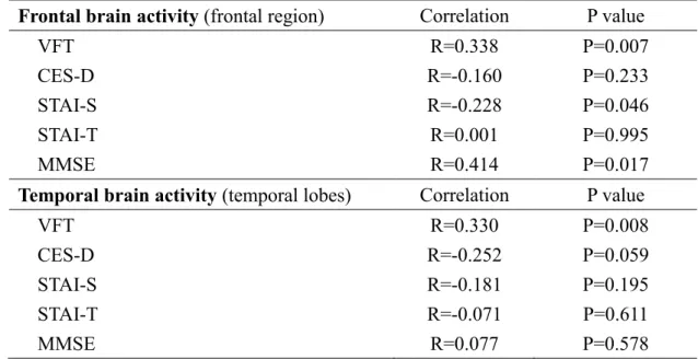
between frontal brain activity and STAI-S (R=-0.228, P=0.046), MMSE (R=0.414, P=0.017) and VFT (R=0.338, P=0.007), but not with CES-D (R=-0.160, P=0.233).
Conclusion: Frontal brain activity assessed by NIRS is reduced, and is associated with high anxiety status and low cognitive function, in HF patients.
Introduction
The prevalence of depression and/or anxiety disorders has been reported to be several times higher in patients with heart failure (HF) than in the general population,1-4 and a substantial proportion of HF patients also have coexisting cognitive problems.5-8 Comorbid mood disorders are associated with increased morbidity, mortality, and medical costs in HF pateints,9-15 but are underdiagnosed and undertreated.16 Cognitive impairment is one of the most common comorbidities in patients with HF,8 and is associated with poor quality of life and self-care, as well as increased morbidity and mortality.7,17
It has been recently reported that reduced cerebral blood flow (CBF) may be associated with altered autonomic, mood, cognitive regulatory sites, and language and speech regulation sites in HF patients.18-20 The neural damage appears on examination by several magnetic resonance imaging (MRI) procedures, and is reflected as regional loss of tissue or injury, as measured by manual assessment,21 voxel-based-morphometry,18 quantitative T2-relaxometry19 and diffusion tensor imaging22 procedures.
Recently, the development of near-infrared spectroscopy (NIRS) has enabled noninvasive and bedside measurements of regional cerebral blood volume in terms of relative concentrations of oxyhemoglobin (oxy-Hb) and deoxyhemoglobin (deoxy-Hb),
with a high time resolution. The concentrations of oxy-Hb and deoxy-Hb are assumed to reflect the regional cerebral blood volume.23-25 In addition, oxy-Hb increases and deoxy- Hb decreases in NIRS have been shown to reflect cortical activation by simultaneous measurements with other methodologies,24 presenting cerebral perfusion and is used as functional brain monitoring.23 A positive correlation has been confirmed between oxy- Hb concentration by NIRS and blood-oxygen-level-dependent signaling by functional MRI.26,27 Further, NIRS has recently been used to investigate the neurocognitive processes associated with neurological (Alzheimer’s disease, Parkinson’s disease, epilepsy, and traumatic brain injury) and psychiatric disorders (depression, bipolar disorder, anxiety disorders, and schizophrenia).28 The frontal NIRS signal has been proposed as a supportive tool in assisting the diagnosis of major psychiatric disorders with depressive symptoms in addition to evaluation of brain activity.24,29,30 Compared to positron emission tomography, single photon emission computed tomography, and functional MRI, NIRS has the advantages of requiring minimal equipment and being easy to use.
In the present study, we aimed to 1) evaluate and compare frontal brain activity using NIRS in HF patients and control subjects, and 2) determine the associations between frontal brain activity and depressive symptoms, anxiety status, and cognitive
function.
Methods
The data that support the findings of this study are available from the corresponding author upon reasonable request.
Subjects and study protocol
This is a cross-sectional study with 28 age-matched control subjects and 35 HF patients who came to Fukushima Medical University Hospital between May 2018 and June 2019.
The diagnosis of HF was made by several cardiologists based on the HF guidelines.31,32 Study subjects underwent echocardiography, carotid artery ultrasonography, laboratory testing, psychological testing and NIRS. The verbal fluency task (VFT) is used to test frontal region function and is commonly used with NIRS analysis.33,34 The control subjects were without history of HF, physical findings of HF, or structural cardiac abnormalities which were detected by echocardiography. The study protocol was approved by the ethical committee of Fukushima Medical University (#823), and the investigation conforms to the principles outlined in the Declaration of Helsinki. All subjects provided written informed consent to participate in the study. Patients with carotid artery stenosis, cerebral infarction, dementia, or patients receiving treatment for
schizophrenia, depression, or bipolar disorder were excluded. We evaluated several comorbidities that often coexist and are associated with adverse prognosis in HF patients.35 Regarding the psychological testing, the Center for Epidemiologic Studies Depression Scale (CES-D) was used to evaluate depressive symptoms,36-38 the State-Trait Anxiety Inventory-state (STAI-S) and STAI-trait (STAI-T) were used to evaluate anxiety status and trait,39 and the Mini-Mental State Examination (MMSE) was used to evaluate cognitive function.40 We compared the findings of CES-D, STAI-S, MMSE, VFT and NIRS findings between the HF patients and control subjects, and determined the associations between frontal brain activity and depressive symptoms, anxiety status and cognitive ability.
Blood samples were obtained from all subjects at Fukushima Medical University Hospital. B-type natriuretic peptide (BNP) levels were measured using a specific immunoradiometric assay (Shionoria BNP kit, Shionogi, Osaka, Japan).
Echocardiography and carotid artery ultrasonography were performed blindly by experienced sonographers using standard techniques.35,41 The LVEF was calculated using Simpson’s method in a four-chamber view.35,41 All measurements were performed using ultrasound systems (ACUSON Sequoia, Siemens Medical Solutions USA, Inc.,
Mountain View, CA, USA). In the present study, HF with LVEF ≥45% was defined as HFpEF, and HF with LVEF <45% was defined as HFrEF.
Measurement of NIRS
A VFT was widely used as an activating task during NIRS analysis as previously reported.33,34 In the current study, oxy-Hb, deoxy-Hb, and total hemoglobin were measured with a 52-channels NIRS machine (Hitachi ETG4000, Hitachi Medical Corp., Tokyo, Japan) using two wavelengths of near-infrared light (695 and 830 nm). NIRS measurement was performed by attaching 52 channels to the head (Figure 1).29,30 The 52- channel device attached to subjects’ foreheads, and the most of lower and forward channels were placed along the line connecting T3-Fpz-T4, based on the international 10- 20 system in a sitting position. This device was connected symmetrically around the prefrontal cortex. The main measured channels were as follows: right temporal lobe (channels 1-3, 11-14, 22-24, 32-35, and 43-45), left temporal lobe (channels 8-10, 18-21, 29-31, 39-42, and 50-52) and frontal region (channels 25-28, 36-38, and 46-49).
Compliance with the scalp measurement sites of the international 10-20 system allows prediction of the measurement sites on the brain surface with relatively high accuracy.
Especially, an increase in cerebral oxy-Hb concentration in the frontal region in response
to the VFT is considered as markers of frontal brain activity.30,42,43 NIRS signal changes were measured during a 10-s pre-task baseline period, a 60-s activation period, and a 55- s post task baseline period. The sampling rate of oxy-Hb concentration data was 0.1 s.
The obtained data were analyzed using the “integral mode”: the pre-task baseline was set as the mean over a 10-s period just before the task period, and the post-task baseline was fixed as the mean over the last 5 s of the post-task period. Linear fitting between the pre- and post-task baselines was applied to the data between the two baselines. The average oxy-Hb concentration during the VFT that was performed for 60 s was used for the analysis. An automatic artifact-rejection procedure was used and individual data were excluded when there were fewer than 6 remaining channels from frontal region and bilateral temporal lobes.30 Data are expressed as waveforms and topographic maps. The Intraclass Correlation Coefficient (ICC) of the mean oxy-Hb concentration during the task segment was calculated for the 52 channels. The single-measure ICC was 0.5309, and the average measure ICC was 0.6936, which are both reliable, as previously reported.44 NIRS analyses were performed using MATLAB R2011 (Math Works Inc., Natick, MA), and Prism 6.0 software (GraphPad Software, Inc., San Diego, CA).
Activation task, VFT
An outline of the VFT procedure is as follows.33,34 Changes in hemoglobin oxygenation occur in people performing the VFT. Artifacts must be eliminated by having the subject sit in a chair, relax, and move as little as possible during the test. The subject is first prompted by a voice saying, “Start /a/, /i/, /u/, /e/, /o/” to repeat the utterance “/a/, /i/, /u/, /e/, /o/” for 30 s. The baseline activity recorded during this meaningless utterance is used to remove the effect of vocalization on brain activity from the data. The subject is next prompted by a voice to vocalize as many words as possible that start with a certain letter. This is done in three 20-s sets. The subjects are verbally prompted to vocalize words starting with a certain letter to increase the difficulty of the task. The exercise is scored by recording the number of words uttered every 20 s. Finally, the subject is prompted by a voice saying, “Stop /a/, /i/, /u/, /e/, /o/” to stop the task and repeat “/a/, /i/, /u/, /e/, /o/” for 70 s.
Statistical analysis
Categorical variables are expressed as numbers and percentages. The chi-square test was used for comparisons of categorical variables and followed by Fisher’s exact test when/if appropriate. Normality was confirmed using the Shapiro-Wilk test in each group.
Parametric variables are presented as mean ± SD, and non-parametric variables (e.g.
BNP, NIRS finding) are presented as a median and interquartile range. Parametric variables were compared using Student’s t-test, whereas non-parametric variables were compared using the Mann-Whitney U test. To compare continuous variable among the HFrEF, HFpEF and control subjects, Kruskal-Wallis test was used. We performed regression analysis to determine brain activity confounding factors. Correlations between each NIRS finding and physiological questionnaire were assessed using Spearman’s correlation analysis. A P value of <0.05 was considered statistically significant for all comparisons. All analyses were performed using a statistical software package (SPSS ver.
24.0, IBM, Armonk, NY, USA).
Results
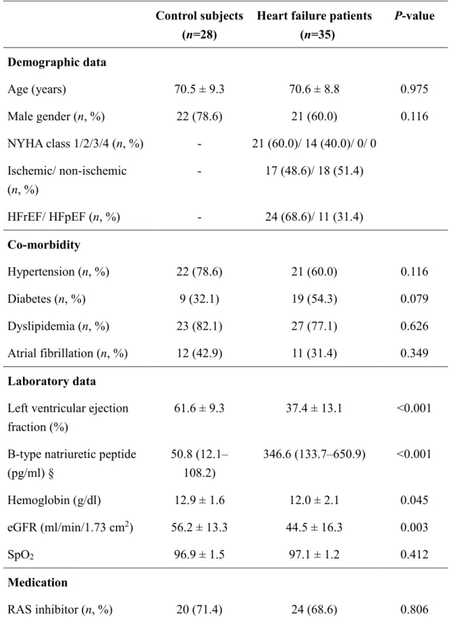
The comparisons of clinical features between the control subjects and HF patients in the present study are shown in Table 1. BNP was significantly higher, and hemoglobin, estimated glomerular filtration rate, and left ventricular ejection fraction were significantly lower in the HF patients than in the control subjects. In addition, we found no significant difference in age, sex, percutaneous oxygen saturation or medication, except for inotropic agent, between the two groups. Regarding psychological testing, VFT
and MMSE were significantly lower, and STAI-S was significantly higher in the HF patients than in the control subjects. In contrast, CES-D and STAI-T did not significantly differ between the groups.
Changes in mean oxy-Hb concentrations in the HF and control groups are shown in Figure 2. The horizontal axis represents time, and the vertical axis represents changes in mean oxy-Hb concentrations (mM mm) during VFT. Figure 3 shows a topographic map of the differences in mean oxy-Hb concentrations. The mean oxy-Hb concentrations of right temporal lobe (channels 2, 13, 14, 32, 34, 35, 43 and 45), left temporal lobe (channels 8, 10, 18-21, 29-31, 39-42, and 50-52) and frontal region (channels 25-28, 36, 38, 46, 47 and 49) were significantly lower in the HF group than in the control subjects.
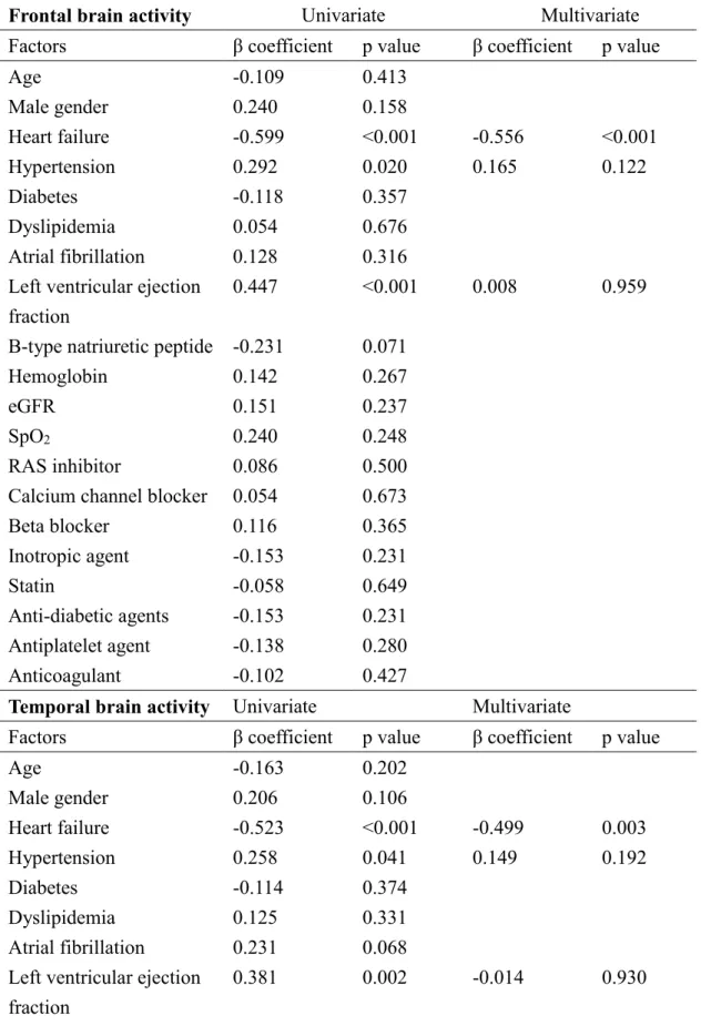
Next, we focused frontal and temporal brain activity (integral values of mean oxy-Hb concentrations in the frontal region and temporal lobes). Frontal and temporal brain activity was compared between the groups and are presented in Figure 4. Frontal and temporal brain activity were significantly lower both in the HFrEF and HFpEF than in the control subjects. In the multiple regression analysis to determine brain activity confounding factors (Table 2), HF was independently associated with frontal brain activity (β=-0.556, p<0.001) and temporal brain activity (β=-0.499, p=0.003). In addition, as shown in Table 3, there were significant correlations between frontal brain activity
and STAI-S (R=-0.228, P=0.046), MMSE (R=0.414, P=0.017) and VFT (R=0.338, P=0.007), but not with CES-D and STAI-T. On the other hand, there was no significant correlations between temporal brain activity and CES-D, STAI-S, or MMSE, except for VFT (R=0.330, P=0.008).
Discussion
In the present study, NIRS was used to evaluate the brain activity of HF patients. NIRS showed that frontal and temporal brain activity (an increase in cerebral oxy-Hb concentration in the frontal region and temporal lobes in response to the VFT), cognitive function (MMSE), and language ability (VFT) were lower, and anxiety status (STAI-S) was higher in the HF patients compared to the control subjects, despite no significant differences in SpO2 and depressive symptoms (CES-D) between the two groups. In addition, frontal brain activity was associated with STAI-S, MMSE and VFT, but not with CES-D and STAI-T. To the best of our knowledge, the current study appears to be the first to evaluate brain activity and psychological status in HF patients using NIRS.
Regional CBF reduction in HF patients appears in multiple brain sites, and those regions include vascular beds over the frontal, parietal, and occipital cortices, as well as
the hippocampus, thalamus, and cerebellar areas; the majority of these brain sites also show brain tissue injury, as reported by previous studies using functional MRI.18-20 HF induces brain structural abnormalities that are associated with depressive symptoms and cognitive impairment.18,45,46 Multiple brain autonomic regulatory sites have been reported to show reduced CBF in HF patients, and include the hippocampus, thalamus, corona radiata and cerebellar sites. The affected structures also show abnormal functional MRI signal responses to autonomic and cardiovascular challenges in HF.47 In the present study, with NIRS, mean oxy-Hb concentrations were lower in the HF group than in the control group in many of the 52 channels. The decrease in the mean oxy-Hb concentration in the frontal region was similar to the results seen in patients with depression.48,49 Frontal hypoperfusion and frontal dysfunction have been observed in patients with depression,50,51 which may be further associated with cognitive impairment.52,53
With respect to mood disorder, brain sites associated with mood regulation include the prefrontal cortex, cingulate, insula, hippocampus, amygdala, and cerebellar areas.19 These brain sites have been associated with structure change in patients with depression only;54 however, the majority of these areas also showed reduced CBF in HF patients.20 The amygdala is also involved in anxiety regulation, and the bilateral amygdalae showed reduced CBF.20 In addition, prefrontal cortex is related with amygdala
mutually, and might be associated with anxiety symptoms.55,56 Reduced CBF in these regions likely contributes to tissue changes, and thus, has the potential to modify levels of depressive and anxiety symptoms in HF patients. A decrease in the oxy-Hb concentration with NIRS reflects a decrease in frontal brain function in patients with depression or in a depressed state.48,49 Although we could not fully explain the reason why frontal brain activity determined by NIRS was associated with anxiety status (STAI- S), but not with depression (CES-D), diagnostic sensitivity of CES-D may have affected these results. Since patients with diagnosed depression were excluded, and mean CES-D was low (i.e. 10-11), CES-D might not be necessarily appropriate for evaluating depressive symptom in the present study subjects.
With respect to cognitive impairment, HF patients exhibit patterns of cortical alterations that overlap with cortical atrophy observed in Alzheimer's disease, including lateral temporal and parietal regions.45,57-61 Several brain sites including the hippocampus and prefrontal cortex regulate short-term memory and decision-making. Higher white matter hyperintensity volume is risk factors associated with dementia in older community-based residents.62 In subject without HF, increased left ventricular mass index corresponds to altered white matter microstructure, particularly among older adults with clinical symptoms of prodromal dementia.63 Cardiac function determined by
compromised global longitudinal strain relates to worse episodic memory among older adults who are free of clinical dementia.64 In previous reports in HF patients, CBF reductions appeared in the prefrontal cortex, a structure which plays critical roles in cognitive actions including executive decision-making.7,8,17,57 HF show cerebral grey matter loss, and is associated with cognitive impairment.45,58 Hippocampal blood flow abnormality associated with cognitive impairment in HF patients.60,61 Resent report presented that the degree of medial temporal lobe atrophy determined by magnetic resonance imaging was strongly associated with the severity of cognitive impairment, whereas the extent of white matter hyperintensities was similar in patients and controls.59 Medial temporal lobe atrophy but not white matter lesion load seems to be related to cognitive impairment.59 Concordant with the present study, cerebral oxygenation is correlated with cognitive function assessed by MMSE in patients with chronic kidney disease.65
Study limitations
The present study has several limitations. First, as a prospective cohort study of a single center with a relatively small number of patients, the present results may not be representative of the general population. Second, since NIRS can evaluate only a shallow
layer of the brain, deep layers (e.g. hippocampus) or detail of regional areas could not be evaluated. Although functional MRI is used to accurately evaluate regional CBF in HF patients, high costs and a large-scale device or contraindication (e.g. implantable device) in MRI interfere with simple and repeatable examination. NIRS is superior to MRI for easy-to-repeat measurements. Third, because of artifact, some NIRS signals in temporal areas could not be fully detected in some study subjects. NIRS signals during VFT may be influenced by skin blood flow. Fourth, although we excluded presence of carotid artery stenosis or cerebral infarction, there may have been changes in cerebral oxy-Hb due to arteriosclerotic changes. Fifth, general condition may have affected the results of several physiological tests. Sixth, associations between brain activity determined by NIRS and each score of psychological testing (e.g. depression, cognitive function and anxiety) were roughly examined. These associations might be preliminary data. Mechanistic Therefore, the present results should be viewed as preliminary, and further studies with a larger population are needed.
Conclusions
Frontal brain activity assessed by NIRS was reduced, and might be associated with high anxiety status and low cognitive function in HF patients.
Acknowledgments: Akiomi Yoshihisa, MD, PhD1, Soichi Kono, MD, PhD,2 Tomofumi Misaka MD, PhD1,Takashi Kaneshiro MD, PhD1, Masayoshi Oikawa, MD, PhD1, Itaru Miura, MD, PhD 2 Hirooki Yabe, MD, PhD 2 and Yasuchika Takeishi, MD, PhD 1
1Department of Cardiovascular Medicine, 2Department of Neuropsychiatry, Fukushima Medical University, Fukushima, Japan
Sources of founding: None declared.
Disclosures: None declared.
References:
1. Easton K, Coventry P, Lovell K, Carter LA, Deaton C. Prevalence and measurement of anxiety in samples of patients with heart failure: Meta-analysis. J Cardiovasc Nurs. 2016;31:367-379
2. Vaccarino V, Kasl SV, Abramson J, Krumholz HM. Depressive symptoms and risk of functional decline and death in patients with heart failure. J Am Coll Cardiol. 2001;38:199-205
3. Rutledge T, Reis VA, Linke SE, Greenberg BH, Mills PJ. Depression in heart failure a meta-analytic review of prevalence, intervention effects, and associations with clinical outcomes. J Am Coll Cardiol. 2006;48:1527-1537
4. Konstam V, Moser DK, De Jong MJ. Depression and anxiety in heart failure. J Card Fail. 2005;11:455-463
5. Angermann CE, Ertl G. Depression, anxiety, and cognitive impairment : Comorbid mental health disorders in heart failure. Curr Heart Fail Rep. 2018;15:398-410 6. Cannon JA, Moffitt P, Perez-Moreno AC, Walters MR, Broomfield NM, McMurray
JJV, Quinn TJ. Cognitive impairment and heart failure: Systematic review and meta- analysis. J Card Fail. 2017;23:464-475
7. Hajduk AM, Kiefe CI, Person SD, Gore JG, Saczynski JS. Cognitive change in heart failure: A systematic review. Circ Cardiovasc Qual Outcomes. 2013;6:451-460
8. Vogels RL, Scheltens P, Schroeder-Tanka JM, Weinstein HC. Cognitive impairment in heart failure: A systematic review of the literature. Eur J Heart Fail. 2007;9:440- 449
9. Alhurani AS, Dekker RL, Abed MA, Khalil A, Al Zaghal MH, Lee KS, Mudd-Martin G, Biddle MJ, Lennie TA, Moser DK. The association of co-morbid symptoms of depression and anxiety with all-cause mortality and cardiac rehospitalization in patients with heart failure. Psychosomatics. 2015;56:371-380
10. Sayers SL, Hanrahan N, Kutney A, Clarke SP, Reis BF, Riegel B. Psychiatric comorbidity and greater hospitalization risk, longer length of stay, and higher hospitalization costs in older adults with heart failure. J Am Geriatr Soc. 2007;55:1585-1591
11. Angermann CE, Gelbrich G, Stork S, Schowalter M, Deckert J, Ertl G, Faller H, Competence Network Heart F. Somatic correlates of comorbid major depression in patients with systolic heart failure. Int J Cardiol. 2011;147:66-73
12. Faris R, Purcell H, Henein MY, Coats AJ. Clinical depression is common and significantly associated with reduced survival in patients with non-ischaemic heart failure. Eur J Heart Fail. 2002;4:541-551
13. Jiang W, Krishnan R, Kuchibhatla M, Cuffe MS, Martsberger C, Arias RM, O'Connor CM, Investigators S-C. Characteristics of depression remission and its relation with cardiovascular outcome among patients with chronic heart failure (from the sadhart- chf study). Am J Cardiol. 2011;107:545-551
14. Jiang W, Kuchibhatla M, Clary GL, Cuffe MS, Christopher EJ, Alexander JD, Califf RM, Krishnan RR, O'Connor CM. Relationship between depressive symptoms and long-term mortality in patients with heart failure. Am Heart J. 2007;154:102-108 15. Sherwood A, Blumenthal JA, Trivedi R, Johnson KS, O'Connor CM, Adams KF, Jr.,
Dupree CS, Waugh RA, Bensimhon DR, Gaulden L, Christenson RH, Koch GG, Hinderliter AL. Relationship of depression to death or hospitalization in patients with heart failure. Arch Intern Med. 2007;167:367-373
16. Celano CM, Villegas AC, Albanese AM, Gaggin HK, Huffman JC. Depression and anxiety in heart failure: A review. Harv Rev Psychiatry. 2018;26:175-184
17. Zuccala G, Marzetti E, Cesari M, Lo Monaco MR, Antonica L, Cocchi A, Carbonin P, Bernabei R. Correlates of cognitive impairment among patients with heart failure:
Results of a multicenter survey. Am J Med. 2005;118:496-502
18. Woo MA, Macey PM, Fonarow GC, Hamilton MA, Harper RM. Regional brain gray matter loss in heart failure. Journal of applied physiology. 2003;95:677-684
19. Woo MA, Kumar R, Macey PM, Fonarow GC, Harper RM. Brain injury in autonomic, emotional, and cognitive regulatory areas in patients with heart failure. J Card Fail. 2009;15:214-223
20. Roy B, Woo MA, Wang DJJ, Fonarow GC, Harper RM, Kumar R. Reduced regional cerebral blood flow in patients with heart failure. Eur J Heart Fail. 2017;19:1294- 1302
21. Vogels RL, van der Flier WM, van Harten B, Gouw AA, Scheltens P, Schroeder-Tanka JM, Weinstein HC. Brain magnetic resonance imaging abnormalities in patients with heart failure. Eur J Heart Fail. 2007;9:1003-1009
22. Woo MA, Palomares JA, Macey PM, Fonarow GC, Harper RM, Kumar R. Global and regional brain mean diffusivity changes in patients with heart failure. J Neurosci Res. 2015;93:678-685
23. Strangman G, Boas DA, Sutton JP. Non-invasive neuroimaging using near-infrared light. Biol Psychiatry. 2002;52:679-693
24. Suto T, Fukuda M, Ito M, Uehara T, Mikuni M. Multichannel near-infrared spectroscopy in depression and schizophrenia: Cognitive brain activation study. Biol Psychiatry. 2004;55:501-511
25. Miura I, Kono S, Oshima S, Kanno-Nozaki K, Mashiko H, Niwa S, Yabe H. Near-
infrared spectroscopy and plasma homovanillic acid levels in bipolar disorder: A case report. Neuropsychiatr Dis Treat. 2014;10:507-511
26. Strangman G, Culver JP, Thompson JH, Boas DA. A quantitative comparison of simultaneous bold fmri and nirs recordings during functional brain activation.
Neuroimage. 2002;17:719-731
27. Sassaroli A, de BFB, Tong Y, Renshaw PF, Fantini S. Spatially weighted bold signal for comparison of functional magnetic resonance imaging and near-infrared imaging of the brain. Neuroimage. 2006;33:505-514
28. Irani F, Platek SM, Bunce S, Ruocco AC, Chute D. Functional near infrared spectroscopy (fnirs): An emerging neuroimaging technology with important applications for the study of brain disorders. Clin Neuropsychol. 2007;21:9-37 29. Takizawa R, Kasai K, Kawakubo Y, Marumo K, Kawasaki S, Yamasue H, Fukuda M.
Reduced frontopolar activation during verbal fluency task in schizophrenia: A multi- channel near-infrared spectroscopy study. Schizophr Res. 2008;99:250-262
30. Takizawa R, Fukuda M, Kawasaki S, Kasai K, Mimura M, Pu S, Noda T, Niwa S, Okazaki Y, Joint Project for Psychiatric Application of Near-Infrared Spectroscopy G.
Neuroimaging-aided differential diagnosis of the depressive state. Neuroimage. 2014;85 Pt 1:498-507
31. McMurray JJ, Adamopoulos S, Anker SD, Auricchio A, Bohm M, Dickstein K, Falk V, Filippatos G, Fonseca C, Gomez-Sanchez MA, Jaarsma T, Kober L, Lip GY, Maggioni AP, Parkhomenko A, Pieske BM, Popescu BA, Ronnevik PK, Rutten FH, Schwitter J, Seferovic P, Stepinska J, Trindade PT, Voors AA, Zannad F, Zeiher A, Task Force for the D, Treatment of A, Chronic Heart Failure of the European Society of C, Bax JJ, Baumgartner H, Ceconi C, Dean V, Deaton C, Fagard R, Funck-Brentano C, Hasdai D, Hoes A, Kirchhof P, Knuuti J, Kolh P, McDonagh T, Moulin C, Popescu BA, Reiner Z, Sechtem U, Sirnes PA, Tendera M, Torbicki A, Vahanian A, Windecker S, McDonagh T, Sechtem U, Bonet LA, Avraamides P, Ben Lamin HA, Brignole M, Coca A, Cowburn P, Dargie H, Elliott P, Flachskampf FA, Guida GF, Hardman S, Iung B, Merkely B, Mueller C, Nanas JN, Nielsen OW, Orn S, Parissis JT, Ponikowski P, Guidelines ESCCfP. Esc guidelines for the diagnosis and treatment of acute and chronic heart failure 2012: The task force for the diagnosis and treatment of acute and chronic heart failure 2012 of the european society of cardiology. Developed in collaboration with the heart failure association (hfa) of the esc. Eur J Heart Fail. 2012;14:803-869
32. Yancy CW, Jessup M, Bozkurt B, Butler J, Casey DE, Jr., Drazner MH, Fonarow GC, Geraci SA, Horwich T, Januzzi JL, Johnson MR, Kasper EK, Levy WC, Masoudi FA,
McBride PE, McMurray JJ, Mitchell JE, Peterson PN, Riegel B, Sam F, Stevenson LW, Tang WH, Tsai EJ, Wilkoff BL, American College of Cardiology F, American Heart Association Task Force on Practice G. 2013 accf/aha guideline for the management of heart failure: A report of the american college of cardiology foundation/american heart association task force on practice guidelines. J Am Coll Cardiol. 2013;62:e147-239
33. Fujiwara T, Kono S, Katakura K, Abe K, Takahashi A, Gunji N, Yokokawa A, Kawashima K, Suzuki R, Wada A, Miura I, Yabe H, Ohira H. Evaluation of brain activity using near-infrared spectroscopy in inflammatory bowel disease patients. Sci Rep. 2018;8:402
34. Klumpp H, Deldin P. Review of brain functioning in depression for semantic processing and verbal fluency. Int J Psychophysiol. 2010;75:77-85
35. Sato Y, Yoshihisa A, Kimishima Y, Kiko T, Watanabe S, Kanno Y, Abe S, Miyata M, Sato T, Suzuki S, Oikawa M, Kobayashi A, Yamaki T, Kunii H, Nakazato K, Ishida T, Takeishi Y. Subclinical hypothyroidism is associated with adverse prognosis in heart failure patients. Can J Cardiol. 2018;34:80-87
36. Shafer AB. Meta-analysis of the factor structures of four depression questionnaires:
Beck, ces-d, hamilton, and zung. J Clin Psychol. 2006;62:123-146
37. LS. R. The ces-d scale: A self-report depression scale for research in the general population. Appl Psychol Meas. 1977;1:385-401
38. Weissman MM, Sholomskas D, Pottenger M, Prusoff BA, Locke BZ. Assessing depressive symptoms in five psychiatric populations: A validation study. Am J Epidemiol. 1977;106:203-214
39. Jiang W, Kuchibhatla M, Cuffe MS, Christopher EJ, Alexander JD, Clary GL, Blazing MA, Gaulden LH, Califf RM, Krishnan RR, O'Connor CM. Prognostic value of anxiety and depression in patients with chronic heart failure. Circulation. 2004;110:3452- 3456
40. Folstein MF, Folstein SE, McHugh PR. "Mini-mental state". A practical method for grading the cognitive state of patients for the clinician. J Psychiatr Res. 1975;12:189- 198
41. Yoshihisa A, Takiguchi M, Shimizu T, Nakamura Y, Yamauchi H, Iwaya S, Owada T, Miyata M, Abe S, Sato T, Suzuki S, Oikawa M, Kobayashi A, Yamaki T, Sugimoto K, Kunii H, Nakazato K, Suzuki H, Saitoh S, Takeishi Y. Cardiovascular function and prognosis of patients with heart failure coexistent with chronic obstructive pulmonary disease. J Cardiol. 2014;64:256-264
42. Takahashi A, Kono S, Wada A, Oshima S, Abe K, Imaizumi H, Fujita M, Hayashi M,
Okai K, Miura I, Yabe H, Ohira H. Reduced brain activity in female patients with non-alcoholic fatty liver disease as measured by near-infrared spectroscopy. PLoS One. 2017;12:e0174169
43. Abe K, Wada A, Oshima S, Kono S, Takahashi A, Kanno Y, Imaizumi H, Hayashi M, Okai K, Niwa SI, Yabe H, Ohira H. Reduced frontal activation during verbal fluency task in chronic hepatitis c patients with interferon-based therapy as measured by near-infrared spectroscopy. Hepatol Res. 2017;47:E55-E63
44. Kakimoto Y, Nishimura Y, Hara N, Okada M, Tanii H, Okazaki Y. Intrasubject reproducibility of prefrontal cortex activities during a verbal fluency task over two repeated sessions using multi-channel near-infrared spectroscopy. Psychiatry Clin Neurosci. 2009;63:491-499
45. Almeida OP, Garrido GJ, Beer C, Lautenschlager NT, Arnolda L, Flicker L. Cognitive and brain changes associated with ischaemic heart disease and heart failure. Eur Heart J. 2012;33:1769-1776
46. Suzuki H, Sumiyoshi A, Matsumoto Y, Duffy BA, Yoshikawa T, Lythgoe MF, Yanai K, Taki Y, Kawashima R, Shimokawa H. Structural abnormality of the hippocampus associated with depressive symptoms in heart failure rats. Neuroimage. 2015;105:84- 92
47. Ogren JA, Macey PM, Kumar R, Fonarow GC, Hamilton MA, Harper RM, Woo MA.
Impaired cerebellar and limbic responses to the valsalva maneuver in heart failure.
Cerebellum. 2012;11:931-938
48. Matsuo K, Kato T, Fukuda M, Kato N. Alteration of hemoglobin oxygenation in the frontal region in elderly depressed patients as measured by near-infrared spectroscopy. J Neuropsychiatry Clin Neurosci. 2000;12:465-471
49. Matsuo K, Kato N, Kato T. Decreased cerebral haemodynamic response to cognitive and physiological tasks in mood disorders as shown by near-infrared spectroscopy.
Psychol Med. 2002;32:1029-1037
50. Narita H, Odawara T, Iseki E, Kosaka K, Hirayasu Y. Psychomotor retardation correlates with frontal hypoperfusion and the modified stroop test in patients under 60-years-old with major depression. Psychiatry Clin Neurosci. 2004;58:389-395 51. Videbech P, Ravnkilde B, Gammelgaard L, Egander A, Clemmensen K, Rasmussen
NA, Gjedde A, Rosenberg R. The danish pet/depression project: Performance on stroop's test linked to white matter lesions in the brain. Psychiatry Res. 2004;130:117-130
52. Castaneda AE, Tuulio-Henriksson A, Marttunen M, Suvisaari J, Lonnqvist J. A review on cognitive impairments in depressive and anxiety disorders with a focus on
young adults. J Affect Disord. 2008;106:1-27
53. Gualtieri CT, Johnson LG, Benedict KB. Neurocognition in depression: Patients on and off medication versus healthy comparison subjects. J Neuropsychiatry Clin Neurosci. 2006;18:217-225
54. Fitzgerald PB, Laird AR, Maller J, Daskalakis ZJ. A meta-analytic study of changes in brain activation in depression. Hum Brain Mapp. 2008;29:683-695
55. Gold AL, Shechner T, Farber MJ, Spiro CN, Leibenluft E, Pine DS, Britton JC.
Amygdala-cortical connectivity: Associations with anxiety, development, and threat.
Depress Anxiety. 2016;33:917-926
56. Britton JC, Grillon C, Lissek S, Norcross MA, Szuhany KL, Chen G, Ernst M, Nelson EE, Leibenluft E, Shechner T, Pine DS. Response to learned threat: An fmri study in adolescent and adult anxiety. Am J Psychiatry. 2013;170:1195-1204
57. Alosco ML, Hayes SM. Structural brain alterations in heart failure: A review of the literature and implications for risk of alzheimer's disease. Heart Fail Rev. 2015;20:561-571
58. Sabayan B, van Buchem MA, Sigurdsson S, Zhang Q, Harris TB, Gudnason V, Arai AE, Launer LJ. Cardiac hemodynamics are linked with structural and functional features of brain aging: The age, gene/environment susceptibility (ages)-reykjavik study. Journal of the American Heart Association. 2015;4:e001294
59. Frey A, Sell R, Homola GA, Malsch C, Kraft P, Gunreben I, Morbach C, Alkonyi B, Schmid E, Colonna I, Hofer E, Mullges W, Ertl G, Heuschmann P, Solymosi L, Schmidt R, Stork S, Stoll G. Cognitive deficits and related brain lesions in patients with chronic heart failure. JACC. Heart failure. 2018;6:583-592
60. Suzuki H, Matsumoto Y, Ota H, Sugimura K, Takahashi J, Ito K, Miyata S, Furukawa K, Arai H, Fukumoto Y, Taki Y, Shimokawa H. Hippocampal blood flow abnormality associated with depressive symptoms and cognitive impairment in patients with chronic heart failure. Circ J. 2016;80:1773-1780
61. Woo MA, Ogren JA, Abouzeid CM, Macey PM, Sairafian KG, Saharan PS, Thompson PM, Fonarow GC, Hamilton MA, Harper RM, Kumar R. Regional hippocampal damage in heart failure. Eur J Heart Fail. 2015;17:494-500
62. Wu A, Sharrett AR, Gottesman RF, Power MC, Mosley TH, Jr., Jack CR, Jr., Knopman DS, Windham BG, Gross AL, Coresh J. Association of brain magnetic resonance imaging signs with cognitive outcomes in persons with nonimpaired cognition and mild cognitive impairment. JAMA Netw Open. 2019;2:e193359
63. Moore EE, Liu D, Pechman KR, Terry JG, Nair S, Cambronero FE, Bell SP, Gifford KA, Anderson AW, Hohman TJ, Carr JJ, Jefferson AL. Increased left ventricular
mass index is associated with compromised white matter microstructure among older adults. Journal of the American Heart Association. 2018;7
64. Kresge HA, Khan OA, Wagener MA, Liu D, Terry JG, Nair S, Cambronero FE, Gifford KA, Osborn KE, Hohman TJ, Pechman KR, Bell SP, Wang TJ, Carr JJ, Jefferson AL.
Subclinical compromise in cardiac strain relates to lower cognitive performances in older adults. Journal of the American Heart Association. 2018;7
65. Miyazawa H, Ookawara S, Ito K, Ueda Y, Yanai K, Ishii H, Mutsuyoshi Y, Kitano T, Shindo M, Aomatsu A, Hirai K, Hoshino T, Morishita Y. Association of cerebral oxygenation with estimated glomerular filtration rate and cognitive function in chronic kidney disease patients without dialysis therapy. PLoS One. 2018;13:e0199366
Figure legends Figure 1
The 52-channel device attached to subjects’ foreheads, and the most of lower and forward channels were placed along the line connecting T3-Fpz-T4, based on the international 10- 20 system in a sitting position.
Figure 2
Comparison of changes in mean oxy-Hb concentrations in the heart failure (red) and control subjects (blue).
Figure 3
Topographic map of the differences in mean oxy-Hb concentration changes between the HF and control subjects. The mean oxy-Hb concentrations of right temporal lobe (channels 2, 13, 14, 32, 34, 35, 43 and 45), left temporal lobe (channels 8, 10, 18-21, 29- 31, 39-42, and 50-52) and frontal region (channels 25-28, 36, 38, 46, 47 and 49) were significantly lower in the HF group than in the control subjects.
Figure 4
Comparisons of frontal brain activity (integral values of mean oxy-Hb concentrations in the frontal region) and temporal brain activity (integral values of mean oxy-Hb concentrations in the temporal lobes) between both HFrEF and HFpEF, and control subjects.
Table 1. Comparisons of clinical features between the control subjects and heart failure patients
Control subjects (n=28)
Heart failure patients (n=35)
P-value
Demographic data
Age (years) 70.5 ± 9.3 70.6 ± 8.8 0.975
Male gender (n, %) 22 (78.6) 21 (60.0) 0.116
NYHA class 1/2/3/4 (n, %) - 21 (60.0)/ 14 (40.0)/ 0/ 0 Ischemic/ non-ischemic
(n, %)
- 17 (48.6)/ 18 (51.4)
HFrEF/ HFpEF (n, %) - 24 (68.6)/ 11 (31.4)
Co-morbidity
Hypertension (n, %) 22 (78.6) 21 (60.0) 0.116
Diabetes (n, %) 9 (32.1) 19 (54.3) 0.079
Dyslipidemia (n, %) 23 (82.1) 27 (77.1) 0.626
Atrial fibrillation (n, %) 12 (42.9) 11 (31.4) 0.349 Laboratory data
Left ventricular ejection fraction (%)
61.6 ± 9.3 37.4 ± 13.1 <0.001
B-type natriuretic peptide (pg/ml) §
50.8 (12.1–
108.2)
346.6 (133.7–650.9) <0.001
Hemoglobin (g/dl) 12.9 ± 1.6 12.0 ± 2.1 0.045
eGFR (ml/min/1.73 cm2) 56.2 ± 13.3 44.5 ± 16.3 0.003
SpO2 96.9 ± 1.5 97.1 ± 1.2 0.412
Medication
RAS inhibitor (n, %) 20 (71.4) 24 (68.6) 0.806
Calcium channel blocker (n, %)
14 (50.0) 15 (42.9) 0.572
Beta blocker (n, %) 18 (64.3) 24 (68.6) 0.720
Inotropic agent (n, %) 0 11 (31.4) <0.001
Statin (n, %) 17 (60.7) 19 (54.3) 0.608
Anti-diabetic agents (n, %) 8 (28.6) 15 (42.9) 0.242 Antiplatelet agent (n, %) 13 (46.4) 15 (42.9) 0.777
Anticoagulant (n, %) 11 (39.3) 18 (51.4) 0.337
Psychological testing
VFT 11.1 ± 4.6 8.6 ± 2.9 0.010
CES-D 10.2 ± 9.6 11.6 ± 8.0 0.566
STAI-S 29.7 ± 11.3 42.1 ± 9.6 0.043
STAI-T 39.0 ± 9.3 40.5 ± 10.7 0.560
MMSE 28.4 ± 1.4 26.4 ± 3.0 0.019
NYHA, New York Hart Association; HFrEF, heart failure with reduced ejection fraction;
HFpEF, heart failure with preserved ejection fraction; eGFR, estimated glomerular filtration rate; SpO2, percutaneous oxygen saturation; RAS, rennin-angiotensin- aldosterone system; CES-D, The Center for Epidemiologic Studies Depression Scale;
STAI-S, State-Trait Anxiety Inventory-state; STAI-T, State-Trait Anxiety Inventory-trait;
MMSE, Mini-Mental State Examination; VFT, verbal fluency task.
§ Data are presented as median (interquartile range).
Table 2. Multiple regression analysis to determine brain activity confounding factors
Frontal brain activity Univariate Multivariate Factors β coefficient p value β coefficient p value
Age -0.109 0.413
Male gender 0.240 0.158
Heart failure -0.599 <0.001 -0.556 <0.001
Hypertension 0.292 0.020 0.165 0.122
Diabetes -0.118 0.357
Dyslipidemia 0.054 0.676
Atrial fibrillation 0.128 0.316 Left ventricular ejection
fraction
0.447 <0.001 0.008 0.959
B-type natriuretic peptide -0.231 0.071
Hemoglobin 0.142 0.267
eGFR 0.151 0.237
SpO2 0.240 0.248
RAS inhibitor 0.086 0.500
Calcium channel blocker 0.054 0.673
Beta blocker 0.116 0.365
Inotropic agent -0.153 0.231
Statin -0.058 0.649
Anti-diabetic agents -0.153 0.231 Antiplatelet agent -0.138 0.280
Anticoagulant -0.102 0.427
Temporal brain activity Univariate Multivariate
Factors β coefficient p value β coefficient p value
Age -0.163 0.202
Male gender 0.206 0.106
Heart failure -0.523 <0.001 -0.499 0.003
Hypertension 0.258 0.041 0.149 0.192
Diabetes -0.114 0.374
Dyslipidemia 0.125 0.331
Atrial fibrillation 0.231 0.068 Left ventricular ejection
fraction
0.381 0.002 -0.014 0.930
B-type natriuretic peptide -0.042 0.745
Hemoglobin 0.051 0.691
eGFR 0.151 0.239
SpO2 0.136 0.517
RAS inhibitor 0.081 0.530
Calcium channel blocker -0.103 0.422
Beta blocker 0.114 0.373
Inotropic agent -0.127 0.321
Statin 0.095 0.460
Anti-diabetic agents -0.124 0.332 Antiplatelet agent -0.102 0.428
Anticoagulant -0.184 0.149
NYHA, New York Hart Association; HFrEF, heart failure with reduced ejection fraction; HFpEF, heart failure with preserved ejection fraction; eGFR, estimated glomerular filtration rate; SpO2, percutaneous oxygen saturation; RAS, rennin- angiotensin-aldosterone system.
Table 3. Correlation analyses with integral values of mean oxy-Hb concentrations in the frontal region and temporal lobes and physiological parameters
Frontal brain activity (frontal region) Correlation P value
VFT R=0.338 P=0.007
CES-D R=-0.160 P=0.233
STAI-S R=-0.228 P=0.046
STAI-T R=0.001 P=0.995
MMSE R=0.414 P=0.017
Temporal brain activity (temporal lobes) Correlation P value
VFT R=0.330 P=0.008
CES-D R=-0.252 P=0.059
STAI-S R=-0.181 P=0.195
STAI-T R=-0.071 P=0.611
MMSE R=0.077 P=0.578
CES-D, The Center for Epidemiologic Studies Depression Scale; STAI-S, State-Trait Anxiety Inventory-state; MMSE, Mini-Mental State Examination; VFT, verbal fluency task.
The 52-channel device attached to subjects’ foreheads, and the most of lower and forward channels were placed along the line connecting T3-Fpz-T4, based on the international 10- 20 system in a sitting position.
Comparison of changes in mean oxy-Hb concentrations in the heart failure (red) and control subjects (blue).
Topographic map of the differences in mean oxy-Hb concentration changes between the HF and control subjects. The mean oxy-Hb concentrations of right temporal lobe (channels 2, 13, 14, 32, 34, 35, 43 and 45), left temporal lobe (channels 8, 10, 18-21, 29- 31, 39-42, and 50-52) and frontal region (channels 25-28, 36, 38, 46, 47 and 49) were significantly lower in the HF group than in the control subjects.
Comparisons of frontal brain activity (integral values of mean oxy-Hb concentrations in the frontal region) and temporal brain activity (integral values of mean oxy-Hb concentrations in the temporal lobes) between both HFrEF and HFpEF, and control subjects.