INTRODUCTION
Hypoxia is an important factor in the progression of solid tumors and has been associated with vari-ous indicators of tumor angiogenesis as well as me-tastasis. The presence of widespread hypoxia in tu-mors has been associated with reduced survival
following radiotherapy, surgery and chemotherapy. In radiotherapy, for example, hypoxic tumor cells are significantly less responsive to radiotherapy than their well-oxygenated counterparts because the oxygen-derived free radicals needed to potentiate ionizing radiation-induced DNA damage are reduced or absent in hypoxic cells (1). In a number of hu-man malignant tumors, hypoxia is associated with poor outcome irrespective of the treatment modal-ity used (2-5).
HIF-1 and HIF-2 are two closely related protein complexes that activate transcription of target genes in response to hypoxia. HIF-1 consists of a
heterodi-ORIGINAL
Prognostic significance of HIF-2 expression on tumor
infiltrating macrophages in patients with uterine
cer-vical cancer undergoing radiotherapy
Takashi Kawanaka
1, Akiko Kubo
1, Hitoshi Ikushima
1, Toshiaki Sano
2,
Yoshihiro Takegawa
3, and Hiromu Nishitani
1 1Department of Radiology,2
Department of Human Pathology, Institute of Health Biosciences, The University of Tokushima Graduate School, and3
Department of Radiologic Technology, School of Health Sciences, The University of Tokushima, Tokushima, Japan
Abstract : Hypoxia-inducible factor (HIF)-2α, a basic helix-loop-helix (bHLH)-PAS pro-tein, is the principal regulator of the hypoxic transcriptional response. An immunohisto-chemical study reported strong HIF-2α expression in the cytoplasm of tumor infiltrative macrophages (TIMs). Thus we assessed the expression of HIF-2α in human cervical cancer tissue before radiation therapy and its relationship to the clinical outcome. Seventy three patients with histologically proven primary advanced squamous cell carcinoma of the uter-ine cervix underwent radiotherapy in Tokushima University Hospital after biopsy speci-mens were taken. Among 73 specispeci-mens stained for HIF-2α, 53 (72.6%) exhibited HIF-2α immunoreactivity in the TIMs. In only 5 of 73 cases, HIF-2α immunoreactivity was ob-served in the nuclei of tumor cells. The HIF-2α positive cell count ratio in TIMs was as-sociated with disease-free survival (DFS) with the worst DFS (p=0.024) being in cases in the group with a high positive cell count ratio. A high HIF-2α positive cell count ratio in TIMs increased the risk of local recurrence (p=0.0142). These findings might suggest that the ratio of the HIF-2α positive cell in TIMs may be a new predictive indicator for prog-nosis before radiation therapy for uterine cervical cancer. J. Med. Invest. 55 : 78-86, Feb-ruary, 2008
Keywords : hypoxia, HIF-2 alpha, macrophage, uterine cervical cancer
Received for publication October 24, 2007 ; accepted December 11, 2007.
Address correspondence and reprint requests to Takashi Kawanaka, M.D., Department of Radiology, Institute of Health Biosciences, The University of Tokushima Graduate School, Kuramoto-cho, Tokushima 770-8503, Japan and Fax : +81-88-633-7174.
mer of HIF-1 α and HIF-1β, identical to the previ-ously identified aryl hydrocarbon nuclear translo-cator (ARNT). Both are members of a family of tran-scription factors, termed bHLH/PAS proteins, which control a variety of critical embryogenic and physi-ological events. An alternative dimerization partner for ARNT, which also transactivates genes via HIF DNA recognition sites, has been identified and termed endothelial PAS domain protein 1 (EPAS-1), HIF-1α-like factor (HLF), and mouse HIF-related factor (HRF). In keeping with its functional homol-ogy with HIF-1α, this protein has been termed HIF-2α. HIF-2 consists of a heterodimer of HIF-2α and HIF-1β. Hypoxia, or genetic alterations of the hypoxia signalling cascade (6, 7) leading to the con-stitutive expression of HIF, could promote intense and chaotic neovascularization that facilitates tu-mour spread. It has now been firmly established that HIF has important roles in tumour progression. Several immunohistochemical analyses have indi-cated that HIF-1α and HIF-2α are overexpressed in primary and metastatic human cancers, and that the level of expression, either as a result of tumour hypoxia or genetic alterations, is correlated with tu-mour angiogenesis and patient mortality (7, 8).
HIF-1α and HIF-2α protein expression was mainly observed in nuclei of tumor cells in various human cancer cells. Talks, et al . reported strong HIF-2α ex-pression in the cytoplasm of tumor infiltrative macro-phages (TIMs) (9). To our knowledge, there is no published report of HIF-2α expression in TIMs in
human uterine cervical cancer. Therefore, we as-sessed the expression of HIF-2α in human cervical cancer and the relationship between its expression and clinical outcomes.
MATERIALS AND METHODS
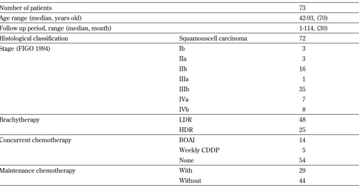
PatientsA retrospective analysis was done on all records of patients with histologically proven primary advanced squamous cell carcinoma of the cervix in the De-partment of Radiology, Tokushima University Hos-pital. Between December 1992 and March 2002, 73 patients with primary advanced squamous cell carci-noma of the cervix underwent radiotherapy. The clinical stage distribution according to the Interna-tional Federation of Gynecology and Obstetrics (FIGO) criteria (10) was as follows : Stage IB = 3 (4.1%), Stage IIA = 3 (4.1%), Stage IIB = 16 (21.9%), Stage IIIA = 1 (1.4%), Stage IIIB = 35 (50%), Stage IVA = 7 (9.6%) and Stage IVB = 8 (11.0%). Patients were staged by use of chest radiography, intravenous pyelography (IVP), blood chemistry, cystoscopy, and rectosigmoidoscopy. Table 1 summarizes pa-tient characteristics. Follow up and survival infor-mation was obtained from hospital records.
All patients had biopsy specimens taken pre-treatment from the exposed tumor on the cervix of the uterus. All samples were immediately formalin-fixed and embedded in paraffin.
Table 1. Patients characteristics and treatment methods
Number of patients 73
Age range (median, years old) 42-93, (70) Follow up period, range (median, month) 1-114, (30) Histological classification Squamouscell carcinoma 72
Stage (FIGO 1994) Ib 3 IIa 3 IIb 16 IIIa 1 IIIb 35 IVa 7 IVb 8 Brachytherapy LDR 48 HDR 25
Concurrent chemotherapy BOAI 14 Weekly CDDP 5
None 54
Maintenance chemotherapy With 29
Radiotherapy Technique
For patients with primary advanced carcinoma of the cervix, radiotherapy consisting of a combi-nation of intracavitary brachytherapy and exter-nal beam irradiation has been used in our depart-ment. Before August 1997, low dose rate intracav-ity brachytherapy (LDR) was delivered with a Cs-137 source. From September 1997, high dose rate intracavity brachytherapy (HDR) was delivered with an Ir-192 source.
External radiation therapy was performed with 6 MV X-rays using the anterior and posterior parallel opposing field technique. Five fractions weekly, with 1.8 or 2.0 Gy per fraction, were delivered to the mid-plane of the pelvis. The area of external radiation therapy included both the primary tumor and the regional lymph nodes. The fraction size was 1.8-2 Gy and total dose to the whole pelvis was 36-52 Gy (me-dian 48 Gy) in the LDR group with midline shield-ing at 15-40 Gy (median 38 Gy), whereas in the HDR group the fraction size was 1.8-2 Gy and the total 40-50 Gy (median 45 Gy), with midline shield-ing at 20-45 Gy (median 43 Gy).
In the LDR group, we delivered the intracavitary irradiation either before or after external radiation therapy using a T.A.O manual afterloading applica-tion (11) with a Cs-137 source. The patient received 44-55 Gy (median 48 Gy) to point A with the exter-nal radiation therapy being designed to bring the dose to a total of 50-70 Gy through use of a midline shield. In the HDR group, intracavitary irradiation was delivered after external irradiation using a Modi-fied Manchester Applicator, an Ir-192 source with a remotely controlled after-loading system (RALS) without general anesthesia. Once per week, at 6 Gy per fraction, a total of 12-30 Gy was delivered to point A.
Adjuvant therapy
Fourteen patients with locally advanced cancer received intra-arterial chemotherapy using the bal-loon occluded arterial infusion (BOAI) method and 5 patients received weekly cisplatin (CDDP) infu-sion as concurrent chemotherapy. A total of 29 pa-tients underwent oral administration of fluorouracil or tegafur-uracil as maintenance chemotherapy. Maintenance chemotherapy was performed continu-ously for two years following radiation therapy.
Immunohistochemical study
A mouse monoclonal antibody (Mab),
anti-EPAS-1/HIF-2α (EP190b ; Novus Biologicals, Littleton, CO, USA), was generated and characterized as re-ported previously (9, 12). Immunostaining was per-formed according to the Labeled StreptAvidin-Biotin (LSAB) method (Dako, Carpinteria, CA, USA), us-ing a streptavidin-biotin-horseradish peroxidase complex. Briefly, deparaffinized and rehydrated sec-tions were treated with 0.3% hydrogen peroxide in methanol for 30 min at room temperature to block endogenous peroxidase activity. Antigen retrieval was achieved by a 750 W microwave in 10 mM cit-rate buffer (pH 6.0) for 15 min. After blocking with 10% goat serum in PBS, sections were incubated with the first antibody, and the reaction was carried out at 4!!overnight. All initial antibodies used for this study were tested for optimal dilutions, and a moderate dilution was determined for the best differentiation of tumor samples. We used 1 : 400-diluted EPAS-1 Mab (1.125 μg/ml). For immuno-histochemical detection of macrophages, dilution of the Mabs anti-human CD68 (KP1, Novus Biologi-cals, Littleton, CO, USA) was done at 1 : 800. Reac-tions were visualized using the 3,3’-diaminobenidine substrate chromogen system (DAB) (K3468, Dako, Carpinteria, CA, USA). All IHC slides were counter-stained by Mayer’s hematoxylin.
Quantification of the Immunohistochemical study
A quantification method for the TIMs and HIF-2α in TIMs was based on a previous study (13, 14). To summarize, for focal macrophage quantification, all slides were first observed at low power magnifi-cation (
!
10) to select three areas of the highest concentration of CD68. The numbers of positively stained cells in the three areas were counted at high power magnification (!
200). The average number of stained cells in the three ‘hotspot’ areas was con-sidered as the CD68 count. The number of HIF-2α positively stained cells in the same area of the three hotspots was counted at high power magnification (!
200). The average count was considered as the HIF-2α count. The average of the HIF-2α count was divided by the average of the CD68 count, and the HIF-2α/CD68 ratio was obtained.Assessment was performed in blinded fashion and independently by two investigators (T. K., A. K.). We used the average data from the two investiga-tors. Conflicting scores were resolved by discussion over the microscope.
Statistical analysis
package (Abacus Concepts, Berkeley, CA, USA) was used for all statistical analysis and generation of survival curves. χ2tests examined the relationships
between categorical tumor variables. Spearman-rank correlations were used to investigate relation-ships between continuous patient and tumor vari-ables. Survival curves were plotted using the method of Kaplan and Meier, and the Log-rank test was used to evaluate differences between life tables. P values less than 0.05 were considered significant.
RESULTS
HIF-2"expressionIn only 5 of 73 samples, HIF-2α immunoreactivity was observed in nuclei of tumor cells. All of these ex-pressions were observed to be very weak at a high power field. No tumor cell contained immunoreac-tive cytoplasm (Fig. 1).
CD68 immunoreactivity was detected in mono-nuclear cells that were distributed mainly in the in-terstitial tissue and slightly in the tumor paren-chyma. These mononuclear cells were identified as TIMs (Fig. 1). HIF-2α immunoreactivity in colocali-zation with CD68 immunoreactive cells was found in 53 (72.6%) of the 73 samples. All the positive HIF-2α immunoreactivity in interstitial cells considered as TIMs was in the cytoplasm and was even stronger than that in tumor cells. The number of HIF-2α counts in 73 samples ranged from 0 to 79 with a median value of 14 (mean=18.7, SD=16.8), which was used as the cutoff point for categorical analy-sis. A group of 39 fell into the high HIF-2α count category (53%). The number of CD68 counts in 73 samples ranged from 0 to 169 with a median value of 57 (mean=62.4, SD=35.1). The high CD68 count group contained 38 samples (52%) using the cut-off point of 57. A positive correlation was found between increasing CD68 count (as a continuous variable) and increasing HIF-2α count (Spear-man Rho=0.54, p!0.0001). The HIF-2α/CD68 ra-tio ranged from 0% to 100% with a median value of 23.2% (mean=29.2%, SD=25.7).
HIF-2"expression and clinical futures
In this study, the cumulative overall 5-year local control rate according to stage was 100% in stage I, 89% in stage II, 78% in stage III, 43% in stage IVa and 25% in stage IVb. The cause-specific 5-year sur-vival rate was 100% in stage I, 79% in stage II, 58% in stage III, 43% in stage IVa and 13% in stage IVb. These data show similar results to previous reports on radiotherapy outcomes of uterine cervical cancer from other institutes (15-18). Table 2 shows the dis-A
A BB
C
C DD
Fig. 1. Immunohistochemical study of HIF-2α and CD68 at hu-man invasive uterine cervical cancer (squamous cell carcinoma) On (A) HIF-2α and (B) CD68 in low power field (
!
10), the uter-ine cervical squamous carcinoma show common positive cells observed in mainly interstitial tissue. On (C) HIF-2α and (D) CD68 in high power field (!
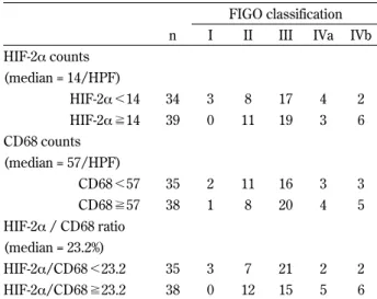
200), both immunoreactivity was de-tected in common mononuclear cells that are distributed mainly in interstitial tissue and slightly in tumor parenchyma. In this case, a few tumor cells show HIF-2α immunoreactivity in nuclei.Table 2. Distribution of HIF-2α, CD68 and HIF-2α/CD68 in FIGO classification
FIGO classification n I II III IVa IVb HIF-2α counts (median = 14/HPF) HIF-2α !14 34 3 8 17 4 2 HIF-2α "14 39 0 11 19 3 6 CD68 counts (median = 57/HPF) CD68!57 35 2 11 16 3 3 CD68"57 38 1 8 20 4 5 HIF-2α / CD68 ratio (median = 23.2%) HIF-2α/CD68 !23.2 35 3 7 21 2 2 HIF-2α/CD68 "23.2 38 0 12 15 5 6
tribution of HIF-2α count, CD68 count and HIF-2α/ CD68 ratio in the FIGO classification. There were relatively balanced distributions of classified HIF-2α count, CD68 count and HIF-2α/CD68 ratio in the FIGO classification.
The HIF-2α count, CD68 count and HIF-2α/ CD68 ratio were compared with DFS and cause-specific survival (CSS). The HIF-2α count and CD68
count failed to achieve significance for either DFS or CSS (Fig. 2 and 3). Only the HIF-2α/CD68 ratio was associated with DFS (p=0.024) with cases in the high category group having the worst DFS (Fig. 4). However the HIF-2α/CD68 ratio was not asso-ciated with CSS. The distribution of the HIF-2α/ CD68 ratio is shown in Table 3. There was no corre-lation between the HIF-2α/CD68 ratio and age,
con-Fig. 2. Kaplan-Meier curves of DFS and CSS based on HIF-2α expression in TIMs
Fig. 3. Kaplan-Meier curves of DFS and CSS based on CD68 expression in TIMs
current or maintenance chemotherapy, brachyther-apy method, pretreatment hemoglobin level or CD68 count. HIF-2α immunoreactivity in the nu-clei of tumor cells had no relationship with DFS or CSS. (Data not shown)
In 19 of 73 patients tumors have recurred during our observation period. The high HIF-2α/CD68 ra-tio group had a significantly increased risk of local recurrence (p=0.0142) (Table 4).
DISCUSSION
To our knowledge, this is the first report about HIF-2α in squamous cell carcinoma of the uter-ine cervix. Major findings in this survey were that most of the HIF-2α immunoreactivity in squamous cell carcinoma of the uterine cervix was detected in TIMs and the percentage of HIF-2α immunore-activity in TIMs significantly correlated with DFS.
It is well established that, when exposed to low-ered levels of oxygen, tissues compensate in a va-riety of ways, ranging from systemic adjustments caused by increased erythropoietin production to tissue-specific effects of increased VEGF expression
and the largely cellular effects of increased glyco-lysis (19). All of these adaptations to hypoxia are regulated wholly or in part by the HIF complex. The oxygen-regulated components of this complex are the HIF-α subunits. To date, three members of the HIF-α family have been cloned : HIF-1α, HIF-2α, and HIF-3α. Of the three HIF-α subunits, the most extensively characterized subunit regarding func-tion is HIF-1α. Its hypoxia-induced stabilizafunc-tion and the following induction of a number of target genes seems to be a general response in most, if not all, cells. In addition, these proteins have at least some unique functions as revealed by their different tar-get genes (20-24). By contrast, HIF-3α is involved in downregulation of the hypoxic response via an alternatively spliced transcription factor, which may function as an inhibitor of HIF-1α (25, 26).
HIF-1α and HIF-2α expression are a significant prognostic factor of various cancers treated by ra-diotherapy (12, 27-30). In uterine cervical cancer, some papers (31-34) reported that overexpression of HIF-1α in pretreatment uterine cervical cancer cells served as a predictive marker for poor prog-nosis after treatment by radiotherapy. However, there has been no study examining the relation-ship between the expression of HIF-2α and uter-ine cervical cancer. In this study, weak HIF-2α im-munoreactivity in tumor cells was observed in only five biopsy sections and was not associated with prognosis. This difference in immunoreactivity be-tween HIF-1α and HIF-2α in cervical cancer cell seems to indicate that there are individual variations in the hypoxic response. Findings relating to the role of TIMs in cervical cancers have been contro-versial. It has been noted that there are higher
Table 3. Distribution of HIF-2α/CD68 ratio
HIF-2α/CD68 ratio (%) Over all (n=73) !23.2 (n= 35) "23.2 (n= 38) P value Age (mean!SD) 70.3!11.2 71.5!12.0 70.8!10.8 p=0.794 Concurrent chemotherapy With 19 11 8
p=0.317 without 54 24 30
Brachytherapy LDR 48 25 23
p=0.227
HDR 25 10 15
Maintenance chemotherapy With 29 15 14
p=0.590 without 44 20 24
Pretreatment hemoglobin level,
mean!SD (/mm3) 11.4!1.9 11.1!2.1 11.6!1.8 p=1.094
CD68 count,
mean!SD(/HPF) 61.5!35.6 58.2!39.7 64.6!31.6 p=0.437
Table 4. Treatment failure pattern
HIF-2α/CD68 ratio (%) !23.2 (n= 35) "23.2 (n= 38) Local only 2 7
Distant (With local) 5 (1) 5 (1)
macrophage counts in invasive carcinomas as op-posed to cervical squamous intraepithelial lesions (35). In turn, squamous intraepithelial lesions con-tain more macrophages than the normal cervix (36). Davidson, et al . (37) looked at 75 cases of carci-noma, staining the tissue with CD68 and two endo-thelial markers, and evaluated the cases by light mi-croscopy. Based on previous reports of an associa-tion between TIM density, increased angiogenesis, and poorer outcome in breast carcinoma (38), these authors sought to evaluate the same in cervical can-cer. No correlation was found between TIM count and tumor stage, grade, or survival, nor with mi-crovessel count.
Leek, et al . reported that HIF-2α expression in TIMs was not associated with DFS but was mar-ginally associated with overall survival in cases in the high expression group which had worse survival in invasive breast cancer (39). To our knowledge, this was the only report which mentioned a corre-lation between HIF-2α expression in TIMs and clini-cal outcome. However in this study, HIF-2α expres-sion in TIMs was associated with DFS. Addition-ally there was a significant tendency for an increased risk of local recurrence in cases with a high HIF-2α expression in the TIM group. This recurrence pat-tern suggests that the role of HIF-2α expression in TIMs is more important in regional tumor devel-opment than in distant metastasis. It seems that HIF-2α secreted by TIMs is a very important key factor in tumor survival. Thus if we can control TIM behavior, it must then contribute to remission of the tumor.
In conclusion, we evaluated HIF-2α expression in uterine cervical cancer. Most of the HIF-2α expres-sion was observed in TIMs and a high HIF-2α ex-pression ratio in TIMs was associated with DFS and local recurrence. These results demonstrated that macrophages recruited into tumor tissue play a role in promoting tumor growth and that HIF-2α expression in TIMs is an important prognostic fac-tor, especially for predicting future local recurrence after radiotherapy in patients with advanced uter-ine cervical cancer.
ACKNOWLEDGMENTS
We thank Ms. Noriko Amou, Department of Hu-man Pathology, Institute of Health Biosciences, The University of Tokushima Graduate School, for ex-cellent advice for Immunohistochemical technique.
REFERENCES
1. Vaupel P, Kelleher DK, Hockel M : Oxygen status of malignant tumors : pathogenesis of hypoxia and significance for tumor therapy. Semin Oncol 28 : 29-35, 2001
2. Hockel M, Knoop C, Schlenger K, Vorndran B, Baussmann E, Mitze M, Knapstein PG, Vaupel P : Intratumoral pO2 predicts survival in advanced cancer of the uterine cervix. Ra-diother Oncol 26 : 45-50, 1993
3. Hockel M, Schlenger K, Aral B, Mitze M, Schaffer U, Vaupel P : Association between tu-mor hypoxia and malignant progression in ad-vanced cancer of the uterine cervix. Cancer Res 56 : 4509-4515, 1996
4. Fyles AW, Milosevic M, Pintilie M, Hill RP : Cervix cancer oxygenation measured follow-ing external radiation therapy. Int J Radiat On-col Biol Phys 42 : 751-753, 1998
5. Brizel DM, Scully SP, Harrelson JM, Layfield LJ, Bean JM, Prosnitz LR, Dewhirst MW : Tu-mor oxygenesion predicts for the likelihood of distant metastasis in human soft tissue sar-coma. Cancer Res 56 : 941-943, 1996
6. Isaacs JS, Jung YJ, Mole DR, Lee S, Torres-Cabala C, Chung YL, Merino M, Trepel J, Zbar B, Toro J, Ratcliffe PJ, Linehan WM, Neckers L : HIF overexpression correlates with bialle-lic loss of fumarate hydratase in renal cancer : novel role of fumarate in regulation of HIF sta-bility. Cancer Cell 8 : 143-153, 2005
7. Kim WY, Kaelin WG : Role of VHL gene mu-tation in human cancer. J Clin Oncol 22 : 4991-5004, 2004
8. Semenza GL : Targeting HIF-1 for cancer ther-apy. Nat Rev Cancer 3 : 721-732, 2003
9. Talks KL, Turley H, Gatter KC, Maxwell PH, Pugh CW, Ratcliffe PJ, Harris AL : The expres-sion and distribution of the hypoxia-inducible factors HIF-1alpha and HIF-2alpha in normal human tissues, cancers, and tumor-associated macrophages. Am J Pathol 157 : 411-421, 2000 10. Pecorelli S, Benedet JL, Creasman WT, Shepherd JH : FIGO staging of gynecologic cancer. 1994-1997 FIGO Committee on Gyne-cologic Oncology. International Federation of Gynecology and Obstetrics. Int J Gynaecol Ob-stet 65 : 243-249, 1999
11. Tazaki E, Arai T, Oryu S : An applicator for in-tracavitary radiation therapy of carcinoma of the uterine cervix. Jpn J of Clin Radiol 10 :
768-775, 1965
12. Koukourakis MI, Giatromanolaki A, Skarlatos J, Corti L, Blandamura S, Piazza M, Gatter KC, Harris AL : Hypoxia inducible factor (HIF-1a and HIF-2a) expression in early esophageal cancer and response to photodynamic therapy and radiotherapy. Cancer Res 61 : 1830-1832, 2001
13. Maeda K, Chung YS, Ogawa Y, Takatsuka S, Kang SM, Ogawa M, Sawada T, Onoda N, Kato Y, Sowa M : Thymidine phosphorylase/ platelet-derived endothelial cell growth factor expression associated with hepatic metastasis in gastric carcinoma. Br J Cancer 73 : 884-888, 1996
14. Matsumura M, Chiba Y, Lu C, Amaya H, Shimomatsuya T, Horiuchi T, Muraoka R, Tanigawa N : Platelet-derived endothelial cell growth factor/thymidine phosphorylase expres-sion correlated with tumor angiogenesis and macrophage infiltration in colorectal cancer. Cancer Lett 128 : 55-63, 1998
15. Barillot I, Horiot JC, Pigneux J, Schraub S, Pourquier H, Daly N, Bolla M, Rozan R : Carcinoma of the intact uterine cervix treated with radiotherapy alone : a French coopera-tive study : update and multivariate analysis of prognostics factors. Int J Radiat Oncol Biol Phys 38 : 969-978, 1997
16. Logsdon MD, Eifel PJ : Figo IIIB squamous cell carcinoma of the cervix : an analysis of prog-nostic factors emphasizing the balance between external beam and intracavitary radiation ther-apy. Int J Radiat Oncol Biol Phys 43 : 763-775, 1999
17. Lanciano RM, Won M, Coia LR, Hanks GE : Pretreatment and treatment factors associated with improved outcome in squamous cell car-cinoma of the uterine cervix : a final report of the 1973 and 1978 patterns of care studies. Int J Radiat Oncol Biol Phys 20 : 667-676, 1991 18. Pedersen D, Sogaard H, Overgaard J, Bentzen
SM : Prognostic value of pretreatment factors in patients with locally advanced carcinoma of the uterine cervix treated by radiotherapy alone. Acta Oncol 34 : 787-795, 1995
19. Semenza GL : Regulation of mammalian O2 homeostasis by hypoxia-inducible factor 1. Annu Rev Cell Dev Biol 15 : 551-578, 1999 20. Hu CJ, Wang LY, Chodosh LA, Keith B, Simon
MC : Differential roles of hypoxia-inducible fac-tor 1 alpha (HIF-1 alpha) and HIF-2 alpha in
hy-poxic gene regulation. Mol Cell Biol 23 : 9361-9374, 2003
21. Brusselmans K, Bono F, Maxwell P, Dor Y, Dewerchin M, Collen D, Herbert JM, Carmeliet P : Hypoxia-inducible factor-2 alpha (HIF-2al-pha) is involved in the apoptotic response to hypoglycemia but not to hypoxia. J Biol Chem 276 : 39192-39196, 2001
22. Park SK, Dadak AM, Haase VH, Fontana L, Giaccia AJ, Johnson RS : Hypoxia-induced gene expression occurs solely through the action of hypoxia-inducible factor 1alpha (HIF-1alpha) : role of cytoplasmic trapping of HIF-2alpha. Mol Cell Biol 23 : 4959-4971, 2003
23. Carmeliet P, Dor Y, Herbert JM, Fukumura D, Brusselmans K, Dewerchin M, Neeman M, Bono F, Abramovitch R, Maxwell P, Koch CJ, Ratcliffe P, Moons L, Jain RK, Collen D, Keshert E : Role of HIF-1alpha in hypoxia-mediated apoptosis, cell proliferation and tu-mour angiogenesis. Nature 394 : 485-490, 1998 24. Sowter HM, Raval RR, Moore JW, Ratcliffe PJ, Harris AL : Predominant role of hypoxia-inducible transcription factor (Hif)-1alpha ver-sus Hif-2alpha in regulation of the transcrip-tional response to hypoxia. Cancer Res 63 : 6130-6134, 2003
25. Makino Y, Kanopka A, Wilson WJ, Tanaka H, Poellinger L : Inhibitory PAS domain protein (IPAS) is a hypoxia-inducible splicing variant of the hypoxia-inducible factor-3alpha locus. J Biol Chem 277 : 32405-32408, 2002
26. Hara S, Hamada J, Kobayashi C, Kondo Y, Imura N : Expression and characterization of hypoxia-inducible factor (HIF)-3alpha in hu-man kidney : suppression of HIF-mediated gene expression by HIF-3alpha. Biochem Biophys Res Commun 287 : 808-813, 2001
27. Kaanders JH, Wijffels KI, Marres HA, Ljungkvist AS, Pop LA, van den Hoogen FJ, de Wilde PC, Bussink J, Raleigh JA, van der Kogel AJ : Pi-monidazole binding and tumor vascularity pre-dict for treatment outcome in head and neck cancer. Cancer Res 62 : 7066 -7074, 2002 28. Aebersold DM, Burri P, Beer KT, Laissue J,
Djonov V, Greiner RH, Semenza GL : Expres-sion of hypoxia-inducible factor-1alpha : a novel predictive and prognostic parameter in the ra-diotherapy of oropharyngeal cancer. Cancer Res 61 : 2911-2916, 2001
29. Hui EP, Chan AT, Pezzella F, Turley H, To KF, Poon TC, Zee B, Mo F, Teo PM, Huang DP,
Gatter KC, Johnson PJ, Harris AL : Coexpres-sion of hypoxia-inducible factors 1alpha and 2alpha, carbonic anhydrase IX, and vascular en-dothelial growth factor in nasopharyngeal car-cinoma and relationship to survival. Clin Can-cer Res 8 : 2595-2604, 2002
30. Koukourakis MI, Giatromanolaki A, Sivridis E, Simopoulos C, Turley H, Talks K, Gatter KC, Harris AL : Hypoxia-inducible factor (HIF1A and HIF2A), angiogenesis, and chemoradio-therapy outcome of squamous cell head-and-neck cancer. Int J Radiat Oncol Biol Phys 53 : 1192-1202, 2002
31. Hutchison GJ, Valentine HR, Loncaster JA, Davidson SE, Hunter RD, Roberts SA, Harris AL, Stratford IJ, Price PM, West CM : Hypoxia-inducible factor 1alpha expression as an intrin-sic marker of hypoxia : correlation with tumor oxygen, pimonidazole measurements, and out-come in locally advanced carcinoma of the cer-vix. Clin Cancer Res 10 : 8405-8412, 2004 32. Ishikawa H, Sakurai H, Hasegawa M, Mitsuhashi
N, Takahashi M, Masuda N, Nakajima M, Kitamoto Y, Saitoh J, Nakano T : Expression of hypoxic-inducible factor 1alpha predicts metastasis-free survival after radiation therapy alone in stage IIIB cervical squamous cell car-cinoma. Int J Radiat Oncol Biol Phys 60 : 513-521, 2004
33. Bachtiary B, Schindl M, Potter R, Dreier B, Knocke TH, Hainfellner JA, Horvat R, Birner P : Overexpression of hypoxia-inducible factor 1alpha indicates diminished response to radio-therapy and unfavorable prognosis in patients receiving radical radiotherapy for cervical can-cer. Clin Cancer Res 9 : 2234-2240, 2003 34. Burri P, Djonov V, Aebersold DM, Lindel K,
Studer U, Altermatt HJ, Mazzucchelli L, Greiner RH, Gruber G : Significant correlation of hypoxia-inducible factor-1alpha with treatment outcome in cervical cancer treated with radical radio-therapy. Int J Radiat Oncol Biol Phys 56 : 494-501, 2003
35. Davidson B, Goldberg I, Kopolovic J : Inflam-matory response in cervical intraepithelial neo-plasia and squamous cell carcinoma of the uter-ine cervix. Pathol Res Pract 193 : 491-495, 1997 36. al-Saleh W, Delvenne P, Arrese JE, Nikkels AF, Pierard GE, Boniver J : Inverse modulation of intraepithelial Langerhans’ cells and stromal macrophage/dendrocyte populations in human papillomavirus-associated squamous intraepi-thelial lesions of the cervix. Virchows Arch 427 : 41-48, 1995
37. Davidson B, Goldberg I, Gotlieb WH, Lerner-Geva L, Ben-Baruch G, Agulansky L, Novikov I, Kopolovic J : Macrophage infiltration and angiogenesis in cervical squamous cell carcinoma -clinicopathologic correlation. Acta Obstet Gy-necol Scand 78 : 240-244, 1999
38. Leek RD, Lewis CE, Whitehouse R, Greenall M, Clarke J, Harris AL : Association of macro-phage infiltration with angiogenesis and prog-nosis in invasive breast carcinoma. Cancer Res 56 : 4625-4629, 1996
39. Leek RD, Talks KL, Pezzella F, Turley H, Campo L, Brown NS, Bicknell R, Taylor M, Gatter KC, Harris AL : Relation of hypoxia-inducible factor-2 alpha (HIF-2 alpha) expres-sion in tumor-infiltrative macrophages to tumor angiogenesis and the oxidative thymidine phos-phorylase pathway in Human breast cancer. Cancer Res 62 : 1326-1329, 2002