Evaluation of peritoneal lavage cytology at
the time of operation for gastric cancer
その他の言語のタイ
トル
胃癌手術時の腹腔洗浄細胞診の評価
イガン シュジュツジ ノ フククウ センジョウ サ
イボウシン ノ ヒョウカ
著者
Terata Nobukuni, Mizutani Kohnosuke, Kodama
Masashi, Okabe Hidetoshi
journal or
publication title
滋賀医科大学雑誌
volume
10
page range
65-72
year
1995-07
URL
http://hdl.handle.net/10422/3155
滋賀医大誌, 10, 65-72, 1995
Evaluation of Peritoneal I。avage Cytology
at the Time of Operation for Gastric Cancer
Nobukuni Terata, Kohnosuke Mizutani,
Masashi Kodama, and *Hidetoshi Okabe
First Department of Surgery and *Department of Laboratory Medicine, Shiga University of Medical Science
Abstract: Cytologic examination of peritoneal lavage fluid obtained during surgery was performed in 120 patients with gastric cancer to assess the prognostic value of a positive
cytology. Positive cytology was obtained in 19.2% of all cases, while 30.5% of cases with overt microscopic serosal invasion were positive. Degree of macroscopic or microscopic invasion to the gastric serosa, macroscopic peritoneal dissemination and stage of cancer were associated with a positive cytology. Patients with intraperitoneal free cancer cells demonstrated a significantly poorer prognosis than those without (p<0.001). In cases of positive lavage cytology, all patients except two died with peritoneal dissemination within 2 years after operation. Peritoneal metastasis was the most frequent site of recurrence. Therefore, this technique may provide a useful indicator of prognosis and for determining subpopulations of patients that might receive adjuvant chemotherapy for micrometastasis.
Key words: peritoneal lavage cytology, peritoneal dissemination, gastric cancer
INTRODUCTION
Considerable recent progress in treatment has improved long-term survival in gastric can-cer patients. This success is largely attributed to the performance of extensive lymph node removal. Despite these efforts, postoperarive recurrence is still frequent. Disseminated peri-toneal metastasis is the most frequent type of recurrence and a critical factor in the prognosis
of patients with gastric cancer. This must develope from residual microscopic cancer cells
despite surgery with curative intent. However, satisfactory attention has not been paid to can-cer invasion into the gastric serosa, a phenome-non likely closely related to the development of
peritoneal metastasis. We therefore conducted a
cytologic examination for free cancer cells ob-tained by washing the peritoneal cavity. Since no effective method of treatment is now availa-ble for established peritoneal dissemination, cytology of lavaged saline from the Douglas pouch may be useful as a predictor of prognosis and an indicator for adjuvant therapies.Accepted for publication December 1, 1994.
Correspondence : Nobukuni Terara, Department of Surgery I , Shiga University of Medical Science, Otsu, Shiga 520-21, Japan
N. Terata
MATERIALS AND METHODS
From January 1986 to December 1992 m-traoperative, peritoneal lavage cytology was performed m 120 patients with gastric cancer. Gastrectomy was performed in 106 patients and laparotomy only m 14 patients.
The methods of peritoneal lavage and cytologic examination are performed as follows: Immediately after opening the peritoneal cavity, Douglas pouch was irrigated with 100 ml of sa-line, and this was aspirated by syringe with a Nelaton tube after mild stirring by hand. The aspirated fluid was centnfuged, and the sediment was smeared and stained by the Papamcolaou method and PAS reaction for microscopic examination. Evaluation was per-formed according to the Papanicolaou's class-ification in which class Illb, IV and V were con-sidered positive for malignant cells.
To explore the clinical significance of cytological examination of peritoneal lavage fluid, its correlation with other parameters of gastric cancer such as the histologic stage, grade of macroscopic serosal invasion by tumor, histologic extent of gastric wall invasion, mac-roscopic grade of peritoneal dissemination, his-tologic type, macroscopic Borrmann's classifica-tion, tumor size and survival rate, was analysed statistically.
Microscopic wall invasion (t), macroscopic
peritoneal metastasis (p) and staging were de-fined in the General Rules for Gastric Cancer Study in Surgery and Pathology published by the Japanese Research Society for Gastric Can-cerl'. The degree of macroscopic serosal inva-sion (S) was also investigated according to the criteria of the previous edition2'for purposes of comparison.
Survival was analyzed according to the
ac-tuanal and Kaplan-Meier method. The
differ-ence in survival rate was assessed statisticallyusing the generalized Wilcoxon method. The
significance of the incidence of positive cytology was determined with the Fisher exact test and the chi square analysis. A probability value smaller than 0.05 was considered significant.
RESULTS
Peritoneal lavage cytology was examined in relation to several other factors. These included staging, degree of macroscopic invasion, extent of gastric wall invasion, macroscopic peritoneal dissemination, histology, macroscopic type, tumor size and survival.
1) Staging
Free cancer cells were demonstrated in
Douglas cavity in 23 of 120 (19.2%) patients. Table 1 shows the results of cytology at
lap-Table 1. Staging and Peritoneal Lavage Cytology S tag e P ap a n ic ola ou 's c la ssifica tion
In c id en ce of p o sitiv e § cy tolog y II Ilia n ib IV l a, I b 13 11 3 .6% II 12 12 0 Ilia, Illb 11 12 3 1.4 一 IV a, IV b ll 3 4.4
* : p<0.01 by Fisher exact test
隻 : According to Papanicolaou's classification, Class IIIb, IV and V were considered positive for malignant cells
Peritoneal Lavage Cytology for Gastric Cancer
arotomy in 120 patients in relation to their
stage. Stage III (31.4%) and IV (34.4%) showed a
significantly higher positive cytology rate as compared to other stages (p<0.01 by Fisherexact test).
2) Degree of macroscopic serosal invasion The relationship between peritoneal lavage cytology and the grade of macroscopic serosal invasion by the primary lesion is shown in Table 2. The incidence of free caccer cells in the peritoneal cavity generally correlated with increasing degree of serosal invasion - SO
(3.7%), SI (0%), S2 (20%, S3 41.7% (SO, SI vs
S2, S3: p<0.01 by Fisher exact test). 3) Extent of gastric wall invasion
In term of the microscopic depth of wall invasion, it was null in tl (m and sm), only one patient without any serosal involvement (t2: mp
and ss) and 30.5% (22/72) in patients with overt serosal involvement 20.4% t3 (se), 52.2% t4 (si)) (Table 3). The difference in incidence of positive cytology between cases with and without serosal involvement was statistically significant (p<0.01).
4) Macroscopic peritoneal dissemination
The incidence of positive cytology in pa-tients with macroscopic peritoneal dissemination
was 4.5% (4/ in PO patients, but averaged
59.4% (19/32) in groups PI, P2 and P3 combined
(Table 4). The greater the increase in the
P-Table 2. Degree of Macroscopic Serosal Invasion and Peritoneal Lavage Cytology
G ra de of m a crosc op ic
P ap a n ic ola ou 's c la ssifica tion In cid en ce of p o sitiv e c ytolog y serosa l
inv a sio n (S ) IT l a lib IV V
0 * 13 11 3.7 %
1 12 0 %
2 18 17 2 0 *
3 10 4 1.7 %
*p<0.01 by Fisher's exact test *0 : mtact mucosa
1 : serosal invasion suspected in small area 2 : definite invasion of the serosa
3 '蝣invasion of organs adjacent to primary serosal lesion
Tab一e 3. Extent of Gastric Wall Invasion and Peritoneal Lavage Cytology
E
x
t
e
n
to
f
g
a
s
t
r
i
cwa
l
l
i
n
v
a
s
i
o
n
P
a
p
a
n
i
c
o
l
a
u
'
sc
l
a
s
s
i
f
i
c
a
t
i
o
n
I
n
c
i
d
e
n
c
eo
f
p
o
s
i
t
i
v
e
c
y
t
o
l
o
g
y
I
T
I
l
i
a
I
l
l
b
I
V
V
t
1
§
(
m.
s
m)
l
l
U7
3
.
4
% ⊃
0
「
t
2
(
mp
,
s
s
)
1
5
1
1
t
3s
e
1
3
2
3
2
0
.
4
% ⊃
」
t
4s
i
4
1
0
* : p<0.01 by Fisher exact test
§ : tl Lamina propria(m), submucosa(sm)
t2 Muscularis propria (mp), subserosa(ss) t3 Penetrates serosa (se)
N. TE:RATA
Tab一e 4. Macroscopic Peritoneal Dissemination and Peritoneal Lavage Cytology
Ma
c
r
o
s
c
o
p
i
c
p
e
r
i
t
o
n
e
a
l
d
i
s
s
e
mi
n
a
t
i
o
n
P
a
p
a
n
i
c
o
l
a
u
'
sc
l
a
s
s
i
f
i
c
a
t
i
o
n
I
n
c
i
d
e
n
c
eo
f
p
o
s
i
t
i
v
e
c
y
t
o
l
o
g
y
I
I
I
l
i
a
f
f
lb
I
V
V
p
0
3
6
4
1
4
.
5
% 「
p
1
3
3
0
1
0
5
5
0
.
5
%
p
2
3
2
0
0
1
3
4
4
.
4
%
p
3
1
0
1
2
0
7
6
3
.
6
%
*p<0.01 by Fisher exact test pO : no macroscopic dissemination
pi : small number of disseminated lesions in perigastric, upper abdomen (above transverse colon)
p2 : moderate number of lesions in entire abdomen p3 : numerous lesions in entire abdomen
Table 5. Histology and Peritoneal Lavage Cytology
H istology
P apanicolau's classification Incidence of positive cytology IT Ilia lib IV V pap§ 1 2 0 0 0 0 0 -0 tubl 6 5 0 0 1 2 21.4% tub2 14 15 13.5% p0r 21 20 10 21.4% m uC 1 2 2 1 0 0 16.7% Sig 1 2 1 0 0 1 20.0%
§ pap : papillary adenocarcinoma,
tubl : tubular adenocarcinoma, well differentiated type tub2 : tubular adenocarcinoma, moderately differentiated type
por : poorly differentiated adenocarcmoma muc : mucinous adenocarcinoma
sig : signet-ring cell carcinoma
number, the larger was the number of cases of positive cytology. There was a significant dif-ference between cases with and without macro-scopic dissemination (p<0.01). It is noteworthy that these four who did not have macroscopic peritoneal dissemination were gastric cancer patients with positive cytologies.
5) Histologic type
There was no relation between the his-tologic differentiation and the incidence of posi-tive peritoneal cytology (Table 5).
6) Macroscopic type
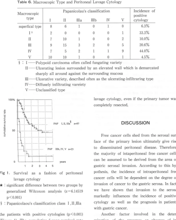
According to the Borrmann's classification,
the incidence of positive lavage cytology was
33.3 % in type IV (Table 6). There was no
cor-relation between this classification and lavage cytology.
7) Tumor Size
The relationship between the incidence of mtrapentoneal free cancer cells and tumor size in 106 patients is shown in Table 7. The rate of detection increased sharply with tumor size greater than 8 cm in diameter (p<0.01).
8) Survival
Those patients with normal cytologic find-ings had a significantly better prognosis than
Peritoneal Lavage Cytology for Gastric Cancer
Table 6. Macroscopic Type and Peritoneal Lavage Cytology
M a crosco p ic P ap a n ic olau 's c lassifica tio n In cid enc e o f p ositiv e cy to log y typ e II Ilia Illb IV V
sup erfical type 8 6 1 0 1 0 6.3 % I § 2 0 0 0 0 1 33.3 %
II 10 10.0 %
u I 15 20.6 %
IⅤ 7 5 2 1 1 9 44.0 %
Ⅴ 10 10 4.5 %
§ : I -- Polypoid carcinoma often called fungating variety
Iト- Ulcerating lesion surrounded by an elevated wall which is demarcated
sharply all around against the surrounding mucosaIII一一Ulcerative variety, described often as the ulcerating-infiltraring type
IV- - Diffusely infiltrating vareiety
V一日Unclassified type
lavage cytology, even if the primary tumor was completely resected, E S S5 9 j b j i B A i A j n s a A i j B i n a i n o PAP I,II,Ilia§ n=97 PAP Mb,IV,V n=23 」 」 ニ_」 ye ars 1 2 3 4 5
Fig 1. Survival as a fashion of peritoneal lavage cytology
★ : sigm丘cant difference between two groups by
generalized Wilcoxon analysis (z=6.14519
p<0.001)
§ '蝣Papanicolaou's classification class 1 ,1日IIa the patients with positive cytologies (p<0.001)
(Figure 1). The cumulative 5-year survival rate in patients with positive peritoneal lavage
cyto-logies was 8.7%, while m those without it was
46.7
All but two patients died as a result of peri-toneal dissemination within 2 years of operation (mean, 5.0±4.76 months), in cases of positive
DISCUSSION
Free cancer cells shed from the serosal sur-face of the primary lesion ultimately give rise to disseminated peritoneal disease. Therefore, the majority of intaperitoneal free cancer cells can be assumed to be derived from the area of gastric serosal invasion. According to this hy-pothesis, the incidence of intraperitoneal free cancer cells will be dependent on the degree of invasion of cancer to the gastric serosa. In fact, we have shown that invasion to the serosa markedly influences the incidence of positive cytology as well as the prognosis in patients with gastric cancer.
Another factor involved in the deter-mination of the presence or absence of in-traperitoneal free cancer cells is the size of the tumor mass. The rate of detection of free malignant cells increased t0 42.9% in patients
with a tumor greater than 8 cm in diameter.
In the present study, 23 out of 120 (19%)
patients with gastric cancer had a positivecyto-N. Terata
Table 7. Tumor Size and Peritoneal Lavage Cytology
T um or size P apanicolaou 's classification Incidence of positive cytology (cm ) la Illb IV V < 2 3 7 1 0 0 0 2- 4 ll 4- 6 10 10 6 - 8 9 9 1 0 1 2 8- 10 3 3 1 0 0 4 36.4% > 10 5 5 2 0 1 4 29.4% *p<0.01 by cm square test
logy. This value is slightly smaller than findings in previous reports by Nakajima et al (30%)4
but is slightly greater values suggested by
Hirano et al (4.0%)5) and Koga et al (13.5%)6). The difference in the percentage of positive cytologies among these several reports may be due to somewhat different patients populations, the method of peritoneal lavage and the proce-dure for aspirating lavage fluid. A high inci-dence of positive cytology tends to depend on lavage of both upper abdomen and Douglas pouch31, rather than Douglas pouch5'6'.
Moreover, in spite of serosal invasion by
the tumor, not so high incidence of positive
cytologies (only 30.5% in our paper) can be
ob-tained. Such a negative diagnosis does not al-ways result from technical failure as already described above, because the possibility that positive cytology is derived from characteristics of cancer cell itself, cannot be ruled out. Some investigators have reported parallel correlation between positive cytologic findings and his-tologic type4'7'or macroscopic classification of serosal surface8'. According to these authors, tumor cells of pooly differentiated carcinomas, which invaded the serosal layer of the stomach, seems to detach and to be liberated into the peritoneal cavity more easily than those of well differentiated carcinomas. But these factors cannot sufficiently account for low incidence of positive cytology in serosal invasion cases. Thisquestion is at present difficult to assess.
We demonstrated four cases in which
in-traperitoneal free cancer cells were detected by peritoneal cytologic exploration despite a grossly normal peritoneum at the time of opera-tion. These subclimcal micrometastases are a potential source of future recurrence. Actually, two of these four patients died in 4 and 10 months, respectively, while one remains alive with abdominal wall metastasis 46 months after operation, and the remaining patient is alive without recurrence. This alive patient without recurrence received intravenous mitomycin C 3 days after operation and 5-fluorouracil per os for 6 years. Except for these two patients that remain alive, all other patients (21 patients with positive cytology) died within 2 years. Thus, the majority of patients with free peritoneal cancer cells detected at the time of surgery will not escape postoperative peritoneal recurrence. However, patients who have only a few in-traperitoneal free cancer cells may escape peri-toneal recurrence if prophylactic manipulations are used during surgery. Peritoneal lavage cyto-logy will facilitate the effective selection of patients for postoperative adjuvant therapy.
A positive cytologic specimen was observed in one case in which the serosal layer of the stomach was not involved microscopically. This patient was described above because he did not
Peritoneal Lavage Cytology for Gastric Cancer have macroscopic peritoneal dissemination.
There have been several recent reports in which positive cytology was not accompanied by serosal invasion. Imada et al.7) have reported one case out of 225 in which cancer invasion
was contained within the mp, and Miwa et al9).
have reported four cases with mp invasion. These authors have suggested that these tive cytologies may in some cases be false posi-tive. For example, macrophages and mesothelial cells may sometimes be mistaken for malignant cellslO. This is also the possibility that the malignant cells may be derived from metastasis in lymph nodes or primary tumors of other
or-gansll. We should also consider that cases with
exceptionally long survival, despite positivecytology, has been reported. Miwa et al. have
stated that one can not conclude that a positive cytology is always associated with a poor prog-nosis9).
Previous reports3'6'9'have shown that the prognosis in surgically treated patients with gastric carcinoma is significantly affected by the presence or absence of intraperitoneal free
cancer cells at the time of surgery. We have
shown that the postoperative 5-year survivalrate was only 8.7% in patients with microscopic
evidence of intraperitoneal free cancer cells,compared with 46.7% m patients without
detec-table tumor cells in the peritoneal washings. Thus, the data suggest that cytology of lavaged saline from Douglas pouch can, although requir-ing a somewhat complicated procedure, be used as a prospective indicator for the risk of pos-toperative peritoneal metastasis. Based on these data, we are now conducting a study of prophy-lactic intraperitoneal administration of cytocidal anticancer drugs to prevent the recurrence of disseminated malignant cells.REFERENCES
1) Japanese Research Society for Gastric Can-cer (1993) The general rules for the gastric cancer study, 12th edition, Kinbara Shup-pan, Tokyo (in Japanese).
2) Japanese Research Society for Gastric Can-cer (1981) The general rules for gastric can-cer study in surgery and pathology. llth edition, Jap J Surg ll, 127-139.
3) Nakajima, T., Harashima, S., Hirata, M., and Kaiitani, T. (1978) Prognostic and ther-apeutic values of peritoneal cytology in gas-trie cancer, Acta Cytologica 22, 225-229. 4) Iitsuka, K., Kanesima, S., Tanida, 0.,
Takeuchi, T., and Koga, S. (1979) In-trapentoneal free cancer cells and their vi-ability in gastric cancer, Cancer 44, 1476-1480.
5) Hirono, M., Matsuki, K., Nakagami, K.,
Niimoto, M., and Hattori, T. (1981)
Com-parative studies on cytological and his-tological metastasis in gastric cancer. Jap J Surg ll, 330-336.6) Koga, S., Kaibara, NリIitsuka, Y., Kudo, H.,
Kimura, A., and Hiraoka, H. (1984) Prog-nostic significance of intraperitoneal free cancer cells in gastric cancer patients, J Cancer Res Clin Onco1 108, 236-238.
7) Imada, T., Abe, M., Noguchi, Y., Tamura, S., Yamamoto, Y., Akaike, M., Amano, T. and Matsumoto, A. (1989) Studies on the
peritoneal dissemination of gastric cancer with a special reference to lavage cytology and histological evaluation, Jap J Gastroen-terol 22, 2353・2357 (in Japanese).
Nakai, M., Yonemura, Y., Matsumoto, H., Tsugawa, K., Kimura, H., Takegawa, S., Kamata, T., Ooyama, S., Kosaka, T., Yamaguchi, A., Miwa. K., Miyazaki, I. (1991) Relationship between gross classifica-tion of serosal surface and histological
find-N. Terata
ings in advanced gastric cancers-with refer-ence to peritoneal lavage cytology- Jap J Clin Sure 52, 1991-1994 (in Japanese). 9) Miwa, K., Yamagishi, M., Kitamura, H.,
Hagino, S., Matsuki, N., Yamaguchi, A., Asano, K., Noguchi, M., Takashima, S., Takeshita, Y., Miyazaki, I., Tanimoto, K. and Matsubara, F. (1990) Evaluation of
peri-toneal lavage cytology at operation in 247 primary gastric cancer patients, J Jap Soc Cancer Ther 15, 1131-1136 English
ab-stract).
10 Sakai, Y. (1973 Cytology of pleural and peritoneal effusion, Jap J Clin Path01 21,
749-752 (in Japanese).
ll) Kurosu, Y., Endo, K., Tomita, R., Amano, S., Matsumoto, S., Nakanism, H., Fuka-machi, S., Tanii, K., Mizuno, T., and
Monta, Kリ(1989) Intraperitoneal free
can-cer cells and continuous hyperthermic pen-toneal per fusion in gastric cancer, Nichidai Zasshi 48, 481-487 (English abstract).