Original
(JTok川 山edhiv) 84 ( 4)122~ 128 (2014)
Changes t
o
P
e
r
i
t
o
n
e
a
l
S
u
r
r
o
g
a
t
e
Markers i
n
CAPD
P
a
t
i
e
n
t
s
Treated w
i
t
h
N
e
u
t
r
a
l
pH
,
Low-GDP P
e
r
i
t
o
n
e
a
l
D
i
a
l
y
s
i
s
S
o
l
u
t
i
o
n
f
o
r
Long P
e
r
i
o
d
s
Chieko HIGUCHI and Hiroshi SAKURA
Department of Medicine, Tokyo Women's Medical University Medical Center East (Accepted J une11, 2014) Introduction: Long-term peritoneal dialysis (PD) is known to injure the peritoneum. One cause of such dam -age is the bioincompatibility of conventionallow-pH solutions containing glucose degradation products. New PD solutions with neutral pH and low levels of glucose degradation products have been available for peritoneal dialy -sis in J apan since2000.We investigated changes to several peritoneal parameters in long-term PD-treated pa -tients using the new solution. Materials and Methods: Participants were 78 patients who had undergone treat -ment with the new solution since starting PD between March 2001and J uly2012and who continued PD for over 12months. We measured the dialysate-to・plasmaratio of creatinine (D/Pcr) and several surrogate markers of peritoneal injury (cancer antigen(CA) 125, mesothelial cell area, hyaluronic acid (HA)) using overnight peritoneal effluent.We studied the relationships between these surrogate markers, and the influence of clinical factors on these peritoneal surrogate markers. Resu1ts: Concentrations ofCA125 in peritoneal effluent tended to decrease, and mesothelial cell area in effluent tended to increase with increased duration of treatment.Using the MIXED procedure, these surrogate markers correlated significantly with duration of treatment.No changes in levels of HA in peritoneal effluent or in D/Pcr were observed during the treatment period. Conc1usion: From these re -su1ts, mesothelial cell injury may increase in patients on long-term treatment even with the use of new PD solu
-tlOns.
Key W ords: neutral pH, low-GDP peritoneal dialysis solution, peritoneum, CA125, mesothelial cell area
Introduction
Injury to the peritoneum occurs with long-term use of peritoneal dialysis (PD) in patients. Pathologi -cal changes to the peritoneum have been reported to include mesothelial detachment, fibrotic changes under the submesothelial compact zone and neoan -giogenesisl).These morphological changes are con -sidered to be the major causes of hyperpermeabil -ity and ultrafiltration failure, resulting in the techni -cal failure of PD2 ).One of the causes of these perito -neal changes is the bioincompatibility of conven -tional solution, due to its low pH and the presence of glucose degradation products (GDP), among other
factors3
).In Japan, new PD solutions that are neutral
pH and contain low levels of GDP have been avail -able for PD treatment since2000, and all PD pa
-tients have been changed from conventional solu同
tions to these new PD solutions as of20054 ). Numer-ous reports have described the influence of the new PD solutions on the peritoneum in PD patients5 )-7). However, observation periods for these reports have ranged from several months to 2 years. The present study investigated changes to several peri -toneal parameters in long-term PD-treated patients using the new solution in our hospita.l Materials and Methods Patients A total of109 patients were treated with the new PD solution when starting PD treatment at Tokyo Women's Medical University Medical Center East between March 2001 and J uly2012.Among these,
participants in the present study were 78patients (53men, 25 women) who continued PD treatment for more than12months. Mean ( :tstandard devia -
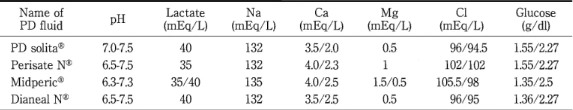
-122-Table 1 Composition of each PD solution Name of pH Lactate Na PD fluid (mEq/L) (mEq/L) PD solita⑧ 7.0-7.5 40 132 Perisate N⑧ 6.5-7.5 35 132 Midperic③ 6.3-7.3 35/40 135 Dianea1 N⑧ 6.5-7.5 40 132 tion) age at the start of PD treatment was 56.7:!: 14.5 years (range, 24-89 years), and mean duration of PD treatment was 37.2:!: 22.2 months (range, 12-99 months). Etiology of end-stage renal disease (ESRD) was glomerulonephritis in 25 patients, diabetes in 24, nephrosclerosis in 8, polycystic kidney disease in 4, gout in 2, and other in 15. During PD treatment, the frequency of peritonitis was as follows: 0 epi -sodes in 36 patients, 1 episode in 15, 2 episodes in 14, 3 episodes in 4, 4 episodes in 5, and 6 episodes in 2. Patient outcomes were as follows: 16 patients con -tinued continuous ambulatory peritoneal dialysis (CAPD) treatment, 25 patients changed to hemo-dialysis (HD) treatment or PD + HD combined ther -apy, 22 patients moved to another hospital (because the physician in charge moved to another hospital or the patient moved to another area), 9 patients died, and 6 patients received renal transplantation. Patients were treated using several kinds of new PD solution (Table 1). Numbers of patients treated with each PD solution were: Perisate N③, 27 pa -tients; Balance③, 16 patients; Midperic⑧, 3 patients; and Dianeal N③,32 patients. Study design E旺luentcancer antigen (CA) 125 levels have been considered likely to depend on mesothelial cell mass or turnover8 ).This study therefore used effluent CA1251evel for mesothelial cell mass. We also stud -ied the mesothelial cell area in overnight peritoneal effluent, as Yamamoto et al and Izumotani et al re -ported this as a useful marker of peritoneal meso -thelial cel1injurl)lO). Peritoneal inflammation occurs consequent to chronic exposure to bio-incompatible PD fluid and induces fibrotic changes under the submesothelial compact zone. Hyaluronic acid (HA) levels in perito -neal effluent are used as a marker of peritoneal in -Ca (mhE4qg/L) C1 G1ucose (mEq/L) (mEq/L) (g/d1) 3.5/2.0 0.5 96/94.5 1.5512.27 4.0/2.3 102/102 1.5512.27 4.0/2.5 1.5/0.5 105.5/98 1.35/2.5 3.5/2.5 0.5 96/95 1.36/2.27 flammation11)in CAPD patients. This study there -fore also analyzed HA levels in peritoneal effluent as a marker of peritoneal inflammation. Dialysate -to回plasmaratio of creatinine (D/Pcr) has been re -ported to increase during long-term PD treatment using conventional PD solution凶 .In our patients, we measured peritoneal function during PD treat -ment using D/Pcr.
We measured CA125 and HA levels in overnight peritoneal effluent at several points during PD treatment.Samples of peritoneal effluent from the overnight bag were collected and frozen at -800
C for later analysis. Analysis of CA1251evels was per -formed by chemiluminescent enzyme immunoassay (Rumiparusu CA125II; Fujirebio, Tokyo,
J
apan). HAlevels were measured by latex-enhanced immuno-turbidimetric assay (Erupiae-su; Mitsubishi Chemi -cal Medience, Tokyo,
J
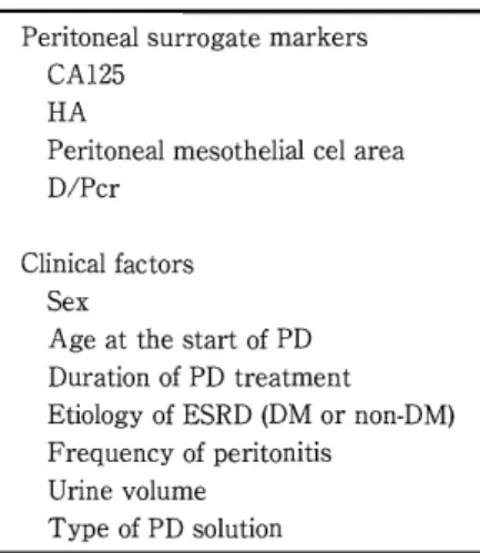
apan). Analysis of mesothelial cell area in the present study was performed by Dr. Yamamoto. D/Pcr was determined at several time points during PD treatment. Furthermore, we analyzed clinical data consid -ered to influence peritoneal changes. We adopted clinical factors such as sex, age at start of PD, PD treatment duration, etiology of ESRD (diabetes mel -litus (DM) or non-DM), frequency of peritonitis, re -sidual renal function (urine volume), and the kind of PD solution for analysis (Table 2). The influences of clinical factors on the peritoneal surrogate markers were examined. All subjects enrolled in this research provided in -formed consent prior to participation, and all study protocols were approved by our institutional com -mittee on human research (registration number 2690). Statistical analysis To elucidate relationships between clinical fac-Table 2 Study variables Peritoneal surrogate markers CA125 HA Peritoneal mesothelial cel area D/Pcr Clinical factors Sex Age at the start of PD Duration of PD treatment Etiology of ESRD (DM or non-DM) Frequency of peritonitis Urine volume Type of PD solution tors and peritoneal surrogate markers, we per -formed multiple regression analysis using a step -wise method with JMP version 11.0 software (SAS Institute J apan, Tokyo). In the stepwise method, peritoneal surrogate
markers were applied to dependent variables and clinical factors were applied to independent vari -ables. We used stepwise forward regression, with
exclusion of covariates showing univariate p values greater than 0.2. Due to the retrospective nature of the study, the number of times measurements were
taken differed between each patient.For surrogate markers identified as independent contributing fac -tors by multiple regression analysis, we examined
the relationship between variables using the MIXED procedure with SAS software (SAS Insti -tute, Cary, NC, USA). Values of p< 0.05 were consid -ered significant. Results Distribution of surrogate markers of perito -neal injury in peritoneal effluent during PD treat -ment periods CA125 levels in effluent have been considered a surrogate marker of mesothelial cell mass, and are
known to decrease with increasing duration of treatment in patients treated using conventional di -alysis solution13 ).In this study, the concentration of CA 125 in peritoneal effluent tended to decrease during PD treatment (Fig. 1a). Mesothelial cell area has been reported to in -crease during treatment in CAPD patients using conventional PD solution14).In our patients, meso -thelial cell area in effluent also tended to increase with the duration of treatment (Fig. 1 b). E妊luentlevels in HA have been considered a sur -rogate marker of peritoneal inflammation and are reported to increase with the PD treatment period among patients using conventional PD solution15). However, HA concentration in this study was un -changed during the period of treatment with the new PD solution. D /Pcr has been increasing with the PD treat -ment period in patients using conventional PD solu -tion12 ).No changes in D/Pcr were observed during the PD treatment period in the present study (Fig.2). Influence of clinical factors on peritoneal sur -rogate markers We analyzed the influence of clinical factors on peritoneal surrogate markers using multiple regres圃 sion analysis. Table 3 shows the results of multiple regression analysis. Duration of PD treatment was an inde -pendent factor on CA125 and mesothelial cell area in peritoneal e旺luent. We studied the dependence of CA 125 and meso -thelial cell area on the duration of treatment by the MIXED procedure using SAS, and these surrogate
markers showed a significant correlation with the duration of PD treatment (p
=
0.0024, p=
0.013). Discussion A statistical study by the J apanese Society for Di -alysis Therapy reported a mean duration for PD treatment in J apan of 3.18 i:3.39 years as of the end of 201016 ).Morphological changes to the peritoneal membrane have been observed with increasing du -ration of PD, including fibrotic changes to the sub -mesothelial compact zone, loss of mesothelial cells and neoangiogenesisI).These changes have been re -ported to correlate with increasing small-solute per -meability2)and a reduction in peritoneal ultrafiltra -tion capacity, and may be partially responsible for the shortened technical survival of PD. These dele -terious changes occur under conditions of chronic exposure to conventional bioincompatible PD solu -tion. In vitro and in vivo evidence suggests that these morphological changes are induced by low pH-124-CA125(U/ml) 100
a
.
:
.
.
.
•
•
80 60 20 40,
・
.
(months) 100 90 80 70 60 50 40 30 20 Mesothelial cell area (μm21 500 10o
o
b•
•
•
-•
•
-. . ・ .
h Jア
.
P
・
T
•
-an・
-. ・ -. ・ ・
'
.
.
‘ ー
••
. -A・
'
・ ・
' 4 1.
4
,
••
, 句 ‘
' '
-' ・ ・
4 4 け l l .-a
、 ・ ・
. 旬 、 ・
h o-.
A
白 色 " ' ・
4 ω
.
・
•
•
•
450 400 350 300 250 200 100 (months) Fig. 1 Scattergram of CA125 concentrations and mesothelial cell surface area in perito -neal effluent in each patient duringPD treatment a) CA125 levels tended to decrease with increasing duration ofPD treatment. b)Mesothelial cell surface area tended to increase with increasing duration ofPD treat -ment. 90 80 70 60 50 40 30 20 10 150 o levels were increased in patients treated with new PD solution and decreased in patients with conven -tional solution20 ).Such results suggested the biocom -patibility of new PD solutions, but study periods for these studies were only several months at best. No and the presence of glucose, GDP, and lactate3).Since2000, new PD solutions have been available on the market and a, ll conventional solutions in
J
apan were changed to new PD solutions from 2005.previous studies have reportedCA 125levels in ef -fluent with the use of new PD solutions for longer periods such as over 2 years. The present study re -vealed thatCA 125levels in effluent declined with long-term PD therapy even when using new PD so -lutions. However, Breborowicz et aFllhad reported that theCA125 level in effluent was not correlated with number of cells in the monolayer of mesothe -lial cultures, and they concluded thatCA125 is not a reflection of mesothelial cell mass. We considered that further research about the meaning ofCA 125 in effluent should be needed. In this study, mesothelial cell area increased with time on PD. The increment in mesothelial cell area in peritoneal effluent reportedly correlates with the degree of peritoneal injury9).Maekawa et al showed
-125-Various reports have described the impact of these new PD solutions on clinical outcomes, includ -ing peritoneal membrane function5)-7). However, follow-up periods for these reports have generally been under 24 months. In the present study, we analyzed changes to several peritoneal parameters in long-term PD-treated patients using the new so -lution in our hospita.l In our study, the concentration ofCA125 in peri -toneal effluent decreased significantly during PD treatment L.ongitudinal study of PD treatment with dialysis solution strated decliningCA 125levels in effluent with in -creasing duration of treatmene3 ).Several studies have found higherCA125 levels in effluent with the use of new PD solutions compared with conven -tional PD solutions17 )-19). In a crossover study, CA125 demon-also has con ven tional
a
HA (ng/ml) 600 500 400 300 J_
.
・
・
ν
.
I.~-
•
!・・'
200 i恵
三
与
・
f
.
、
s
Z
2
句
・
.
, .
.
・
.
・
1
、
1 0 : ? &診
戸
仰
、
、
.
・
3
J
a
a
y
e
•
•
o
20 40 60 80 100 (months) b D/Pcr 1.1 1•
.
・
- ・
. h '
4 .•
. 、
'
. . 4 ‘ ,.
4
•
•
••
、
. a v.
4
、
・
.
. 4 .•
・
'
.
e
a-・ -・ ﹁ -・
・ ' ・ ・ ・
1 . 4・ ' ・ . ,
' ' ・ 4 l a -ι r・
h・
-“ ' ' ・ 1 拘 恥 ・¥ ,
.
-d ω
4 4・
h '・
1・
p -H ' u h '・
'
p
,
3
4 . .
' ' d・ ・ ・
i 1. げ
? ぶ
v u
-r
, 寸 l 寸 l 寸 l 寸 l ﹁ n ヨ 0 0 守 ' r O F 3 n u n u n u n u n u•
•
• • •
•
• •
0.4•
0.3o
20 40 60 80 100 (months)Fig. 2 Scattergram of HA levels in PD effluent and D/Pcr during PD treatment a) HA; b)D/Pcr.
HA levels in PD effluent and D/Pcr remained unchanged with increasing duration of PD treatment.
Table 3 Results of regression coefficient and P values between peritoneal surrogate markers and clini -cal factors CA125 HA Mesothelial cell area D/Pcr Sex regression coefficient -0.027 -0.309 0.063 -0.174 p value 0.926 0.731 0.817 0.492 Age at start of PD regression coefficient 0.046 -0.016 -0.308 -0.001 p value 0.641 0.982 0.374 0.866 Duration of PD treatment regression coefficient 0.173 -0.202 0.535 -0.001 p value 0.013* 0.663 0.011 * 0.188 DM/non DM regression coefficient 0.400 -0.309 0.322 -0.303 p value 0.161 0.227 0.234 0.240 Frequency f peritonitis regression coe旺icient -0.965 3.517 -0.470 0.011 p value 0.279 0.603 0.882 0.277 Urin volume regression coefficient 0.006 -0.039 0.004 0.001 p value 0.078 0.068 0.711 0.641 Type of PD solution regression coefficient -3.192 0.146 0.313 0.05 p value 0.995 0.558 0.234 0.839
Duration of PD treatment was independently associated with CA125 and mesothelial cell area in peritoneal effluen.t
*: p<0.05.
-126-that mesothelial cell area increased during treat -ment in CAPD patients using conventional PD solu -tion14l
. Mesothelial cell area was lower with new PD
solutions than with conventional solution according to several reports山 2).In the
J
apan Balance study,the effect of the new PD solution in lowering meso-thelial cell area was recognized only within the first
9 months of the 27-month follow-up period. In the present study, mesothelial cell area increased dur -ing prolonged PD treatment.Given these resu1ts of increased mesothelial cell area and decreased CA 125 levels in effluent during PD treatmen,t mesothelial cell injury may occur during long-term PD treatment even with the use of new PD solu -tions. HA levels in peritoneal effluent are considered as a marker of peritoneal inflammationll)in CAPD p仕 tients. Effluent levels of HA reportedly show signifi -cant positive correlations with time on CAPD among patients using conventional PD solution15 ).In the present study, effluent HA levels were un -changed during the PD treatment period. Based on these resu1ts, we considered that peritoneal inflam -mation in patients using the new PD solution may be milder than that with conventional PD solutions. However, this study did not conduct direct compari圃
sons with parameter from patients using conven -tional PD solution, and further research in this area is warranted.
In the present study, duration of PD treatment was not independently associated with D /Pcr. D / Pcr has been reported to increase during long-term (84-month) PD treatment using conventional PD so -lution12).Several studies using new PD solutions
have suggested no differences in D /Pcr compared to conventional PD solution23
)24).However, follow-up
periods for those reports were under 2 years. In the present study, D/Pcr among patients treated with new PD solutions was unchanged during long-term PD treatment.Our results suggest that the effect of new PD solutions on changes in solute transport with long-term PD treatment may di妊erfrom that of conventional PD solution. Conclusion In PD patients treated with new PD solutions for long periods, concentrations of CA 125 in PD efflu -ent decreased, and mesothelial cell area in perito -neal effluent increased with treatment period. Given these results, mesothelial cell injury may in -crease in patients receiving long-term treatmen,t even with the use of new PD solutions.
Acknowledgment The authors are grateful to Dr Satoru Shimizu for as -sisting in the statistical analysis. The authors have no conflicts of interest to dec1are. References 1)Williams jD, Craig Kj, Topley N et al: Morpho -logical changes in the peritoneal membrane of pa -tients with renal disease.J Am Soc Nephrol 13: 470-479,2002 2) Krediet RT, Lindholm B, Rippe B: Pathophysiol -ogy of peritoneal membrane failure. Perit Dial Int 20 (Suppl 4): S22-S42, 2000 3) Chaudhary K, Khanna R: Biocompatible perito -nea1dialysis solutions; do we have one? ClinJ Am Soc Nephro15: 723-732, 2010 4) Hi郡lchiC, Nishimura H, Sanaka T: Biocompati -bility of peritoneal dialysis fluid and influence of compositions on peritoneal fibrosis. Ther Apher Diall0:372-379,2006 5) Fusshoeller A, Plail M, Grabensee B et al: Bio -compatibility pattern of a bicarbonate / lactate -buffered peritoneal dialysis fluid in APD: a prospec -tive, randomized study. Nephrol Dial transplant 19: 2101-2106,2004 6) Kim S, Oh j, Kim S et al: Benefits of biocompatible PD fluid for preservation of residual renal function in incident CAPD patients: a 1-year study. Nephrol Dial Transplant 24: 2899-2908, 2009 7) Rippe B, Simonsen 0, Heimburger 0 et al: Long -term clinical e旺'ectsof a peritoneal dialysis fluid with less glucose degradation products. Kidney Int 59:348-357,2001 8) Krediet RT: Dia1ysate cancer antigen 125 concen -tration as marker of peritoneal membrane status in patients treated with chronic peritoeneal dialysis. Perit Dial Int 21: 560-567, 2001 9) Yamamoto T, Izumotani T, Otoshi T et al: Mor -phological studies of mesothelial cells in CAPD ef -fluent and their clinical significance. Am J Kidney Dis 32: 946-952.1998 10) Izumotani T, IshimuraE, Yamamoto T et al: Correlation between peritoneal mesothelia1cell cy -tology and peritoneal histopathology with respect to prognosis in patients on continuous ambulatory peritoneal dialysis. Nephron 89: 43-49, 2001 11) Yung S, Chan TM: Hya1uronan・regulatorand in -itiator of peritoneal inflammation and remodeling. IntJ Artif Organs 30: 477-483, 2007 -127
12) Davies SJ: Longitudina1 re1ationship between sol -ute transport and u1trafi1tration capacity in perito -nea1 dia1ysis patients. Kidney Int 66: 2437 -2445, 2004 13) Ho・DacヂannekeetM M, Hiralall JK, Struijk DG et al: Longitudina1 follow-up of CA125 in peritonea1 e百1uent.Kidney Int 51: 888-893,1997
14) Maekawa K, Yamamoto T, Hino H et al: Re1a四
tionship between duration of peritonea1 dia1ysis and mesothe1ia1 cell area in e1derly patients-Com -parison of neutra1ized dia1ysate with conventiona1 dia1ysate. Kidney and Dia1ysis 61 (Supp1): 237-239,
2006
15) Wakabayashi Y, Yamada K, Miura Y et al: Type III procollagen N-peptide and hya1uronate in serum and dia1ysate of CAPD patients. Nihon Jinzo Gakkai Shi39:408-413,1997
16) Nakai S, Iseki K, Itami N et al: An overview of regu1ar dia1ysis treatment in Japan (As of 31 De -cember 2010). Ther Apher Dia145: 1-47,2012
17) Jones S, Holmes CJ, Krediet RT et al: Bicarbon -ate /lactate-based peritonea1 dia1ysis solution in -creases cancer antigen 125 and decreases hya1uronic acid 1eve1s. Kidney Int 59: 1529-1538,
2001
18) Choi HY, Kim DK, Lee TH et al: The c1inica1 use -fu1ness of peritonea1 dia1ysis fluids with neutra1 pH and 10w glucose degradation product concentra
-tion: An open randomized prospective tria.lPerit Dia1 Int 28: 174-182,2008
19) Haag-Weber M, Kramer R, Haake R et al:
Low-⑧ GDP fluid (Gambroso1 trio "") attenuates dec1ine of residua1 rena1 function in PD patients: a prospec -tive randomized study. Nephro1 Dia1 Transp1ant 25: 2288-2296,2010 20) Williams JD, Topley N, Craig KJ et al:The euro司 ba1ance trial: The e旺ectof a new biocompatib1e peritonea1 dialysis fluid (ba1ance) on the peritonea1 membrane. Kidney Int 66: 408-418, 2004
21) Breborowicz A, Breborowicz M, Pyda M et al: Limitations of CA 125 as an index of peritonea1 mesothe1ia1 cell mass. Nephron C1in Pract 100: c46 -c51,2005
22) Yamamoto T, Hi郡lchiC, N akamoto H et al: The
J apan Ba1ance study: Eva1uation of peritonea1 dia1y -sis fluid with neutra1 pH and 10w 1eve1s of glucose degradation products in peritonea1 dia1ysis pa -tients. J Jpn Soc Dia1 Ther 42: 835-846, 2009 23) Tranaeus A: A 10ng-term study of bicarbonate/ 1actate-based peritonea1 dia1ysis solution - c1inica1 benefits. Perit Dia1 Int 20: 516-523, 2000
24) Fan SLS, Pile T, Punzalan T et al: Randomized controlled study of biocompatib1e peritonea1 dia1y -sis solutions; effect on residua1 rena1 function. Kid -ney Int 73: 200-206, 2008 中性・低GDP透析液長期使用による CAPD患者の腹膜パラメーターの変化 東京女子医科大学東医療センター内科 ヒ グ チ チ エ コ サ ク ラ 樋口千恵子・佐倉 宏 〔緒言〕長期腹膜透析では腹膜が障害されるが,その原因のひとつとして従来透析液の低いPH (酸性)および グルコース分解産物 (GDP) があげられていたこれに対し本邦では中性・低 GDPの新しい透析液が 2000年よ り 使 用 さ れ る よ う に な っ た 今 回 中 性 ・ 低GDP透析液の長期使用による腹膜パラメーターの変化について検討 した〔対象,方法