INTRODUCTION
Accurate diagnosis of lymph node metastasis is very important in deciding the optimal treatment
ORIGINAL
Diagnosis of the presence of lymph node metastasis and
decision of operative indication using
fluorodeoxyglu-cose - positron emission tomography and computed
tomography in patients with primary lung cancer
Hiroaki Toba
a, Kazuya Kondo
a, Hideki Otsuka
b, Hiromitsu Takizawa
a,
Koichiro Kenzaki
a, Shoji Sakiyama
a, and Akira Tangoku
a aDepartment of Thoracic and Endocrine Surgery and Oncology, andb
Department of Radiology, Institute of Health Biosciences, the University of Tokushima Graduate School, Tokushima, Japan
Abstract : Objectives : We evaluate whether integrated fluorodeoxyglucose-positron emis-sion tomography and computed tomography (FDG-PET/CT) scan can diagnose the pres-ence of lymph node metastasis more accurately than computed tomography (CT) scan alone. Methods : Forty-two patients with lung cancer preoperatively underwent integrated PET/CT scan using FDG and CT scan and underwent pulmonary resection and lymph node dissection. We judged cases as lymph node metastasis if the lymph node visually accumulated FDG at PET/CT scan and measured 1 cm or greater in the short axis at CT scan. We retrospectively analyzed whether our judgments in each modality were consis-tent with the pathological diagnosis. Results : Two-hundred and seventeen stations of lymph node were dissected and 21 stations (9.7%%) were histologically diagnosed as posi-tive metastasis. Thirty-two stations of lymph node visually accumulated FDG at PET/CT scan and 17 stations measured 1 cm or greater in the short axis at CT scan. Concerning the diagnosis of lymph node metastasis, PET/CT scan showed significantly higher sensi-tivity than CT scan (81%% vs. 48%%, p=0.024). The false-positive rate was significantly high in PET-positive lymph nodes measuring less than 1 cm in diameter. There were 4 false-negative lymph nodes with both scans. All of these were less than 7 mm in diameter and had a low percentage of metastatic foci in the lymph node. Concerning the diagnosis of N staging, there was no significant difference between PET/CT scan and CT scan (83% vs. 69%%, p=0.124). However, the identification of N2 disease at PET/CT scan was signifi-cantly more accurate than that at CT scan (100%% vs. 38%%, p=0.031). Conclusions : PET/CT is superior to CT scan in lymph node staging. However, because the false-positive rate is high in PET-positive lymph nodes measuring less than 1 cm in diameter, we think that clinical background should be considered and other modalities or histological examina-tions should be undertaken if necessary. J. Med. Invest. 57 : 305-313, August, 2010
Keywords : fluorodeoxyglucose-positron emission tomography and computed tomography scan, computed to-mography scan, lung cancer, lymph node metastasis, diagnosis
Received for publication April 21, 2010 ; accepted July 9, 2010. Address correspondence and reprint requests to Kazuya Kondo, Department of Thoracic and Endocrine Surgery and Oncology, Institute of Health Biosciences, the University of Tokushima Graduate School, Kuramoto - cho 3 - 18 - 15, Tokushima 770 - 8503, Japan and Fax : + 81 - 88 - 633 - 7144.
of lung cancer. Recently, positron emission tomogra-phy and computed tomogratomogra-phy (PET/CT) scan has been undertaken in many facilities and its efficacy for the diagnosis of accurate staging of lung can-cer has been reported (1-2). Gould MK. et al. (3) reported that PET scan was significantly better than computed tomography (CT) scan in a meta-analysis. The PLUS multi-center randomized trial also proved the effectiveness of PET scan in preoperative pa-tients with suspected lung cancer (4). Subsequently, Birm Ö et al. (5) reported the same results in a meta-analysis and, recently, it has been indicated that PET scan should be undertaken for patients with lung cancer with regard to mediastinal and extrathoracic staging, and it is recommended as grade IB by a standardized method ‘Methodology for Lung Cancer Evidence Review and Guideline Development’ in the ACCP guidelines (6).
Since PET/CT scan was introduced in our facil-ity in December 2005, we have used it for lung can-cer patients to diagnose preoperative staging, recur-rence, and to judge the effectiveness of induction therapy. We retrospectively evaluated the effective-ness of PET/CT scan for preoperative lymph node staging for lung cancer patients compared with that of CT scan.
MATERIALS AND METHODS
PatientsBetween December 2005 and April 2007, 42 con-secutive patients with a histologic diagnosis of lung cancer, who underwent a curative operation at Tokushima University Hospital, were retrospectively reviewed. All patients preoperatively underwent both integrated PET/CT scan using fluorodeoxyglucose (FDG) and chest CT scan including chest X-ray and brain MRI as well as operative indication being assessed. The protocol of this study was not sub-mitted to Institutional Review Board because FDG-PET/CT scan and chest CT scan were routine pre-operative examination and this study was retrospec-tive one. The patients underwent pulmonary resec-tion and lymph node dissecresec-tion, which was a radi-cal operation. None of the patients in the study re-ceived preoperative chemotherapy or radiotherapy. The histologic type of lung cancer was adenocarci-noma in 30 patients, squamous cell carciadenocarci-noma in 8, large cell carcinoma in 1, small cell carcinoma in 2, and pleomorphic carcinoma in 1. The pathologic N stages were N0 in 32, N1 in 2, N2 in 8, and N3 in 0
(Table 1). The lymph nodes were classified accord-ing to the original lymph node map of lung cancer (7).
FDG-PET/CT scan protocol and image interpre-tation
The patients were required to fast for 6 hours and avoid strenuous work or exercise for 24 hours before PET/CT scan. Sixty minutes after the intrave-nous injection of FDG (3.7 MBq/kg body weight), PET/CT scan was performed with an Aquiduo (Toshiba Medical Systems, Tokyo, Japan). Patients were imaged from the skull base to the mid-thigh level. The technical parameters used for the CT scan were a detector row configuration of 16
!
1.25 mm, a helical pitch of 15, a gantry rotation speed of 0.5 s, peak voltage of 120 kVp, a tube load of 50 mA and slice thickness of 2 mm. An emission scan was acquired immediately following the CT scan for 2 min per bed position in 3D mode. The images were usually reconstructed using ordered subset expec-tation maximization selecting 4 iterations and 12 subsets, a 128!
128 matrix, and post-smoothing with an 8 mm Gaussian filter. The reconstructedTable 1. Characteristics of the patients
Variable Data NO. of patients 42 Age (yrs) 68.0"9.5 Sex Male 25 Female 17 Tumor location
Right upper lobe 14 Right middle lobe 4
Right lower lobe 9
Left upper lobe 5
Left lower lobe 10
Histology
Adenocarcinoma 30
Squamous cell carcinoma 8 Large cell carcinoma 1 Small cell carcinoma 2 Pleomorphic carcinoma 1 Pathologic N stage N0 32 N1 2 N2 8 N3 0
spatial resolution after smoothing was 9.2 mm. Abnormal FDG uptake was defined as greater than background activity in surrounding normal tis-sue excluding physiologic uptake sites. Regions of interest (ROI) were placed on visible uptake sites and standardized uptake values (SUV) were calcu-lated as
SUV=decay-corrected activity (kBq)/tissue volume (ml)/injected-FDG activity (kBq)/body weight (g). In this study, the maximum SUV was used to mini-mize of partial volume effect and assure reproduci-bility of measurements.
CT scan protocol and image interpretation
Multidetector CT scan was performed with an Aquilion 16 (Toshiba Medical Systems, Tokyo, Japan). The technical parameters used for the CT scan were a detector row configuration of 16
!
0.5 mm, a helical pitch of 15, a gantry rotation speed of 0.5 s, peak voltage of 120 kVp, and a tube load of 100-400 mA and slice thickness of 1 mm. The win-dow level and width were set at -500 Hounsefield units (HU), and 1500 HU.Criteria of lymph node metastasis
We judged the lymph node station as metastasis if FDG was visually accumulated greater than back-ground activity in surrounding normal tissue ex-cluding physiologic uptake sites at PET/CT scan. At the same time, we measured the short-axis di-ameter of the largest lymph node at its station. We also judged the lymph node station as metastasis if the short-axis diameter of the lymph node was greater than 1 cm at axial view of CT scan as in pre-vious reports (8, 9). Two chest surgeons HT and KK analyzed PET/CT scan by consensus while re-ferring to reports made by several experienced nu-clear medicine physicians. Two chest surgeons HT and KK also analyzed CT scan by consensus while referring to reports made by several radiologists. Discrepancies were resolved after consultation.
Histological Examination
The dissected lymph nodes were examined his-tologically using formalin-fixed and paraffin-embed-ded sections and the largest section in each lymph node was cut and examined with hematoxylin and eosin staining with a microscope.
Statistical Analysis
True-positive (TP), true-negative (TN), false-positive (FP), and false-negative (FN) results of
PET/CT scan and CT scan for lymph node metas-tasis were compared with the results of pathologic diagnosis. Sensitivity was calculated as TP/TP+FN, specificity as TN/TN+FP, positive predictive value (PPV) as TP/TP+FP, negative predictive value (NPV) as TN/TN+FN, and accuracy as TP+TN/ total. All data were analyzed for significance by the 2-tailed Student’s t test using the SPSS 11.0 for Windows software (SPSS Japan Inc., Tokyo, Japan).
RESULTS
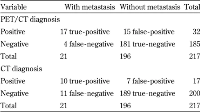
In the 217 dissected lymph node stations, 21 sta-tions (9.7%) were histologically diagnosed as positive metastasis. Thirty-two lymph node stations visually accumulated FDG at PET/CT scan and the mean value of maximum SUV of those lymph node sta-tions was between 1.7 and 8.6 (mean"SD, 3.7" 1.6). PET/CT scan yielded TP in 17 lymph node stations, FP in 15, FN in 4, and TN in 181 (Table 2).
Seventeen lymph node stations measured 1 cm or greater in short axis at CT scan. This yielded TP in 10, FP in 7, FN in 11, and TN in 189 (Table 2). The sensitivity, specificity, accuracy, positive predic-tive value, and negapredic-tive predicpredic-tive value of PET/CT scan were 81%, 92%, 91%, 53%, and 98%, respectively, whereas those of CT scan were 48%, 96%, 91%, 59%, and 94% (Table 3). PET/CT scan showed signifi-cantly higher sensitivity than CT scan (p=0.024).
Table 2. PET/CT and CT analysis with pathologic diagnosis Variable With metastasis Without metastasis Total PET/CT diagnosis
Positive 17 true - positive 15 false - positive 32 Negative 4 false - negative 181 true - negative 185
Total 21 196 217
CT diagnosis
Positive 10 true - positive 7 false - positive 17 Negative 11 false - negative 189 true - negative 200
Total 21 196 217
Table 3. Diagnostic results of PET/CT scan and CT scan Variable PET/CT CT Difference Sensitivity 0.81 0.48 P = .024 Specificity 0.92 0.96 0.08 Accuracy 0.91 0.91 0.86 Positive predictive value 0.53 0.59 0.93 Negative predictive value 0.98 0.94 0.15
Table 4 shows a comparison of lymph node stag-ing by PET/CT scan and CT scan with that by pa-thologic lymph node staging. The accuracy of lymph node staging was 83% at PET/CT scan and 69% at CT scan (p=0.124). However, the identification of N2 disease was 8 of 8 (100%) at PET/CT scan, but 3 of 8 (37.5%) at CT scan. We could diagnose the N2 disease at PET/CT scan more accurately than at CT scan (p=0.0031).
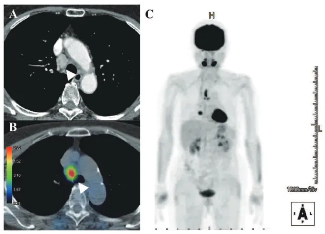
A representative case with a both PET and CT true-positive result is shown in Figure 1. FDG was accumulated in pretracheal lymph node (#3) at PET/CT scan. The size and maximum SUV values were 11.0 mm and 6.2. The lymph node was surgi-cally dissected and diagnosed as metastasis.
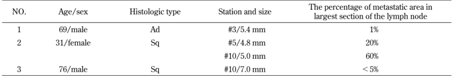
There were 4 false-negative lymph nodes in 3 cases (Table 5). All of these measured less than 7 mm in diameter and showed small metastatic areas.
Table 4. Comparison of N staging with PET/CT scan and CT scan
PET/CT diagnosis CT diagnosis
N0 N1 N2 N3 Total N0 N1 N2 N3 Total Pathologic N Stage N0 26 2 4 0 32 25 5 2 0 32 N1 1 1 0 0 2 1 1 0 0 2 N2 0 0 8 0 8 4 1 3 0 8 N3 0 0 0 0 0 0 0 0 0 0 Total 27 3 12 0 42 30 7 5 0 42
Figure 1. Representative case of true-positive FDG - PET/CT scan and chest CT scan A : Chest CT scan demonstrated the significant swelling of pretracheal lymph node (#3).
B : PET/CT scan demonstrated the accumulation of FDG (arrow) in the pretoracheal lymph node (#3).
C : The view of maximum intensity projection (MIP) of PET/CT scan demonstrated the accumulation of FDG in primary tumor and mediastinal lymph nodes.
A representative case with a PET false-negative re-sult is shown in Figure 2. A 75-year-old male (pa-tient 3 in Table 5) with squamous cell carcinoma received preoperative PET/CT scan. FDG was not accumulated in mediastinal and hilar lymph nodes and clinical N-stage was diagnosed as N0. He un-derwent surgical resection of the primary tumor and lymph node dissection. Histopathologically, right hi-lar lymph node station (#10) showed involvement of tumor cells and pathological N-stage was diag-nosed as N1. The size of the metastatic lymph node
was 7 mm in diameter and the metastatic foci occu-pied less than 5% of the total in the largest section of the lymph node.
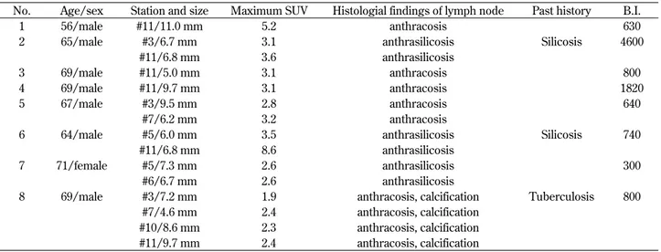
There were 15 false-positive lymph nodes in 8 cases. They were 8 mediastinal and 7 hilar lymph nodes. Median size of the lymph nodes was between 4.6 and 11.0 mm (mean"SD, 7.5"1.9 mm). Maxi-mum SUV was between 1.9 and 5.2 (mean"SD, 3.4"1.6). Their histological characteristics were lymphoid follicular hyperplasia with anthracosis, calcification, and anthrasilicosis (Table 6). All 8
Table 5. False negative PET/CT findings
NO. Age/sex Histologic type Station and size The percentage of metastatic area in largest section of the lymph node
1 69/male Ad #3/5.4 mm 1%
2 31/female Sq #5/4.8 mm 20%
#10/5.0 mm 60%
3 76/male Sq #10/7.0 mm !5%
Ad = Adenocarcinoma, Sq = Squamous cell carcinoma
#3 = Pretracheal lymph nodes, #5 = Subaortic lymph nodes, #10 = Hilar lymph nodes
Figure 2. Representative case of false - negative FDG - PET/CT scan A : FDG was not accumulated in the hilar lymph node (#10) at PET/CT scan.
B : Microscopic appearance of lymph nodes (hematoxylin and eosin staining, original magnification
!
100). A few tumor cells (arrow) were recognized in the largest section of the lymph node.C : The view of maximum intensity projection (MIP) of PET/CT scan demonstrated no accumulation of FDG in the mediastinal and hilar lymph nodes.
cases with false-positive results on FDG-PET/CT scan had smoking history (B.I.=300-4600), 2 cases (25.0%) had silicosis, and one case (12.5%) had had tuberculosis in the past. A representative case with a PET false-positive result is shown in Figure 3.
A 69-year-old male (patient 8 in Table 6) with adeno-carcinoma received preoperative PET/CT scan. FDG was accumulated in mediastinal and hilar lymph node stations (#3, #7, #10, #11) clinical N-stage was diagnosed as N2. Sizes and maximum
Figure 3. Representative case of false - positive FDG - PET/CT scan
A : PET/CT scan demonstrated accumulation of FDG (arrow) in the subcarinal lymph node (#7).
B : Microscopic appearance of lymph nodes (hematoxylin and eosin staining, original magnification
!
100). Follicular lymphoid hy-perplasia with anthrasilicosis and no tumor cells in the lymph nodes were revealed.C : The view of maximum intensity projection (MIP) of PET/CT scan demonstrated the accumulation of FDG in primary tumor and mediastinal and hilar lymph nodes.
Table 6. False positive PET/CT findings
No. Age/sex Station and size Maximum SUV Histologial findings of lymph node Past history B.I.
1 56/male #11/11.0 mm 5.2 anthracosis 630
2 65/male #3/6.7 mm 3.1 anthrasilicosis Silicosis 4600 #11/6.8 mm 3.6 anthrasilicosis
3 69/male #11/5.0 mm 3.1 anthracosis 800
4 69/male #11/9.7 mm 3.1 anthracosis 1820
5 67/male #3/9.5 mm 2.8 anthracosis 640
#7/6.2 mm 3.2 anthracosis
6 64/male #5/6.0 mm 3.5 anthrasilicosis Silicosis 740 #11/6.8 mm 8.6 anthrasilicosis
7 71/female #5/7.3 mm 2.6 anthrasilicosis 300
#6/6.7 mm 2.6 anthrasilicosis
8 69/male #3/7.2 mm 1.9 anthracosis, calcification Tuberculosis 800 #7/4.6 mm 2.4 anthracosis, calcification
#10/8.6 mm 2.3 anthracosis, calcification #11/9.7 mm 2.4 anthracosis, calcification #3 = Pretracheal lymph nodes, #5 = Subaortic lymph nodes, #6 = Paraaortic lymph nodes
#7 = Subcarinal lymph nodes, #10 = Hilar lymph nodes, #11 = Interlobar lymph nodes B.I. = Brinkman Index
SUV values of the lymph nodes (#7) were 4.6 mm in diameter and 2.4. But because the degree of ac-cumulation of FDG in each lymph node station was almost same compared with background activity in surrounding normal tissue in the view of maxi-mum intensity projection (MIP), the possibility of the N0 disease was suggested. He underwent sur-gical resection of the primary tumor and lymph node dissection. Histopathologically, dissected lymph nodes showed follicular lymphoid hyperplasia with anthrasilicosis and no involvement of tumor cells ; pathological N-stage was diagnosed as N0.
Figure 4 shows the relationship between metas-tatic states and the size of PET-positive lymph nodes. If the cut-off value of the size of lymph nodes was set at 1 cm in diameter, 9 (90%) of 10 lymph node stations with PET-positive lymph nodes of more than 1 cm showed metastasis. However, me-tastasis was shown in only 8 (36%) of 22 lymph node stations with PET-positive lymph nodes of less than 1 cm. The positive predictive value of lymph nodes measuring more than 1 cm in diameter was signifi-cantly higher than that of lymph nodes measuring less than 1 cm in diameter (0.90 vs. 0.36, p=0.015).
DISCUSSION
In this study, the sensitivity of PET/CT scan was significantly higher than that of only CT scan (81%
vs. 48%, p=0.024). On the other hand, the specificity and accuracy of both modalities were similar. Con-cerning lymph node staging, there were no signifi-cant differences between FDG-PET/CT scan and CT scan. However, concerning the identification of N2 disease, which has sometimes been a clinical problem of therapeutic strategy, FDG-PET/CT scan was significantly more accurate than CT scan.
Many authors have reported that FDG-PET/CT scan was effective with regard to lymph node stag-ing in patients with lung cancer and the sensitivity and specificity were almost 80-90% and were gener-ally superior to those of CT scan (1-6, 9). Our data was similar to previous reports and the sensitivity of PET/CT scan was significantly superior to CT scan although there was no difference between FDG-PET/CT scan and CT scan with regard to the specificity. It is recommended that FDG-PET/ CT scan should be performed as a non-invasive di-agnostic tool in ACCP guidelines (5) and ESTS guidelines (10). In view of our data, it is thought that the FDG-PET/CT scan is currently the most reliable non-invasive diagnostic tool with regard to the presence of lymph node metastasis and should be performed preoperatively as a routine examina-tion.
In this study, the PPV of FDG-PET/CT scan was 53%. There were 8 cases with 15 false-positive lymph node stations at FDG-PET/CT scan. All false-positive lymph nodes showed lymphoid follicular hy-perplasia with anthracosis, anthrasilicosis, and cal-cification histologically. In addition, all patients with false-positive lymph node stations were smokers.
Several previous studies have reported that the causes of false-positive accumulation of FDG in lymph nodes may be the infiltration of activated macrophages and inflammatory cells in conditions such as active tuberculosis and granuloma (11-13). Lymphoid follicular hyperplasia is a type of chronic lymphadenopathy and Chung JH et al. (14) reported that the overexpression of glucose transporter 1 (Glut 1), which is known to be related to FDG up-take, was recognized in lymph nodes with the find-ing of lymphoid follicular hyperplasia. Therefore, some authors mentioned that the presence of lym-phoid follicular hyperplasia was possibly related to false-positive results on FDG-PET/CT scan (14, 15). Chung JH et al. (14) also mentioned that pa-tients with heavy smoking, chronic airway disease, or tuberculosis might have mediastinal lymphoid follicular hyperplasia. This would be compatible with our findings because our cases with false-positive
Figure 4. The relationships between metastatic state and the size of lymph nodes at
FDG - PET/CT scan. The positive predictive value of lymph nodes of more than 1 cm in diameter was higher than that of those of less than 1 cm in diameter (0.90 vs. 0.36, p = 0.015).
results on FDG-PET/CT scan had a smoking his-tory and concomitant lung diseases such as silico-sis and previous pulmonary tuberculosilico-sis. In our cases, if false-positive cases with those factor were excluded, The specificity, accuracy, positive predic-tive value of PET/CT scan were 98%, 97% and 85% and they were more higher. Furthermore the accu-racy of PET/CT scan was also significantly higher than that of CT scan (p=0.023). So we have to judge the findings of PET/CT scan in consideration of clinical background.
Our study suggested that the PPV of lymph nodes measuring more than 1 cm in diameter was signifi-cantly higher than that of those measuring less than 1 cm in diameter. Therefore, if there is accumula-tion of FDG in lymph nodes of more than 1 cm, we will be able to diagnose metastasis at a high rate. However, if there is accumulation of FDG in lymph nodes measuring less than 1 cm, invasive modali-ties such as mediastinoscopy, endobronchial ultra-sonography transbronchial needle aspiration (EBUS-TBNA), esophageal endoscopic ultrasound-guided fine needle aspiration (EUS-FNA), and video-as-sisted thoracic surgery (VATS) should be consid-ered to decide the indication for curative operation (16).
The negative predictive value (NPV) of FDG-PET/CT scan was high. However, it is difficult to diagnose as false-negative for lymph nodes in which FDG is not found to have accumulated at PET/CT scan. In our false-negative cases, the sizes of these lymph nodes were small, less than 7 mm in diame-ter, or a small metastatic area in the lymph node was histologically identified. As they were inciden-tal lymph node metastases, we could not preopera-tively diagnose the presence of metastasis. There-fore, there are limitations of the diagnostic capac-ity of FDG-PET/CT scan especially in cases with micrometastasis and small lymph nodes. If FDG was not found to have accumulated in the lymph nodes at PET/CT scan, we should diagnose as cN0 and make the decision on therapeutic principle. If incidental lymph node metastases of N1 or N2 sta-tion are found on final pathology examinasta-tion of the resected specimen, we should treat by using surgi-cal resection followed by adjuvant chemotherapy (17).
In conclusion, FDG-PET/CT scan is more ac-curate than CT scan concerning the diagnosis of lymph node metastasis and staging. However, we have to judge the findings in consideration of clini-cal background because false-positive results on
FDG-PET/CT scan are often recognized. We should recognize the limitations of the diagnostic capacity of FDG-PET/CT and select an appropriate therapy. When there is accumulation of FDG in lymph nodes of more than 1 cm in diameter, we should judge as true-positive and decide the appropriate therapeu-tic strategy. When there is accumulation of FDG in the lymph nodes of less than 1 cm in diameter, we have to make their histological diagnoses us-ing EBUS-TBNA, EUS-FNA, mediastinoscopy and VATS if we are obliged to change our therapeutic strategy by the presence of metastasis. When there is no accumulation of FDG in lymph nodes, we should judge as true-negative and consider the sur-gical approach.
REFERENCES
1. Shim SS, Lee KS, Kim BT, Chung MJ, Lee EJ, Han J, Choi JY, Kwon OJ, Shim YS, Kim S : Non-small Cell Lung cancer : Prospective Com-parison of Integrated FDG PET/CT and CT alone for Preoperative Staging. Radiology 236 : 1011-1019, 2005
2. Cerfolio RJ, Bryant AS, Ohja B, Bartolucci AA : The maximum standardized uptake values on positron emission tomography of a non-small cell lung cancer predict stage, recurrence, and survival. J Thorac Cardiovasc Surg 130 : 151-9, 2005
3. Gould MK, Kuschner WG, Rydzak CE, Maclean CC, Demas AN, Shigemitsu H, Chan JK, Owens DK : Test Performance of Positron Emission Tomography and Computed Tomography for Mediastinal Staging in Patients with Non-Small-Cell Lung Cancer. Ann Intern Med 139 : 879-892, 2003
4. van Tinteren H, Hoekstra OS, Smit EF, van den Bergh JH, Schreurs AJ, Stallaert RA, van Velthoven PC, Comans EF, Diepenhorst FW, Verboom P, van Mourik JC, Postmus PE, Boers M, Teule GJ : Effectiveness of positron emission tomography in the preoperative assessment of patients with suspected non-small-cell lung cancer : the PLUS multicentre randomized trial. Lancet 359 : 1388-92, 2002
5. Birm Ö, Kappetein AP, Stijnen T, Bogers AJJC : Meta-Analysis of Positron Emission To-mographic and Computed ToTo-mographic Imag-ing in DetectImag-ing Mediastinal Lymph Node Me-tastases in Nonsmall Cell Lung Cancer. Ann
Thorac Surg 79 : 375-81, 2005
6. Silvestri GA, Gould MK, Margolis ML, Tanoue LT, McCrory D, Toloza E, Detterbeck F. Non-invasive Staging of Non-small Cell Lung Can-cer. Chest 132 : 178-201, 2007
7. Naruke T, Suemasu K, Ishikawa S. Lymph node mapping and curability at various levels of metastasis in resected lung cancer. J Thorac Cardiovasc Surg 76 : 832-9, 1978
8. McLoud TC, Bourgouin PM, Greenberg RW, Kosiuk JP, Templeton PA, Shepard JA, Moore EH, Wain JC, Mathisen DJ, Grillo HC : Bron-chogenic carcinoma : analysis of staging in the mediastinum with CT by correlative lymph node mapping and sampling. Radiology 182 : 319-323, 1992
9. Glazer GM, Gross BH, Aisen AM, Quint LE, Francis IR, Orringer MB : Imaging of the pul-monary hilum : a prospective comparative study in patients with lung cancer. AJR Am J Roent-genol 145 : 245-248, 1985
10. Leyn PD, Lardinois D, van Schil PE, Rami-Porta R, Passlick B, Zielinski M, Waller DA, Lerut T, Weder W : ESTS guidelines for preop-erative lymph node staging for non-small cell lung cancer. Eur J Cardiothorac Surg 32 : 1-8, 2007
11. Gupta NC, Graeber M, Rogers JF, Bishop HA : Comparative efficacy positron emission tomography with FDG and computed to-mographic scanning in preoperative staging
of non-small cell lung cancer. Ann Surg 229 : 286-291, 1999
12. Farrell MA, McAdams HP, Herndon JE, Patz EF : Non-small cell lung cancer : FDG PET for nodal staging in patients with stage I disease. Radiology 215 : 886-90, 2000
13. Roberts PF, Follette DM, von Haag D, Park JA, Valk PE, Pounds TR, Hopkins DM : Factors as-sociated with false-positive staging of lung can-cer by positron emission tomography. Ann Tho-rac Surg 70 : 1154-9 (discussion 9-60), 2000 14. Chung JH, Cho KJ, Lee SS, Baek HJ, Park JH,
Cheon GJ, Choi CW, Lim SM : Overexpression of Glut 1 in Lymphoid Follicles Correlates with False-Positive18F-FDG PET Results in Lung
Cancer Staging. J Nucl Med 45 : 999-1003, 2004
15. Takamochi K, Yoshida J, Murakami K, Niho S, Ishii G, Nishimura M, Nishiwaki Y, Suzuki K, Nagai K : Pitfalls in lymph node staging with positron emission tomography in non-small cell lung cancer patients. Lung Cancer 47 : 235-242, 2005
16. Detterbeck FC, Jantz M, Wallace M, Vansteenkirste J, Silvestri GA : Invasive Medi-astinal Staging of Lung Cancer. Chest 132 : 202-220, 2007
17. Van Meerbeeck JP, Surmont Veerle FM : Stage IIIA-N2 NSCLC : A review of its treatment ap-proaches and future developments. Lung Can-cer 65 : 257-267, 2009