Introduction
Superior vena cava (SVC) syndrome results from ob- struction of SVC. Malignancy accounts for more than 90%
of causes of obstruction. The most common malignant cases are non-small cell lung cancer (50%), small cell lung cancer (25%) and lymphoma (12%) (1,2). On the other hands, adult T-cell leukemia/lymphoma (ATL) usually runs an aggressive course with multiple organs involvement (3). Here, we report a rare case of SVC syndrome as the earliest symptom of ATL.
Case report
A 74-year-old man with a smoking history was admitted
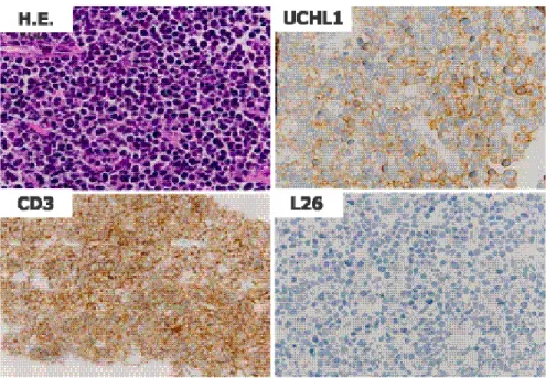
for back and right arm pain. On examination, swelling of the face and arms and prominent spidery telangiectasia on his anterior chest were noted: the jugular veins were nonpulsatile and distended. Chest X-ray and contrast-enhanced computed tomographic imaging of the chest demonstrated a bulky (10.0 x 7.2 cm) tumor with a low-density area in the right upper mediastinum (Figure 1). The lesion invaded the anterior chest wall and compressed the superior vena cava. CT-guided transcutaneous mediastinal biopsy was performed, and diffuse and invasively progressed small round cells and atypical lym- phocytes were found (Figure 2). Immunostaining of the same samples was positive for both UCHL1 and CD3, which indi- cate T-cells. Both L26 and CD79a, which indicate B-cells, were negative. CD79a is not shown. Pathological diagnosis was malignant lymphoma, diffuse, T-cell type. We then in-
Acta Med. Nagasaki56: 59−61Address correspondence:Minoru Fukuda, M.D, Ph.D., Division of Chemotherapy, Japanese Red Cross Nagasaki Genbaku Hospital, 3-15 Mori-machi, Nagasaki, 852-8511, Japan
Phone: +81-(95)-822-3251, Fax: +81-(95)-826-8798, E-mail: [email protected]
Received March 10, 2011; Accepted April 18, 2011
MS#AMN 07084
Case Report
Superior vena cava syndrome caused by adult T-cell leukemia/lymphoma:
a case report
Takaya I
KEDA1, Minoru F
UKUDA2, 3, Yoichi N
AKAMURA1, Kohei M
OTOSHIMA1, Seiji N
AGASHIMA2, Masatoshi M
ATSUO2, Masaomi Y
AMAMURA3, Kuniko A
BE4, Junji I
RIE4, Shigeru K
OHNO11Second Department of Internal Medicine, Nagasaki University Hospital, Nagasaki, Japan
2Department of Medicine, Japanese Red Cross Nagasaki Genbaku Hospital, Nagasaki, Japan
3Department of Internal Medicine, Nagasaki Municipal Hospital, Nagasaki, Japan
4Department of Pathology, Nagasaki Municipal Hospital, Nagasaki, Japan
A 74-year-old man with a smoking history was admitted for back and right arm pain. On examination, swelling of the face and arms were noted, and computed tomographic imaging of the chest demonstrated a bulky (10.0 x 7.2 cm) tumor in the right upper mediastinum. The lesion compressed the superior vena cava (SVC). Despite treatment, the patient died 5 months after the first admission. On autopsy, he was diagnosed that SVC syndrome caused by adult T-cell leukemia/lymphoma (ATL). ATL usually runs an aggressive course with multiple organs involving lymph nodes, liver, spleen, skin, lung, peripheral blood and bone marrow. Although it is extremely rare, SVC syndrome can appear as the earliest symptom of ATL.
ACTA MEDICA NAGASAKIENSIA 56: 59−61, 2011 Keywords: superior vena cava syndrome, adult T-cell leukemia/lymphoma
Takaya Ikeda et al.: SVC syndrome caused by ATL
vestigated the blood sample data, which showed elevation of human T-cell leukemia virus type 1 (HTLV-1) antibody, LDH (1063 IU/l) and soluble IL-2 receptor (4643 U/ml).
Therefore, we clinically diagnosed the patient as having su- perior vena cava (SVC) syndrome caused by adult T-cell leukemia/lymphoma (ATL), lymphoma type (3). There were no abnormal lymphocytes in peripheral blood, no bone marrow suppression, no skin lesions, no lymphnode swel- ling at other sites than mediastinum, and no enlargement of liver and spleen. Despite treatment including chemotherapy consisted by therarubicin, cyclophosphamide, vincristine, irinotecan and carboplatin, and thoracic radiotherapy, the patient died 5 months after the first admission. On autopsy, a sample of renal lesion was examined by southern blot analysis (Figure 3). The HTLV-1 provirus was detected and the final diagnosis of ATL was confirmed.
60
a b
Figure 1. a) Chest X-ray on admission, showing a huge tumor in the right upper mediastinum. b) CT image of the chest on admission, showing a huge tumor with low- density area, invading the anterior chest wall and compressing the superior vena cava.
Figure 2.H.E. staining and immunostaining of the CT-guided transcutaneous mediastinal biopsy samples (high-power view). Both UCHL1 and CD3, which indicate T-cells, were positive. Both L26 and CD79a, which indicate B-cells, were negative. CD79a is not shown.
Figure 3.Southern blot analysis using DNA extracted from the renal tissue at autopsy was positive for HTLV-1 proviral integra- tion. M: size marker (λDNA/Hind III), 1: positive control (mono- clonal integrated DNA), 2: negative control, 3: sample. Restriction enzyme E: Eco RI digestion, P: Pst I digestion.
Takaya Ikeda et al.: SVC syndrome caused by ATL
Discussion
ATL is an aggressive malignancy of CD4+ T-cell lym- phoid caused by HTLV-1 infection (4). Transmission of HTLV-1 by blood transfusion, needle sharing, breast feed- ing, and from male to female (rarely the reverse) by sexual intercourse has been documented (5). The interval between HTLV-1 infection and the onset of lymphoma is long (10- 40 years) and only less than 5% of infected people actually develop to ATL. SVC syndrome is the clinical expression of obstruction of blood flow through the SVC (6). The most common malignant lesions are lung cancer and lymphoma.
Since the tumor cells aggressively infiltrate multiple or- gans, commonly involving lymph nodes, liver, spleen, skin, and lung in ATL, it is extremely rare that the disease appear in only right upper mediastinum and SVC syndrome as the earliest symptom.
The differential diagnosis of ATL includes mycosis fungoides, Sezary syndrome, Hodgkin's lymphoma, and T-cell chronic lymphocytic leukemia. In our case, HTLV-I seropositivity, negative L26 and CD79a staining, and positive UCHL1 and CD3 staining support ATL diagnosis. Detection of integrated HTLV-1 provirus by southern blot analysis on autopsy made the diagnosis definitive.
SVC syndrome due to malignancy produces acute dis- tress and degrades the quality of life during the limited sur- vival (6). Endovascular stents have been used successfully as a relatively new option and provide relief of the symp- toms (7). In our case, we could achieve relief of symptoms
of SVC syndrome by first line chemotherapy and second line chemoradiotherapy. Endovascular stents after second line therapy were not applicable because of systemic pro- gression of ATL. Multiagent chemotherapy usually attains only partial, short lasting response in ATL. Since the dis- ease is not curable at present, prenatal HTLV-1 screening and cessation of breast-feeding by HTLV-1-infected women may suppress disease progression.
In conclusion, we report the first case of SVC syndrome caused by ATL. Although it is extremely rare, SVC syn- drome can appear as the earliest symptom of ATL.
References
1) Wilson LD, Detterbeck FC, Yahalom J. Superior vena cava syndrome with malignant causes.N Engl J Med356:1862-1869, 2007 2) Yellin A, Rosen A, Reichert N, Lieberman Y. Superior vena cava syn-
drome. The myth-the facts.Am Rev Respir Dis141:1114-1118, 1990 3) Shimoyama M and members of The Lymphoma Study Group (1984-
87). Diagnostic criteria and classification of clinical subtypes of adult T-cell leukaemia-lymphoma.British Journal of Haematology79:428- 437, 1991
4) Poiesz BJ, Ruscetti FW, Reitz MS, et al. Isolation of a new type C retrovirus (HTLV) in primary uncultured cells of a patient with Sezary T-cell leukemia.Nature294:268-271, 1981
5) Concalves DU, Proietti FA, Ribas JG, et al. Epidemiology, treatment, and prevention of human T-cell leukemia virus type 1-associated dis- eases.Clin Microbiol Rev23:577-589, 2010
6) Escalante CP. Causes and management of superior vena cava syn- drome.Oncology7:61-68, 1993
7) Koutoulidis V, Ladopoulos Ch, Vlachos L. Stent therapy for malignant superior vena cava syndrome: should be first line therapy or simple ad- junct to radiotherapy.Eur J Radiol47:247-250, 2003
61