Fukushima Medical University
福島県立医科大学 学術機関リポジトリ
This document is downloaded at: 2021-11-08T00:19:44Z
Title Prognostic Significance of Insomnia in Heart Failure( 本文 )
Author(s) 菅野, 優紀
Citation
Issue Date 2018-03-21
URL http://ir.fmu.ac.jp/dspace/handle/123456789/751
Rights © 2016 THE JAPANESE CIRCULATION SOCIETY. This is the peer reviewed version. Published version: Circ J. 2016 Jun 24;80(7):1571-7. doi: 10.1253/circj.CJ-16-0205.
DOI
Text Version ETD
1
Prognostic Significance of Insomnia in Heart Failure
Yuki Kanno, MD
Department of Cardiovascular Medicine, Fukushima Medical University
2
論 文 内 容 要 旨
学位論文題名
心不全患者における不眠症の検討
近年のメタ解析において、不眠症は、冠動脈心疾患や脳卒中の発症、さらには、心血管死 亡と関連し、そのリスクを増加させることが報告されている。その背景として、睡眠不足 や不眠症は肥満症や糖尿病、高血圧や脂質異常症を増加させ心血管疾患のリスクを上昇さ せると考えられている。しかし、不眠症と心不全の予後との関連はいまだ明らかではない。
そこで、我々は、不眠症を合併した心不全患者の特徴と不眠症の予後への影響を明らかに するため検討を行った。2009年から2013年に当院に入院し退院し得た心不全患者連続 1011例を対象に前向き観察研究を行った。心不全に不眠(症状、既往)を伴う:不眠群 519名と不眠を伴わない:非不眠群492名に分類し、2群間における患者背景や退院時の 血液検査、心臓超音波検査、運動耐容能の検査並びに心臓死および心不全増悪による再入 院の心イベントについて比較検討を行った。研究結果は不眠症を合併した心不全患者の特 徴として、高齢で女性が多く、心房細動や慢性腎臓病の合併率が高値であった。血液検査 ではレニン活性、レニン濃度、アルドステロン濃度が高値であった。また、心臓超音波検 査による心機能に差を認めないものの、運動耐容能が低値であり、心イベント発生率は高 値だった。また、不眠症は心不全の予後予測因子であることがわかった。不眠症自体が心 不全の予後に対し悪影響をおよぼしているのか、心不全の状態が悪いために不眠症になっ ているのか、眠剤の予後への影響などは今後の課題である。しかし、不眠が心不全に及ぼ す影響は大きいと考えられるため不眠症を有する心不全患者に対して、引き続き積極的に 介入していかなければならないと考えられた。
This paper was published in Circulation Journal 2016; 80: 1571-1577.
3
Abstract
Background: Insomnia is associated with incident heart failure (HF). However, the clinical significance and impact of insomnia on HF remain unclear.
Methods and Results: Consecutive 1011 patients admitted for treatment of HF were divided into two groups according to the presence of insomnia: HF with insomnia (insomnia group, n=519) and
HF without insomnia (non-insomnia group, n=492). We compared 1) cardiac event rates including
cardiac death and worsening HF and 2) underlying clinical background including laboratory data,
echocardiographic data, and cardio-pulmonary exercise test findings between the two groups. In the
Kaplan-Meier analysis, cardiac event rates were significantly higher in the insomnia group than in
the non-insomnia group (39.1 vs. 23.4%, P<0.001). The insomnia group, as compared to the
non-insomnia group, had 1) higher levels of plasma renin activity (P=0.042), renin concentration
(P=0.007), and aldosterone (P=0.047), 2) lower peak VO
2(14.9 vs. 16.3 ml/kg/min, P=0.002) and
higher VE/VCO
2slope (36.0 vs. 33.5, P=0.001), and 3) similar levels of B-type natriuretic peptide
and left ventricular ejection fraction. Importantly, in the multivariable Cox proportional hazard
analyses after adjusting for potential confounding factors, insomnia was an independent predictor of
cardiac events in HF patients (hazard ratio 1.899, P<0.001).
Conclusions: Insomnia was an independent predictor of cardiac events in HF patients. HF patients
with insomnia exhibited activated renin-angiotensin-aldosterone system and lower exercise capacity.
4
Keywords heart failure, insomnia, sleep disorder, exercise capacity, renin-angiotensin-aldosterone system, prognosis
Introduction
Heart failure (HF) is a major cause of death among the elderly in many countries.
1-4It has recently
been reported that insomnia, which is linked with incidence of HF in the general population (hazard
ratio 4.53, 1.99–10.31)
5, is also associated with an increased risk of incident cardiovascular
disease.
5-9This hyperarousal disorder is accompanied by chronic activation of stress responses with
increased activity in the hypothalamic-pituitary-adrenal axis and sympathetic nervous system leading
to increased secretion of cortisol and up-regulation of the renin-angiotensin-aldosterone system
(RAAS).
5, 10Stress response caused by insomnia is also accompanied by increased heart rate,
decreased heart rate variability, increased blood pressure, secretion of pro-inflammatory cytokines
and catecholamines, and impaired exercise capacity and activity
1, 5, which are risk factors for the
progression of HF and prognostic factors of HF. These risk factors may in turn contribute to
endothelial dysfunction, atherosclerosis, renal dysfunction, and impaired cardiac function. Moreover,
these abnormalities may represent a biologically plausible causal link between insomnia and HF. On
the other hand, insomnia is highly prevalent in patients with chronic disease including HF and is a
significant contributing factor to fatigue and poor quality of life.
11-17However, the prognostic impact of insomnia on HF patients remains unclear. We hypothesize
5
that HF patients with insomnia have poor prognosis accompanied with activated RAAS,
18sympathetic nervous activity and inflammation, impaired cardiac function, and exercise capacity.
To address these issues, we aimed to investigate the impact of insomnia on prognosis of HF
and compare the underlying clinical background in HF patients with or without insomnia (e.g.
clinical features, echocardiographic parameters, exercise capacity, and neurohumoral and
inflammatory factors such as plasma noradrenalin, renin activity, renin concentration, aldosterone,
and C-reactive protein).
Methods
Subjects and study protocol
This was a prospective observational study that enrolled consecutive symptomatic HF patients (n =
1083) who were hospitalized to treat decompensated HF and were discharged from Fukushima
Medical University between 2009 and 2013. The diagnosis of decompensated HF was made by
several cardiologists based on the Framingham criteria.
19Patients with acute coronary syndrome (n =
23), dialysis (n = 14) and already diagnosed depression
20(n = 35) were excluded. Patients (n = 1011)
were divided into two groups according to the presence of insomnia based on symptoms in normal
daily life and/or at discharge, but not at hospitalization, by direct interview using a questionnaire
taken by the attending physicians and medical staffs for patients or caregivers. Insomnia was defined
by several physicians as the usual use of hypnotics 1) (‘Do you take hypnotics more than 3 times per
week’ with the response options yes/ no) or 2) presence of either insomnia symptom of grade 3 or 4
6
accompanied by impairment of daytime function,
5, 21specifically as follows: difficulty initiating
sleep (‘Do you have difficulties falling asleep?’ with the response options 1. Never, 2. Occasionally,
3. Often, 4. Almost every night), difficulty maintaining sleep and/or early morning awakenings (‘Do
you wake up in the early hours unable to get back to sleep?’ with the response options 1. Never, 2.
Occasionally, 3. Often, 4. Almost every night), non-restorative sleep (‘How often do you suffer from
poor sleep?’ with the response options 1. Never or a few times a year, 2. One to two times per month,
3. About once a week, 4. More than once a week), based on modified International Classification of
Sleep Disorders-2 criteria
5, 21supported by the American Academy of Sleep Medicine and the
Japanese Society of Sleep Research, which are widely spread in Japanese clinical practice.
We performed examinations such as general laboratory tests, echocardiography, and
cardio-pulmonary exercise tests at discharge, and compared parameters between the insomnia and
non-insomnia groups. Co-morbidities were also assessed by several attending physicians.
Hypertension was defined as the recent use of antihypertensive drugs, or systolic blood pressure
> 140 mmHg, and/or diastolic blood pressure > 90 mmHg. Diabetes was defined as the recent use of
insulin or antidiabetic drugs, a fasting blood glucose value of > 126 mg/dL, and/or a hemoglobin
A1c value of > 6.5%. Dyslipidemia was defined as the recent use of cholesterol-lowering drugs, a
triglyceride value of > 150 mg/dL, a low-density lipoprotein cholesterol value of > 140 mg/dL,
and/or a high-density lipoprotein cholesterol value of < 40 mg/dL. The estimated glomerular
filtration rate (GFR) was measured by the Modification of Diet in Renal Disease formula.
22Chronic
7
kidney disease was defined as an estimated GFR < 60 ml/min/1.73 m
2.
22Anemia was defined as
hemoglobin of < 12.0 g/dl in females and < 13.0 g/dl in males.
1Preserved left ventricular ejection
fraction (LVEF) was defined as more than 50%.
2The patients were followed up until March 2015 for cardiac events, which were composite end
points of cardiac death and/or worsening HF,
23, 24were adjudicated by several independent
cardiologists. Cardiac death was defined including worsening heart failure, which met the
Framingham criteria
19, and ventricular fibrillation documented by electrocardiogram or implantable
devices. Status and dates of deaths of all patients were obtained from the patients’ medical records or
cardiologists at the patient’s referring hospital. Survival time was calculated from the date of
hospitalization until the date of death or last follow-up. Written informed consent was obtained from
all study subjects. The study protocol was approved by the ethical committee of Fukushima Medical
University. The investigation conforms with the principles outlined in the Declaration of Helsinki.
Reporting of the study conforms to STROBE along with references to STROBE and the broader
EQUATOR guidelines.
25Echocardiography
Echocardiography was performed blindly by an experienced echocardiographer using the standard
techniques.
26The echocardiographic parameters investigated included LVEF, the ratio of early
transmitral flow velocity to mitral annular velocity (mitral valve E/e’), inferior vena cava diameter,
8
right ventricular fractional area change (RV-FAC), and tissue Doppler-derived tricuspid lateral
annular systolic velocity (tricuspid valve S’).
27, 28The LVEF was calculated using a modification of
Simpson’s method. Mitral valve E/E’ was calculated by transmitral Doppler flow and tissue Doppler
imaging. Tissue Doppler imaging was obtained from the average of the lateral and septal annulus
velocities. The RV-FAC, defined as (end diastolic area − end systolic area) ÷ end diastolic
area
×100, is a measure of right ventricular systolic function.
27, 28All recordings were performed on
ultrasound systems (ACUSON Sequoia, Siemens Medical Solutions USA, Inc., Mountain View, CA,
USA).
Cardiopulmonary exercise testing
The patients underwent incremental symptom-limited exercise testing using an upright cycle
ergometer with a ramp protocol before discharge (Strength Ergo 8, Fukuda Denshi Co. Ltd., Tokyo,
Japan). Breath-by-breath oxygen consumption (VO
2), carbon dioxide production (VCO
2), and
minute ventilation (VE) were measured during exercise using an AE-300S respiratory monitor
(Minato Medical Science, Osaka, Japan).
28-30Peak VO
2was measured as an average of the last 30 s
of exercise. Ventilatory response to exercise (expressed as a VE/VCO
2slope) was calculated as the
regression slope relating VE to CO
2from the start of exercise until the respiratory compensation
point (the time at which ventilation is stimulated by CO
2output and end-tidal CO
2tension begins to
decrease).
28, 31The ventilatory anaerobic threshold was calculated with the V-slope method.
9
Statistical analysis
Normally distributed data are presented as mean ± SD, and non-normally distributed data are
presented as median (inter-quartile range). Categorical variables are expressed as numbers and
percentages. The chi-square test was used for comparisons of categorical variables. Data of the two
groups were compared using the independent Student’s
t-test for normally distributed data and theMann-Whitney U test for non-normally distributed data. The Kaplan-Meier method was used for
presenting the event-free rate, and the log-rank test was used for initial comparisons. Univariable and
multivariable Cox proportional hazard analyses were used to analyze predictors of cardiac events
with adjusting confounding factors. To prepare for potential confounding, we considered the
following clinical factors, which are known to affect the risk of cardiac event in HF patients: age,
gender, New York Heart Association functional class III or IV, body mass index, systolic blood
pressure, heart rate, preserved LVEF, B-type natriuretic peptide, sodium, albumin, presence of
hypertension, diabetes, dyslipidemia, atrial fibrillation, chronic kidney disease, anemia and insomnia,
usage of RAAS inhibitors, β-blockers, diuretics, inotropics and device therapy (implantable
cardioverter defibrillator and/or cardiac resynchronization therapy). Among these factors, those
which were independent in predicting cardiac events with a value of
P < 0.05 were included in thefinal adjusted model. A value of
P < 0.05 was considered significant for all comparisons. These10
analyses were performed using a statistical software package (SPSS ver. 21.0, IBM, Armonk, NY,
USA).
Results
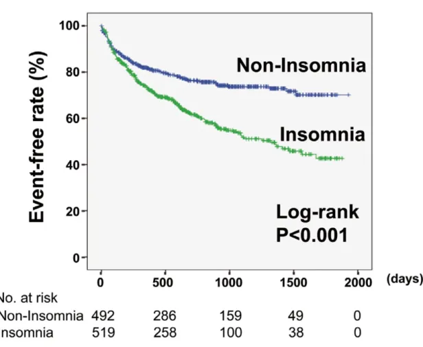
Of all the HF patients, 519 (51.3%) were categorized into the insomnia group as shown in Table 1.
During the follow-up period (mean 801 days, median 748 days), there were 236 worsening HF cases
(163 and 73 in the insomnia group and non-insomnia groups, respectively) and 151 cardiac deaths
(85 and 66 in the insomnia and non-insomnia groups, respectively). As shown in Figure 1, the insomnia group experienced more cardiac events than the non-insomnia group (P < 0.001).
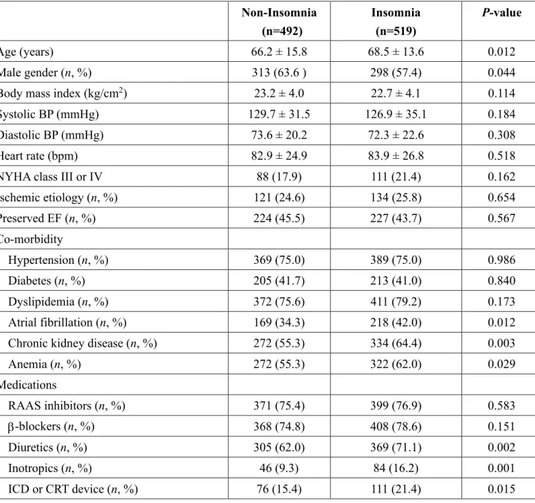
The clinical features of the study subjects are summarized in Table 1. The insomnia group patients were of a higher age, had a higher prevalence of female gender, and had higher usage of
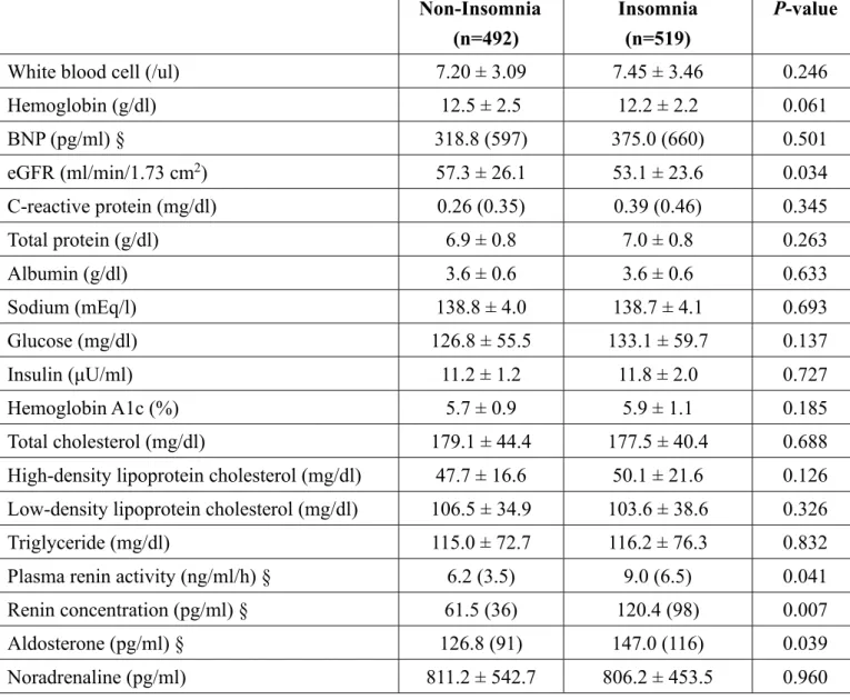
diuretics and inotropics. Comparisons of the laboratory data between the two groups are shown in
Table 2. The insomnia group had lower levels of estimated GFR, and higher levels of plasma renin activity, renin concentration, and aldosterone. In contrast, BNP, C-reactive protein, albumin, sodium,
glucose and lipid parameters, and plasma noradrenaline did not differ between the two groups. The
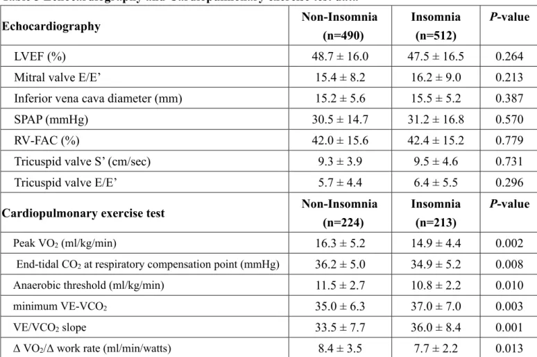
parameters of echocardiography and the cardio-pulmonary exercise test are summarized in Table 3.
Although left and right ventricular systolic function did not differ between the two groups, peak VO
2,
end-tidal CO
2at respiratory compensation point, anaerobic threshold, and ΔVO
2/Δwork rate were
significantly lower in the insomnia group than in the non-insomnia group. The minimum VE-VCO
2and VE/VCO
2slopes were higher in the insomnia group than in the non-insomnia group. Taken
11
together, these data suggest that worse prognosis of HF patients with insomnia may not be related to
cardiac function but to activated RAAS and impaired exercise capacity.
The Cox proportional hazard model was used to examine the prognostic impact of insomnia on
patients with HF (Table 4). We confirmed that the Cox models supported the assumption of proportional odds. In the multivariable analysis, insomnia was an independent predictor of cardiac
events (HR 1.899, 95% CI 1.333–2.705, P < 0.001).
Then, we focused on the relationship between RAAS and cardiac event rates in HF patients with
or without insomnia. In the Cox proportional hazard analysis, plasma renin activity and renin
concentration were predictors of cardiac events only in HF patients with insomnia (plasma renin
activity, HR 1.018, 95% CI 1.003-1.034, P = 0.020; renin concentration, HR 1.001, 95% CI
1.001-1.002, P <0.001), but not in HF patients without insomnia. Aldosterone was not a predictor of
cardiac event in both groups.
Discussion
To the best of our knowledge, the present study is the first to show that HF patients with
insomnia experienced more cardiac events, but their worse prognosis was related to rather activated
RAAS and impaired exercise capacity than cardiac function.
In our study, insomnia was an independent predictor of cardiac events in HF patients after
adjusting for multiple known confounding factors. Thus, our data suggest that insomnia itself may
12
be associated with adverse outcomes in HF patients, or that insomnia as a symptom can be a
potential marker in risk-stratification of HF patients. In addition, the insomnia group exhibited
activated RAAS, impaired renal function, and lower exercise capacity. These mechanisms may in
part explain the poor prognosis of HF patients with insomnia. In contrast, plasma noradrenalin,
C-reactive protein, and echocardiographic parameters did not differ between the two groups.
Although we did not investigate the reason for these results, HF itself and HF treatment may
strongly affect sympathetic activity, inflammation, and cardiac function.
Restorative functions occur during different stage of sleep, with physical restoration
occurring primarily during non-rapid eye movement (NREM) sleep and brain restoration occurring
primarily in rapid eye movement (REM) sleep. Sleep and exercise influence each other through
complex and bilateral interactions that involve multiple physiological and psychological
pathways.
32Insomnia causes inhibition of restorative functions and fatigue, and these are resulting
in impairment of psychomotor and physical performance
1and activity,
5,17which are risk factors for
poor prognosis in HF.
With regard to inflammation, proinflammatory cytokines, interleukin-6, and tumor necrosis
factor α are fatigue-inducing cytokines that negatively influence quality of sleep. Mean 24 h
secretions of these cytokines did not differ between insomnia patients and normal sleepers; however,
there was a significant increase of interleukin-6 from mid-afternoon to evening.
10, 33In addition, the
characteristic circadian secretion of tumor necrosis factor α with a peak close to sleep offset was
13
observed in the normal sleepers, but not in the insomnia patients.
33The hypersecretion and/or
circadian alteration of the cytokine secretion associated with a hypothalamic-pituitary-adrenal axis
activation may explain the fatigue and poor sleep associated with insomnia.
10Another study
reported elevated C-reactive protein levels in insomnia patients.
34In contrast, erythrocyte
sedimentation rate is not associated with incidence of HF in insomnia patients.
9Thus, the
associations between insomnia and inflammation are complex and not fully addressed especially
in HF patients.
Furthermore, symptoms of HF itself, including coughing, orthopnea, paroxysmal nocturnal
dyspnea, and nocturia, often lead to insomnia,
13, 35and insomnia itself may reflect the severity of
HF. In addition, insomnia is also an indicator of depression, which is associated with adverse
prognosis of HF.
20These in turn are associated with poor prognosis of HF patients. On the other
hand, insomnia increases with both the number of chronic illnesses the patient has and the number
of medications taken.
13, 36-38Insomnia could also be partially caused by medications used in the
treatment of HF.
1, 2Melatonin production may be affected by β-blockers, diuretics cause nocturia,
and inotropics affect agitation, all of which result in poor sleep quality.
13In future, functional imaging may be useful to determine the association between HF and
insomnia. Functional neuroimaging studies have shown that transition from wakefulness to sleep is
associated with a decrease of brain activity in specific regions, such as the brain stem, thalamus, and
prefrontal cortex.
39Cerebral abnormalities detected by magnetic resonance imaging and cognitive
14
performance in HF patients have been reported.
40For instance, medial temporal lobe atrophy was
related to cognitive dysfunction, involving memory impairment and executive dysfunction, whereas
total white matter hyperintensities were related to depression resulting in insomnia.
40To date, there are no data evaluating effective treatment for insomnia in HF patients. General
behavioral measures for improved sleep hygiene, such as minimal use of caffeine, cigarettes and
alcohol, maintaining a regular sleep schedule, going to bed only when sleepy, regular exercise, and
avoiding daytime naps, should be explained to the patients.
10It has been recently reported that
exercise training improves sleep quality in HF patients.
41Since HF patients with insomnia had
impaired exercise capacity in present data, cardiac rehabilitation may be more strongly
recommended.
1,41Study limitations
There are several limitations in the present study. Firstly, the number of subjects was relatively small
as the study was performed in a single institution. Further studies with a larger population are needed.
However, diagnosis of cardiac events was accurately made by our experienced cardiologists.
Secondly, we diagnosed insomnia based on patient’s symptoms assessed by interview or medical
history, hence we could not completely exclude the effect of psychiatric disorders, depression and
cognitivty. In addition, we did not consider any changes in any parameters, and baseline data at
admission were used for the analyses. Furthermore, we did not use polysomnography or actigraphy,
15
which are objective tests of sleep disorders. However, these are not routinely performed in patients
with HF and/or insomnia. Thirdly, levels of plasma renin activity and concentrations of renin,
aldosterone, and noradrenaline might be affected by administration of RAAS inhibitors and
β-blockers. Fourthly, although we have conducted multivariable analyses to evaluate associations
between insomnia and prognosis in HF patients, confounding factors cannot be entirely eliminated.
Our results do not establish a cause-effect relationship between the presence of insomnia and
increased cardiac events. Finally, further studies are required to examine the impact of hypnotics on
prognosis of HF patients with insomnia.
Conclusions
Insomnia was a common and independent predictor of cardiac events in HF patients. HF patients
with insomnia exhibited activated RAAS and impaired exercise capacity, and insomnia may be a
potential marker of adverse prognosis in HF patients. Further studies are required to determine
whether controlling insomnia improves the prognosis of such patients.
Acknowledgements
The authors acknowledge the efforts of Drs. Aya Goto and Shinya Ito (Department of Public Health,
Fukushima Medical University) for their invaluable advice of medical statistics, and Ms. Kumiko
Watanabe and Yuko Niimura for their outstanding technical assistance.
16
Funding Sources
This study was supported in part by a grant-in-aid for Scientific Research (No. 25461061) from the
Japan Society for the Promotion of Science, and grants-in-aid from the Japanese Ministry of Health,
Labor, and Welfare, Tokyo, Japan.
Disclosures, Conflict of interest
None.
17
References
1. McMurray JJ, Adamopoulos S, Anker SD, Auricchio A, Bohm M, Dickstein K, et al. ESC guidelines for the diagnosis and treatment of acute and chronic heart failure 2012: The Task Force for the Diagnosis and Treatment of Acute and Chronic Heart Failure 2012 of the European Society of Cardiology. Developed in collaboration with the Heart Failure Association (HFA) of the ESC.
Eur J Heart Fail 2012;14:803-869.
2. Writing Committee M, Yancy CW, Jessup M, Bozkurt B, Butler J, Casey DE, Jr., et al. 2013 ACCF/AHA guideline for the management of heart failure: a report of the American College of Cardiology Foundation/American Heart Association Task Force on practice guidelines. Circulation 2013;128:e240-327.
3. Miura M, Sakata Y, Miyata S, Nochioka K, Takada T, Tadaki S, et al. Prognostic impact of subclinical microalbuminuria in patients with chronic heart failure. Circ J 2014;78:2890-2898.
4. Group JCSJW. Guidelines for treatment of acute heart failure (JCS 2011). Circ J 2013;77:2157-2201.
5. Laugsand LE, Strand LB, Platou C, Vatten LJ, Janszky I. Insomnia and the risk of incident heart failure: a population study. Eur Heart J 2013:1382-1393.
6. Li Y, Zhang X, Winkelman JW, Redline S, Hu FB, Stampfer M, et al. Association between insomnia symptoms and mortality: a prospective study of U.S. men. Circulation 2014;129:737-746.
7. Li M, Zhang XW, Hou WS, Tang ZY. Insomnia and risk of cardiovascular disease: a meta-analysis of cohort studies. Int J Cardiol 2014;176:1044-1047.
8. Sofi F, Cesari F, Casini A, Macchi C, Abbate R, Gensini GF. Insomnia and risk of cardiovascular disease: a meta-analysis. Eur J Prev Cardiol 2014;21:57-64.
9. Ingelsson E, Lind L, Arnlov J, Sundstrom J. Sleep disturbances independently predict heart failure in overweight middle-aged men. Eur J Heart Fail 2007;9:184-190.
10. Basta M, Chrousos GP, Vela-Bueno A, Vgontzas AN. Chronic Insomnia and Stress System. Sleep Med Clin 2007;2:279-291.
11. Redeker NS, Jeon S, Muench U, Campbell D, Walsleben J, Rapoport DM. Insomnia symptoms and daytime function in stable heart failure. Sleep 2010;33:1210-1216.
12. Johansson P, Arestedt K, Alehagen U, Svanborg E, Dahlstrom U, Brostrom A. Sleep disordered breathing, insomnia, and health related quality of life -- a comparison between age and gender matched elderly with heart failure or without cardiovascular disease. Eur J Cardiovasc Nurs 2010;9:108-117.
13. Hayes D, Jr., Anstead MI, Ho J, Phillips BA. Insomnia and chronic heart failure. Heart Fail Rev 2009;14:171-182.
14. Redeker NS, Stein S. Characteristics of sleep in patients with stable heart failure versus a comparison group. Heart Lung 2006;35:252-261.
18
15. Redeker NS, Hilkert R. Sleep and quality of life in stable heart failure. J Card Fail 2005;11:700-704.
16. Brostrom A, Stromberg A, Dahlstrom U, Fridlund B. Sleep difficulties, daytime sleepiness, and health-related quality of life in patients with chronic heart failure. J Cardiovasc Nurs 2004;19:234-242.
17. Erickson VS, Westlake CA, Dracup KA, Woo MA, Hage A. Sleep disturbance symptoms in patients with heart failure. AACN Clin Issues 2003;14:477-487.
18. Ueda T, Kawakami R, Nishida T, Onoue K, Soeda T, Okayama S, et al. Plasma renin activity is a strong and independent prognostic indicator in patients with acute decompensated heart failure treated with renin-angiotensin system inhibitors. Circ J 2015;79:1307-1314.
19. McKee PA, Castelli WP, McNamara PM, Kannel WB. The natural history of congestive heart failure: the Framingham study. N Engl J Med 1971;285:1441-1446.
20. Kato N, Kinugawa K, Yao A, Hatano M, Shiga T, Kazuma K. Relationship of depressive symptoms with hospitalization and death in Japanese patients with heart failure. J Card Fail 2009;15:912-919.
21. American Academy of Sleep Medicine eW, IL, 2005. The International Classification of Sleep Disorders, Diagnostic and Cording Manual, Second Edition (ICSD-II). .
22. Levey AS, Coresh J, Greene T, Stevens LA, Zhang YL, Hendriksen S, et al. Using standardized serum creatinine values in the modification of diet in renal disease study equation for estimating glomerular filtration rate. Ann Intern Med 2006;145:247-254.
23. Zannad F, McMurray JJ, Krum H, van Veldhuisen DJ, Swedberg K, Shi H, et al. Eplerenone in patients with systolic heart failure and mild symptoms. N Engl J Med 2011;364:11-21.
24. Cohn JN, Tognoni G, Valsartan Heart Failure Trial I. A randomized trial of the angiotensin-receptor blocker valsartan in chronic heart failure. N Engl J Med 2001;345:1667-1675.
25. von Elm E, Altman DG, Egger M, Pocock SJ, Gotzsche PC, Vandenbroucke JP, et al.
Strengthening the Reporting of Observational Studies in Epidemiology (STROBE) statement:
guidelines for reporting observational studies. BMJ 2007;335:806-808.
26. Lang RM, Badano LP, Mor-Avi V, Afilalo J, Armstrong A, Ernande L, et al. Recommendations for cardiac chamber quantification by echocardiography in adults: an update from the American Society of Echocardiography and the European Association of Cardiovascular Imaging. J Am Soc Echocardiogr 2015;28:1-39 e14.
27. Rudski LG, Lai WW, Afilalo J, Hua L, Handschumacher MD, Chandrasekaran K, et al. Guidelines for the echocardiographic assessment of the right heart in adults: a report from the American Society of Echocardiography endorsed by the European Association of Echocardiography, a registered branch of the European Society of Cardiology, and the Canadian Society of Echocardiography. J Am Soc Echocardiogr 2010;23:685-713; quiz 786-688.
28. Nakamura Y, Kunii H, Yoshihisa A, Takiguchi M, Shimizu T, Yamauchi H, et al. Impact of
19
peripheral artery disease on prognosis in hospitalized heart failure patients. Circ J 2015;79:785-793.
29. O'Neill JO, Young JB, Pothier CE, Lauer MS. Peak oxygen consumption as a predictor of death in patients with heart failure receiving beta-blockers. Circulation 2005;111:2313-2318.
30. Arena R, Myers J, Guazzi M. Cardiopulmonary exercise testing is a core assessment for patients with heart failure. Congest Heart Fail 2011;17:115-119.
31. Ponikowski P, Francis DP, Piepoli MF, Davies LC, Chua TP, Davos CH, et al. Enhanced ventilatory response to exercise in patients with chronic heart failure and preserved exercise tolerance: marker of abnormal cardiorespiratory reflex control and predictor of poor prognosis.
Circulation 2001;103:967-972.
32. Chennaoui M, Arnal PJ, Sauvet F, Leger D. Sleep and exercise: a reciprocal issue? Sleep Med Rev 2015;20:59-72.
33. Vgontzas AN, Zoumakis M, Papanicolaou DA, Bixler EO, Prolo P, Lin HM, et al. Chronic insomnia is associated with a shift of interleukin-6 and tumor necrosis factor secretion from nighttime to daytime. Metabolism 2002;51:887-892.
34. Meier-Ewert HK, Ridker PM, Rifai N, Regan MM, Price NJ, Dinges DF, et al. Effect of sleep loss on C-reactive protein, an inflammatory marker of cardiovascular risk. J Am Coll Cardiol 2004;43:678-683.
35. Principe-Rodriguez K, Strohl KP, Hadziefendic S, Pina IL. Sleep symptoms and clinical markers of illness in patients with heart failure. Sleep Breath 2005;9:127-133.
36. Foley D, Ancoli-Israel S, Britz P, Walsh J. Sleep disturbances and chronic disease in older adults:
results of the 2003 National Sleep Foundation Sleep in America Survey. J Psychosom Res 2004;56:497-502.
37. Foley DJ, Monjan A, Simonsick EM, Wallace RB, Blazer DG. Incidence and remission of insomnia among elderly adults: an epidemiologic study of 6,800 persons over three years. Sleep 1999;22 Suppl 2:S366-372.
38. Foley DJ, Monjan AA, Brown SL, Simonsick EM, Wallace RB, Blazer DG. Sleep complaints among elderly persons: an epidemiologic study of three communities. Sleep 1995;18:425-432.
39. Nofzinger EA, Buysse DJ, Miewald JM, Meltzer CC, Price JC, Sembrat RC, et al. Human regional cerebral glucose metabolism during non-rapid eye movement sleep in relation to waking. Brain 2002;125:1105-1115.
40. Vogels RL, Oosterman JM, van Harten B, Gouw AA, Schroeder-Tanka JM, Scheltens P, et al.
Neuroimaging and correlates of cognitive function among patients with heart failure. Dement Geriatr Cogn Disord 2007;24:418-423.
41. Suna JM, Mudge A, Stewart I, Marquart L, O'Rourke P, Scott A. The effect of a supervised exercise training programme on sleep quality in recently discharged heart failure patients. Eur J Cardiovasc Nurs 2015;14:198-205.
20
FIGURE LEGENDS
Figure 1. Comparison of cardiac events between the insomnia and non-insomnia groups.
Kaplan-Meier analysis for cardiac events (Insomnia vs. Non-insomnia group) in all HF patients
(n = 1011).
21
Table 1 Comparisons of clinical features
Non-Insomnia (n=492)
Insomnia (n=519)
P-value
Age (years) 66.2 ± 15.8 68.5 ± 13.6 0.012
Male gender (n, %) 313 (63.6 ) 298 (57.4) 0.044
Body mass index (kg/cm
2) 23.2 ± 4.0 22.7 ± 4.1 0.114
Systolic BP (mmHg) 129.7 ± 31.5 126.9 ± 35.1 0.184
Diastolic BP (mmHg) 73.6 ± 20.2 72.3 ± 22.6 0.308
Heart rate (bpm) 82.9 ± 24.9 83.9 ± 26.8 0.518
NYHA class III or IV 88 (17.9) 111 (21.4) 0.162
Ischemic etiology (n, %) 121 (24.6) 134 (25.8) 0.654
Preserved EF (n, %) 224 (45.5) 227 (43.7) 0.567
Co-morbidity
Hypertension (n, %) 369 (75.0) 389 (75.0) 0.986
Diabetes (n, %) 205 (41.7) 213 (41.0) 0.840
Dyslipidemia (n, %) 372 (75.6) 411 (79.2) 0.173
Atrial fibrillation (n, %) 169 (34.3) 218 (42.0) 0.012
Chronic kidney disease (n, %) 272 (55.3) 334 (64.4) 0.003
Anemia (n, %) 272 (55.3) 322 (62.0) 0.029
Medications
RAAS inhibitors (n, %) 371 (75.4) 399 (76.9) 0.583
-blockers (n, %)
368 (74.8) 408 (78.6) 0.151
Diuretics (n, %) 305 (62.0) 369 (71.1) 0.002
Inotropics (n, %) 46 (9.3) 84 (16.2) 0.001
ICD or CRT device (n, %) 76 (15.4) 111 (21.4) 0.015
RAAS, renin-angiotensin-
aldosterone system; ICD, implantable cardioverter defibrillator; CRT,
cardiac resynchronization therapy.
22
Table 2 Laboratory data
Non-Insomnia (n=492)
Insomnia (n=519)
P-value
White blood cell (/ul) 7.20 ± 3.09 7.45 ± 3.46 0.246
Hemoglobin (g/dl) 12.5 ± 2.5 12.2 ± 2.2 0.061
BNP (pg/ml) § 318.8 (597) 375.0 (660) 0.501
eGFR (ml/min/1.73 cm
2) 57.3 ± 26.1 53.1 ± 23.6 0.034
C-reactive protein (mg/dl) 0.26 (0.35) 0.39 (0.46) 0.345
Total protein (g/dl) 6.9 ± 0.8 7.0 ± 0.8 0.263
Albumin (g/dl) 3.6 ± 0.6 3.6 ± 0.6 0.633
Sodium (mEq/l) 138.8 ± 4.0 138.7 ± 4.1 0.693
Glucose (mg/dl) 126.8 ± 55.5 133.1 ± 59.7 0.137
Insulin (μU/ml) 11.2 ± 1.2 11.8 ± 2.0 0.727
Hemoglobin A1c (%) 5.7 ± 0.9 5.9 ± 1.1 0.185
Total cholesterol (mg/dl) 179.1 ± 44.4 177.5 ± 40.4 0.688
High-density lipoprotein cholesterol (mg/dl) 47.7 ± 16.6 50.1 ± 21.6 0.126 Low-density lipoprotein cholesterol (mg/dl) 106.5 ± 34.9 103.6 ± 38.6 0.326
Triglyceride (mg/dl) 115.0 ± 72.7 116.2 ± 76.3 0.832
Plasma renin activity (ng/ml/h) § 6.2 (3.5) 9.0 (6.5) 0.041
Renin concentration (pg/ml) § 61.5 (36) 120.4 (98) 0.007
Aldosterone (pg/ml) § 126.8 (91) 147.0 (116) 0.039
Noradrenaline (pg/ml) 811.2 ± 542.7 806.2 ± 453.5 0.960
BNP, B-type natriuretic peptide; eGFR, estimated glomerular filtration.
§ Data are presented as median (interquartile range).
23
Table 3 Echocardiography and Cardiopulmonary exercise test data
Echocardiography Non-Insomnia
(n=490)
Insomnia (n=512)
P-value
LVEF (%) 48.7 ± 16.0 47.5 ± 16.5 0.264
Mitral valve E/E’ 15.4 ± 8.2 16.2 ± 9.0 0.213
Inferior vena cava diameter (mm) 15.2 ± 5.6 15.5 ± 5.2 0.387
SPAP (mmHg) 30.5 ± 14.7 31.2 ± 16.8 0.570
RV-FAC (%) 42.0 ± 15.6 42.4 ± 15.2 0.779
Tricuspid valve S’ (cm/sec) 9.3 ± 3.9 9.5 ± 4.6 0.731
Tricuspid valve E/E’ 5.7 ± 4.4 6.4 ± 5.5 0.296
Cardiopulmonary exercise test Non-Insomnia (n=224)
Insomnia (n=213)
P-value
Peak VO2 (ml/kg/min)
16.3 ± 5.2 14.9 ± 4.4 0.002
End-tidal CO2 at respiratory compensation point (mmHg)
36.2 ± 5.0 34.9 ± 5.2 0.008
Anaerobic threshold (ml/kg/min)
11.5 ± 2.7 10.8 ± 2.2 0.010
minimum VE-VCO2
35.0 ± 6.3 37.0 ± 7.0 0.003
VE/VCO2 slope
33.5 ± 7.7 36.0 ± 8.4 0.001
Δ VO2/Δ work rate (ml/min/watts)
8.4 ± 3.5 7.7 ± 2.2 0.013
LVEF, left ventricular ejection fraction; Mitral valve E/E’, ratio of the peak transmitral velocity
during early diastole to the peak mitral valve annular velocity during early diastole; SPAP, systolic
pulmonary artery pressure; RV-FAC, right ventricular fractional area change; Tricuspid valve S’,
Doppler-derived tricuspid lateral annular systolic velocity; Tricuspid valve E/E’, ratio of the peak
transtricuspid velocity during early diastole to the peak tricuspid valve annular velocity during early
diastole; VO
2, oxygen consumption; VCO
2, carbon dioxide production; VE, minute ventilation; Peak
VO
2, peak oxygen uptake; minimumVE-VCO
2, and rate of minute ventilation to carbon dioxide
production; VE/VCO
2slope, rate of increase in ventilation per unit increase in carbon dioxide; Δ
VO
2/Δ work rate, rate of increase in VO
2to increase in work rate.
24
Table 4 Cox proportional hazard models of cardiac events in HF (318 events/ n = 1011)
Risk factor Univariate Multivariate
HR 95% Cl
P-valueHR 95% Cl
P-valueAge 1.019 1.011-1.028 <0.001 1.011 0.996-1.025 0.150
Male 0.977 0.780-1.224 0.842
NYHA III or IV 3.777 2.983-4.783 <0.001 2.284 1.497-3.424 <0.001 Body mass index 0.955 0.925-0.987 0.006 0.997 0.953-1.043 0.891 Systolic blood pressure 0.993 0.989-0.997 <0.001 0.996 0.989-1.002 0.195
Heart rate 1.003 0.999-1.007 0.109
Preserved LVEF 0.471 0.371-0.596 <0.001 0.722 0.477-0.994 0.042
Log BNP 2.389 1.866-3.058 <0.001 1.075 0.730-1.582 0.714
Sodium 0.926 0.901-0.952 <0.001 0.965 0.925-1.007 0.097
Albumin 0.622 0.511-0.756 <0.001 0.970 0.701-1.343 0.856
Ischemic etiology 1.295 1.013-1.654 0.039 1.083 0.706-1.661 0.715 Hypertension 0.985 0.763-1.272 0.909
Diabetes 1.507 1.210-1.878 <0.001 1.002 0.701-1.432 0.993 Dyslipidemia 1.151 0.875-1.514 0.315
Atrial fibrillation 1.398 1.121-1.743 0.003 1.187 0.845-1.667 0.323 Chronic kidney disease 2.848 2.189-3.707 <0.001 1.786 1.194-2.671 0.005
Anemia 2.162 1.692-2.763 <0.001 1.301 0.875-1.934 0.194
RAAS inhibitors 0.774 0.602-0.994 0.045 1.005 0.656-1.540 0.980
-blockers