The Insertion of a Removable Partial Denture Increases Unstimulated Salivary Flow Rates in Non-Denture Wearers
(義歯未装着者への部分床義歯の装着は安静時唾液量を増加させる)
日本大学大学院松戸歯学研究科歯学専攻
川原 綾夏
(指導: 河相 安彦 教授)
Abstract
Purpose: The difference in salivary flow rates between non-removable partial denture (RPD) wearers and regular RPD wearers is not well investigated. The purpose of this study was to examine the change in salivary flow rate after the insertion of dentures in elderly patients, especially for those with a shortened dental arch (SDA). Factors associated with the changes were also investigated. Subjective measures of oral dryness were also assessed to investigate changes in the amount of individual salivary flow associated with denture usage.
Methods: Fifty-four participants were enrolled, including subjects not wearing dentures (ND: n = 21) and subjects wearing dentures (WD: n = 33) at the commencement of the study. Unstimulated saliva (US) and stimulated saliva (SS) were collected at baseline and at the 1-month follow up. Follow-up was performed 1 month after insertion for ND subjects, and 1 month after baseline measurements for WD subjects. Maximal occlusal force and an oral dryness questionnaire were administered at baseline and follow-up.
Results: ND subjects showed significantly lower US values at baseline (ND: 0.04 mL/min, WD: 0.12 mL/min, p = 0.007). At follow-up, no significant difference in US was observed between the groups (WD: 0.10 mL/min, ND: 0.08 mL/min, p = 0.57). At follow-up, salivary flow rate and subjective measures improved in the ND group.
Conclusions: Patients without RPDs had lower salivary flow rates than those wearing dentures. Increases in US flow rate and improvements in subjective oral dryness were observed after denture insertion.
Introduction
Removable partial dentures (RPDs) and dental implants are widely implemented to rehabilitate missing teeth and oral function. Especially, RPDs are used with great frequency because of their applicability to various classifications of partially edentulous arches at a lower cost than implant therapy. However, some patients abandon the use of RPDs for various reasons:intolerance of poorly fitting dentures (1);
or because of pain, food trapping, and the feeling of a foreign body in their mouth (1, 2).
In some cases, a denture defect was related (3). As a result of the removability of RPDs, the patient can ultimately decide whether or not they will use them. Yeung (4) reported that approximately half of subjects reported constant usage of their dentures or one of their dentures for 5 to 6 years. To phrase it another way, half of the patients refrain from using their dentures, even though they have missing teeth yet to be treated.
Especially, patients lacking only molars (Kennedy class I/II) are presumed to masticate without dentures because of the above reasons. The oral functions of subjects with shortened dental arch (SDA) did not differ significantly from subjects with an SDA and RPD (5). Masticatory performance was not significantly different among RPD wearers and non-wearers in cases with unilateral and bilateral missing free ends (6).
Recently suggested for patients with missing posterior molar teeth, the SDA concept suggested by Käyser in 1981 provides that molars are not replaced and compromised dentition is restored to the second premolars (5). However, SDA patients demonstrate significantly lower occlusal forces and occlusal contact areas than those with complete natural dentition and RPD patients (7). In addition, RPD wearers showed better masticatory performance and ability and reduced masticatory time than those without prostheses or SDA patients without RPDs (8). Although the evidence supports the advantages of wearing dentures, the pros and cons of the SDA concept still remain unclear, and further parameters should be investigated to clarify this question.
Studies indicate that salivary flow is associated with both age and gender (9-12).
However, no study has investigated the difference in salivary flow rate between non-RPD wearers and regular users. The effects of RPD intervention in non-RPD wearers in terms of salivary flow changes are also unclear. An unstimulated salivary (US) flow rate below 0.1 mL/min and stimulated salivary (SS) flow rate below 0.5 mL/min are classified as hypofunction (13, 14). A low salivary flow rate results in a feeling of oral dryness, and is associated with symptoms such as burning and soreness of the mucosa, especially the tongue; difficulty with mastication, swallowing, and speech; impairment of taste; painful ulcers; difficulty wearing dentures; and increases
in the frequency and/or total intake of fluids (13). These symptoms are related to subjective changes reported by patients; subjective assessment is also important when measuring the effects of RPD intervention in non-RPD wearers.
Thus, the purpose of this study was to examine pre- and post-denture insertion salivary flow rates in non-denture wearing elderly patients to determine the effect of RPDs on salivary flow, especially for patients with an SDA. Subjective measures of oral dryness were also assessed and analyzed to determine if denture use affects subjective oral dryness.
Material and Methods
1. Study design, sampling procedures, and ethical considerations
A non-randomized clinical trial was conducted between January, 2012 and July, 2013 with a 1-month follow-up at Nihon University School of Dentistry Matsudo Hospital, Matsudo, Japan. The sample population consisted of partially edentulous men and women who were outpatients. The study protocol and consent form for this trial were approved by the human ethics committee at the Dental Hospital of Nihon University School of Dentistry at Matsudo (EC11-026).
2. Study subjects
Subjects were eligible if they were aged from 65 years to 80 years, those with an SDA who had at least 12 intact teeth in the anterior region (15), Kennedy class I or II partially edentulous patients (including maxilla, mandible or both) wearing existing dentures, and those without dentures at least 1-month. The study participants possessed an adequate understanding of written and spoken Japanese, and were able to understand and respond to a test questionnaire.
After a preliminary examination, patients were excluded if they exhibited symptoms of orofacial motor disorders, severe oral manifestations of systematic disease, psychoactive drug user or psychological or psychiatric conditions that could influence their response to the study.
All patients who met the criteria and accepted the conditions of the study gave informed consent, and were then grouped into those wearing existing dentures at the study commencement (WD, n = 33; 15 men and 18 women; mean age, 69.9 years) and those without dentures (ND, n = 21; 6 men and 15 women; mean age, 70.7 years).
3. Clinical procedures and follow-up
The hospital prosthodontist treated and delivered the RPDs to the ND patients using a fabrication procure described elsewhere (16). All of the denture design was based on the principles of the cross arch stabilization. The adjustment of the dentures was concluded when both the clinician and patient agreed to terminate the procedure. The ND subjects returned for follow-up visits at 1 month post-delivery, calculated from the day on which the subject left the clinic with their new denture (17, 18). The WD subjects returned for follow-up visits at 1 month, calculated from the day of their preliminary, baseline examination.
4. Outcomes
1) Unstimulated saliva (US) and stimulated saliva (SS)
US and SS were collected between 9:00 AM and noon. Prior to the collection of saliva, the subjects were instructed to refrain from food and beverage intake and smoking for 2 hours. Before the collection of US, the subjects were asked to sit in an upright position and swallow all the saliva in their oral cavity. They were then asked not to swallow for 5 minutes and to subsequently expectorate the accumulated saliva into a disposable cup after the session. SS was collected by the mastication method. For the collection of SS, subjects were asked to sit in an upright position and swallow all the saliva in their oral cavity. They were then asked to chew a piece of salivary gum without taste or odor (HORIBA, Ltd., Kyoto, Japan) for 2 minutes at their own pace and subsequently expectorate the accumulated saliva into a disposable cup (17, 18).
Flow rates of whole saliva were expressed in mL/min. The collected data was assessed as an objective continuous assessment of saliva secretion.
2) Subjective oral dryness
The subjects rated their subjective oral dryness on a 100 mm visual analogue scale (VAS) at baseline and follow-up. The VAS consisted of a horizontal 100-mm line anchored by words representing no dryness on the left of the scale (0) and dryness on the right (100). The questions consisted of five items (Table 1), and data were recorded prior to US collection (19-21).
3) Maximal occlusal force
Maximal occlusal force was measured using a commercially available detection system (DENTAL PRESCALE®: 50H, type R, GC, Tokyo, Japan) consisting of
pressure-sensitive sheets and an analyzing computer (OCCLUZER®: FPD707, Fujifilm, Tokyo, Japan). All subjects were asked to sit upright and occlude on the DENTAL PRESCALE® using their maximal clenching force for 3 seconds. The measurement was carried out for one session. Occlusal force and occlusal contact were analyzed by an OCCLUZER specially designed for the DENTAL PRESCALE® system.
All outcome data were gathered at the baseline and 1-month follow-up visits.
4) Statistical analysis
The differences between the groups in US and SS flow rates, subjective oral dryness, and maximal occlusal force were analyzed using a Wilcoxon rank-sum test. The differences between baseline and follow-up in US and SS flow rates were analyzed using a Wilcoxon signed-rank test. The effects in the groups were adjusted for potential confounding factors (gender, age, number of residual teeth, maximal occlusal force, baseline salivary flow, and patient ratings) for salivary flow rate and for the subjective ratings using a multiple regression analysis. The level of significance was set to 5%, with the respective tests performed using StataTM version 7.0 (College Station, TX, USA).
Results
1. Characteristics of the subjects
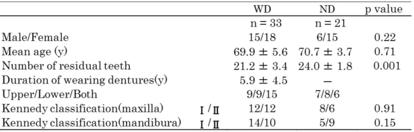
The baseline characteristics of the two groups were generally comparable, but there were significant differences between the groups in the number of residual teeth (WD:
21.2 ± 3.4, ND: 24.0 ± 1.8, p=0.001). All subjects attended the follow-up session, resulting in a 100% follow-up rate (Table 2).
2. Unstimulated salivary flow rate and stimulated salivary flow rate
There was a significant difference between the WD and ND groups in US flow rate at baseline (WD: 0.12 mL/min, ND: 0.04 mL/min, p = 0.007). At follow-up, there was no significant difference between the baseline and follow-up in US flow rates groups in US flow rate (WD: 0.10 mL/min, ND: 0.08 mL/min, p = 0.57) (Fig. 1). US flow rates was a significantly different between the baseline and follow-up in ND group (baseline: 0.04 mL/min, follow-up: 0.08 mL/min, p = 0.02) and WD group showed no difference between the baseline and follow-up (baseline: 0.12mL/min, follow-up: 0.10mL/min, p = 0.33) (Fig. 2). There were no significant differences between the groups in SS flow rate at baseline (WD: 1.20 mL/min, ND: 1.25 mL/min, p = 0.50) or at follow-up (WD: 1.30
mL/min, ND: 1.15 mL/min, p = 0.37) (Fig. 3). SS flow rates showed no difference between the baseline and follow-up in both WD group (baseline: 1.20mL/min, follow-up: 1.30mL/min, p =0.64) and ND group (baseline: 1.25mL/min, follow-up:
1.15mL/min, p = 0.49) (Fig. 4). All figures are illustrated in box and whiskers; the box indicates 75% percentile (upper quartile) and 25% percentile (lower quartile), while the horizontal line across the box indicates the median. The height of the box is the interquartile range. The whiskers are extended from the bottom and top of the box to the smallest and largest datum that is no further from the box than 1.5 times the interquartile range. If any data are as to be lie beyond the whiskers, they are termed outliers.
3. Subjective of oral dryness
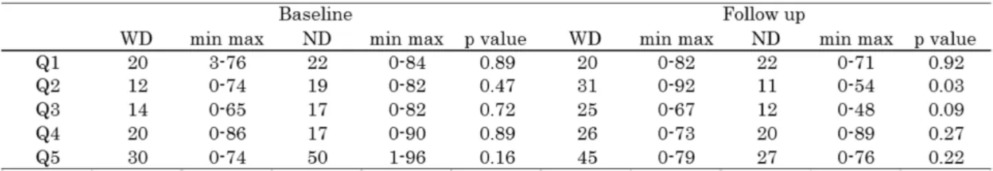
There was a significant difference (WD: 31, ND: 11, p = 0.03) in the responses to the question rating mouth dryness between the WD and ND groups. While not statistically significant, there was a trend towards a reduction of tongue dryness in the ND group (WD: 25, ND: 12, p = 0.09) (Table 3).
4. Maximal occlusal force
There was a no significant difference between the WD and ND groups in maximal occlusal force at baseline (WD: 391.3 N, ND: 323.5 N, p = 0.17) and at the 1 month follow-up (WD: 366.4 N, ND: 376.5 N, p = 0.93).
5. Potential confounders
The effects of differences in salivary flow rate among the groups were adjusted for potential confounding factors (gender, age, number of residual teeth, maximal occlusal force, baseline salivary flow, and patient ratings) and subjective ratings were assessed using a multiple regression analysis. US was significantly affected by the US flow rate at baseline (regression coefficient = 0.57, p < 0.001, Table 4). As for subjective oral dryness, question 1 (“Rate the difficulty you experience while speaking due to dryness,”
regression coefficient = 2.86, p = 0.01) and question 3 (“Rate the dryness of your tongue,”
regression coefficient = 1.74, p = 0.04) were significantly affected by the number of residual teeth. Question 2 (“Rate the dryness of your mouth,” regression coefficient = -17.33, p = 0.02), question 3 (“Rate the dryness of your tongue,” regression coefficient
=-15.43, p = 0.01) and question 5 (“Rate how much amount of saliva in your mouth,”
regression coefficient = -14.46, p = 0.02) were significantly affected by the group.
Discussion
The purpose of this study was to examine the difference in salivary flow rate, occlusal force, and subjective assessments after the insertion of dentures in elderly patients with an SDA and to extract factors that impacted salivary flow rate. The results of this study indicate that the insertion of a denture in SDA patients without prior experience wearing dentures increased the US flow rate. The results also showed that subjective oral dryness was significantly lower in those that received an RPD for the first time.
A US rate below 0.1 mL/min is considered hypofunction (13, 14). In this study, the median US rate for the WD group was 0.12 mL/min and 0.04 mL/min for the ND group at baseline. Obviously, the ND group demonstrated a lower salivary flow rate, which may cause various oral dysfunctions. However, the US rate increased to 0.08 mL/min after the insertion of dentures in ND patients. Wolff et al. showed that the unstimulated whole salivary flow rate increased significantly 2 days after the insertion of replaced dentures and was then decreased at 3 weeks, but remained significantly above the baseline value (18). It was suggested that salivary flow rates might increase temporarily by stimulation from the denture itself. To preclude this stimulation, the follow-up period was determined as 1 month after the termination of denture adjustments.
The regression analysis showed that the US rate at the 1 month follow-up was significantly affected by the baseline US rate. However, group allocation had no effect on the US rate. This suggests that the WD and ND groups became equivalent in terms of salivary flow rate at follow-up, because the ND patients had changed their denture-wearing behavior.
One unanticipated finding was that maximal occlusal force had no effect on US rate.
Yeh et al. (22) showed that bite force was correlated with salivary flow. Matsuda et al.
(17) showed that increases in salivary flow rate improved along with increases in occlusal force after the replacement of complete dentures. However, the findings of the current study do not support these previous results. These differences can be explained in part by the study conditions. Our study subjects possessed certain numbers of remaining teeth, and their teeth maintained an occlusal force at a certain value; thus, the delivered dentures may have had a small influence on the improvement of occlusal force. Therefore, it is possible that the increases in US rate in ND subjects may be caused by unknown denture factors, and also by increased occlusal force. Additionally, the present findings seem to be inconsistent with other research that found female subjects had lower salivary flow rates, because salivary glands of females are smaller
than those of males (9, 10). This may be due to the fact that the US rate at follow up was influenced by the baseline US values, regardless of age or gender.
Although a significant difference in US rate was observed at baseline, this study did not show any significant difference in SS rate between the WD and ND groups at baseline. An SS below 0.5 mL/min is considered hypofunction (13, 14), but the median values in the WD and ND groups were both above 1.0 mL/min at baseline and at follow-up. Although the conditions differed, the findings of the current study may be consistent with those of Percival et al. (23), who found that SS flow rates of parotid saliva were not significantly different in four age groups ranging from their twenties to their eighties, even though histomorphometric examinations of "normal" salivary gland tissue observed decreases in the numbers of acinar cells in elderly individuals (24). The current study indicates that SDA patients without RPDs present reduced US rates, but when they are stimulated, such as during mastication, may increase the amount of saliva and become equivalent, although the baseline US rate still differs.
Thus, when patients masticate without dentures, they may not feel the difficulty of mixing and swallowing the food particles. However, the total duration of mastication during the day is short, and a focus on US flow during the day and night should be emphasized.
Subjective dryness also improved by wearing dentures in those that had no previous denture experience. Question 2 (“rate the dryness of your mouth”), question 3 (“rate the dryness of your tongue”), and question 5 (“rate how much amount of saliva in your mouth”) were significantly different among the groups. The ND group showed lack of subjective dryness in the respective questions, when adjusted for potential factors.
Additionally, question 1 (“rate the difficulty you experience while speaking due to dryness”) and question 3 (“rate the dryness of your tongue”) were significantly affected by the number of residual teeth, in which the WD and ND groups showed a significant difference at baseline. The association between residual teeth and salivary flow is not well understood. In this study, subjective oral dryness tended to increase according to increasing numbers of residual teeth. The reason is unknown, and should be investigated in future research. Nevertheless, a denture intervention in those with no previous denture experience increased the US flow rate and improved subjective oral dryness.
Within the limitations of the present study, several questions remain unanswered, and further research should be performed to investigate the following issues.
Examination of the relationship between salivary flow rate and periodontal disease was not done in this study. Some study analyzed the relationship between salivary flow
rate and periodontal disease, which still unclear with their relationship (25, 26). This study compared a denture wearing group to a non-denture wearing group. However, the effect of RPD intervention is affected by several factors, e.g. denture wearing experience, residual teeth, and other factors. Further research should consider randomly allocating SDA subjects into RPD intervention and non-intervention groups.
Another issue is the follow-up period, because the WD group rated their dryness as higher than the ND group, even though their salivary flow rates were higher. Further studies with a longer term follow-up will need to be undertaken and their findings taken into account. Nevertheless, this study has several important conclusions. These clinical findings may help us to better understand the salivary flow rates of SDA patients, and the findings have important implications for clinical decision making and treatment planning for SDA subjects in terms of the advantages of objective salivary flow increases and improvements in subjective oral dryness.
References
1. Wetherell JD, Smales RJ: Partial denture failures: a long-term clinical survey. J Dent, 8:
333-340, 1980.
2. Nakazawa I: A clinical survey of removable partial dentures. Analysis of follow-up examinations over a sixteen-year period. Bull Tokyo Med Dent Univ, 24: 125-137, 1977.
3. Budtz-Jorgensen E, Isidor F: A 5-year longitudinal study of cantilevered fixed partial dentures compared with removable partial dentures in a geriatric population. J Prosthet Dent, 64: 42-47, 1990.
4. Yeung AL, Lo EC, Clark RK, Chow TW: Usage and status of cobalt-chromium removable partial dentures 5-6 years after placement. J Oral Rehabil, 29: 127-132, 2002.
5. Witter DJ, van Elteren P, Kayser AF, van Rossum MJ: The effect of removable partial dentures on the oral function in shortened dental arches. J Oral Rehabil, 16: 27-33, 1989.
6. Ikebe K, Hazeyama T, Takahashi T, Matsuda KI, Gonda T, Nokubi T: Masticatory performance and prostheses in subjects with shortened dental arches. Nihon Hotetsu Shika Gakkai Zasshi, 51: 710-716, 2007.
7. Aras K, Hasanreisoglu U, Shinogaya T: Masticatory performance, maximum occlusal force, and occlusal contact area in patients with bilaterally missing molars and distal extension removable partial dentures. Int J Prosthodont, 22: 204-209, 2009.
8. Arce-Tumbay J, Sanchez-Ayala A, Sotto-Maior BS, Senna PM, Campanha NH:
Mastication in subjects with extremely shortened dental arches rehabilitated with removable partial dentures. Int J Prosthodont, 24: 517-519, 2011.
9. Fenoll-Palomares C, Munoz Montagud JV, Sanchiz V, Herreros B, Hernandez V, Minguez M, et al.: Unstimulated salivary flow rate, pH and buffer capacity of saliva in healthy volunteers. Rev Esp Enferm Dig, 96: 773-783, 2004.
10. Inoue H, Ono K, Masuda W, Morimoto Y, Tanaka T, Yokota M, et al.: Gender difference in unstimulated whole saliva flow rate and salivary gland sizes. Arch Oral Biol, 51:
1055-1060, 2006.
11. Ben-Aryeh H, Miron D, Szargel R, Gutman D: Clinical science whole-saliva secretion rates in old and young healthy subjects. J Dent Res, 63: 1147-1148, 1984.
12. Navazesh M, Mulligan RA, Kipnis V, Denny PA, Denny PC: Comparison of whole saliva flow rates and mucin concentrations in healthy Caucasian young and aged adults. J Dent Res, 71: 1275-1278, 1992.
13. Sreebny LM, Valdini A: Xerostomia. A neglected symptom. Arch Intern Med, 147:
1333-1337, 1987.
14. Dawes C: How much saliva is enough for avoidance of xerostomia? Caries Res, 38:
236-240, 2004.
15. Kayser AF. Teeth, tooth loss and prosthetic appliances. In: Bengt O, Kayser AF, Carlsson GE. Prosthodontics: principles and management strategies. London:
Mosby-Wolfe; 1996. P. 41.
16. Carr AB, Brown DT: McCracken's Removable Partial Prosthodontics. Twelfth edition, 2011, Elsevier, St. Louis, USA.
17. Matsuda K, Ikebe K, Ogawa T, Kagawa R, Maeda Y: Increase of salivary flow rate along with improved occlusal force after the replacement of complete dentures. Oral Surg Oral Med Oral Pathol Oral Radiol Endod, 108: 211-215, 2009.
18. Wolff A, Ofer S, Raviv M, Helft M, Cardash HS: The flow rate of whole and submandibular/sublingual gland saliva in patients receiving replacement complete dentures. J Oral Rehabil, 31: 340-343, 2004.
19. Campbell WI, Patterson CC: Quantifying meaningful changes in pain. Anaesthesia, 53:
121-125, 1998.
20. Downie WW, Leatham PA, Rhind VM, Wright V, Branco JA, Anderson JA: Studies with pain rating scales. Ann Rheum Dis, 37: 378-381, 1978.
21. Pai S, Ghezzi EM, Ship JA: Development of a Visual Analogue Scale questionnaire for subjective assessment of salivary dysfunction. Oral Surg Oral Med Oral Pathol Oral Radiol Endod, 91: 311-316, 2001.
22. Yeh CK, Johnson DA, Dodds MW, Sakai S, Rugh JD, Hatch JP: Association of salivary flow rates with maximal bite force. J Dent Res, 79: 1560-1565, 2000.
23. Percival RS, Challacombe SJ, Marsh PD: Flow rates of resting whole and stimulated parotid saliva in relation to age and gender. J Dent Res, 73: 1416-1420, 1994.
24. Scott J, Flower EA, Burns J: A quantitative study of histological changes in the human parotid gland occurring with adult age. J Oral Pathol, 16: 505-510, 1987.
25. Syrjala AM, Raatikainen L, Komulainen K, Knuuttila M, Ruoppi P, Hartikainen S:
Salivary flow rate and periodontal infection - a study among subjects aged 75 years or older.
Oral Disease, 17: 387-392, 2011.
26. Ship JA, Fox PC, Baum BJ: How much saliva is enough? 'Normal' function defined.
JADA, 122: 63-69, 1991.
Table & Figures
Fig.1 Between group difference of unstimulated salivary flow rates at baseline and follow up Wilcoxon rank sum test *p< 0.05
Fig.2 Between baseline and follow-up in unstimulated salivary flow rates Wilcoxon signed-rank test *p< 0.05
Fig.3 Between group difference of stimulated salivary flow rates at baseline and follow up
Fig.4 Between baseline and follow-up in stimulated salivary flow rates
Table 1 Subjective oral dryness questions
Table 2 Comparison of baseline characteristics of WD and ND
Table 3 Median between group comparison of subjective oral dryness at baseline and follow up (100mm VAS)
Table 4 Effect of potential influencing factors on unstimulated salivary flow rate at follow up
Table 5 Effect of potential influencing factors on subjective oral dryness at follow up