Multi-institutional Survey of Squamous Cell Carcinoma of the External Auditory Canal in Japan
Kiyoto Shiga, MD ; Ken-ichi Nibu, MD ;
Yasushi Fujimoto, MD; Takahiro Asakage, MD; Akihiro Homma, MD; Hiroki Mitani, MD;
Takenori Ogawa, MD; Kenji Okami, MD; Shigeyuki Murono, MD ; Shigeru Hirano, MD ;
Tsutomu Ueda, MD; Nobuhiro Hanai, MD; Kiyoaki Tsukahara, MD; Ichiro Ota, MD; Seiichi Yoshimoto, MD;
Takeshi Shinozaki, MD ; Shigemichi Iwae, MD ; Katsunori Katagiri, MD; Daisuke Saito, MD;
Naomi Kiyota, MD; Makoto Tahara, MD; Fumiaki Takahashi, PhD; Ryuichi Hayashi, MD
Objectives:This study aimed to evaluate the efficacy of chemoradiotherapy (CRT) for patients with advanced cancer of the external auditory canal (EAC) by analyzing the outcome of the patients.
Methods:This is a multi-institutional retrospective survey, and we reviewed the medical records of the subjects. A total of 181 patients with tumor (T)3 or T4 tumor in 17 institutions were enrolled. Further analysis was performed for 74 patients who underwent CRT under curative intent.
Results: Overall 5-year survival rates of the patients who underwent CRT (n = 74) were 54.6%. Those of the patients who underwent CRT with modified TPF (docetaxel, cisplatin [CDDP], and 5-fluorouracil) regimen (n = 50) and CRT with CDDP regimens (n = 24) were 64.4% and 36.7%, respectively. Significant differences were observed between these two groups.
Conclusion:Given the tendency that head and neck surgeons prefer CRT for advanced larger cancer of the EAC, CRT for advanced EAC cancer using the modified TPF regimen showed good clinical outcomes.
Key Words:Squamous cell carcinoma, external auditory canal, chemoradiotherapy, multi-institutional study.
Level of Evidence:4
Laryngoscope, 131:E870–E874, 2021
INTRODUCTION
Squamous cell carcinoma (SCC) of the temporal bone is an extremely rare disease, and its annual incidence was estimated to be between one and six cases per million of the population.1Because of the rarity of this disease, prospective studies were not conducted for this disease, and there is no high-level evidence thus far. As for the tumor, node, and metastasis (TNM) classification, most tumors have been classified using the modified Pittsburgh Staging System.2According to relatively large-scale case– control studies and meta-analysis of retrospective
studies,3–5 the standard strategies have been sleeve resection of tumors or radiation therapy for T1 tumors, lateral temporal bone resection and postoperative radio- therapy (RT) for T2 tumors, and subtotal resection or total temporal bone resection and postoperative RT for T3 and T4 tumors, respectively. Even if patients had advanced tumor, the first choice of radical therapy for this carcinoma has been thought to be surgical resection, which consists of partial or total resection of the temporal bone. However, expert surgeons are needed for radical surgery because of its rarity and the complex anatomy of
From the Department of Head and Neck Surgery (K.S.,K.K.,D.S.), Iwate Medical University School of Medicine, Yahaba, Japan; Department of Otolaryngology-Head and Neck Surgery (KEN-ICHI.N.), Kobe University Graduate School of Medicine, Kobe, Japan; Department of Otorhinolaryngology (Y.F.), Nagoya University Graduate School of Medicine, Nagoya, Japan; Department of Head and Neck Surgery (T.A.), Tokyo Medical and Dental University, Bunkyo-ku, Japan; Department of Otolaryngology-Head and Neck Surgery, Faculty of Medicine and Graduate School of Medicine (A.H.), Hokkaido University, Sapporo, Japan; Department of Head and Neck Surgery (H.M.), Cancer Institute Hospital of JFCR, Tokyo, Japan; Department of Otolaryngology-Head and Neck Surgery (T.O.), Tohoku University Hospital, Sendai, Japan; Department of Otolaryngology (K.O.), Tokai University School of Medicine, Hiratsuka, Japan; Department of Otolaryngology (S.M.), Fukushima Medical University, Fukushima, Japan; Department of Otolaryngology-Head and Neck Surgery (S.H.), Kyoto Prefectural University of Medicine, Kyoto, Japan; Department of Otorhinolaryngology-Head and Neck Surgery, Graduate School of Biomedical and Health Sciences (T.U.), Hiroshima University, Higashihiroshima, Japan; Department of Head and Neck Surgery (N.H.), Aichi Cancer Center Hospital, Nagoya, Japan; Department of Otorhinolaryngology (K.T.), Head and Neck Surgery, Tokyo Medical University, Shinjuku-ku, Japan; Department of Otolaryngology-Head and Neck Surgery (I.O.), Nara Medical University, Kashihara, Japan; Department of Head and Neck Surgery (S.Y.), National Cancer Center Hospital, Tokyo, Japan; Department of Head and Neck Surgery (T.S.,R.H.), National Cancer Center Hospital East, Kashiwa, Japan; Department of Head and Neck Surgery (S.I.), Hyogo Cancer Center, Akashi, Japan; Department of Medical Oncology/Hematology (N.K.), Kobe University Hospital Cancer Center, Kobe, Japan; Department of Head and Neck Medical Oncology (M.T.), National Cancer Center Hospital East, Kashiwa, Japan; and theDivision of Medical Engineering, Department of Information Science (F.T.), Iwate Medical University, Yahaba, Japan.
Editor’s Note: This Manuscript was accepted for publication on June 24, 2020.
This study was partially supported by a National Cancer Center Research and Development Fund (29-A-3). The authors have no other funding,finan- cial relationships, or conflict of interest to disclose.
Send correspondence to Kiyoto Shiga, MD, PhD, Department of Head & Neck Surgery, Iwate Medical University, 19-1 Uchimaru, Morioka 020-8505, Japan. E-mail: kshiga@iwate-med.ac.jp
DOI: 10.1002/lary.28936 The Laryngoscope
© 2020 The American Laryngological, Rhinological and Otological Society, Inc.
the temporal bone involving critical organs such as the internal carotid artery, cranial nerves, dura mater, and brain. Moreover, although deafness and facial nerve palsy are inevitable because of the organ configuration, subtotal and total temporal bone resection often cause severe com- plications, such as meningitis, leakage of cerebrospinal fluid, encephalitis, and brain infarction.6-8 However, RT and chemotherapy (CT) have been considered to be even less desirable options because they have been found to be less effective in treating cancer in this location than they are in treating other types of cancer such as mucosal head and neck cancer.
A recent study including a meta-analysis revealed that definitive chemoradiotherapy (CRT) may be an effec- tive alternative for surgical treatment, although there was no control study that compared CRT and conven- tional therapy.9Several reports have described relatively good outcomes of the patients with advanced the external auditory canal (EAC) cancers who underwent concurrent chemoradiation therapy (CCRT).10–12 They concluded that CCRT using the modified TPF (docetaxel, cisplatin [CDDP], and 5-fluorouracil [5-FU]) regimen is safe and effective as the initial treatment for patients with advanced cancers of the EAC. Moreover, long-term adverse events were mainly stenosis of the EAC and hearing impairment due to long-term effects by radiation therapy, suggesting that CCRT using TPF regimen was tolerable but had some influence on the quality of life of patients with SCC of the EAC.13
This study aimed to reveal the role of CRT for EAC cancer by analyzing the outcome of the patients with advanced EAC cancer in the institutions belonging to the Japan Clinical Oncology Group (JCOG) Head and Neck Cancer Study Group (JCOG-HNCSG). Moreover, we ana- lyzed the influence of the CT regimens used in CRT for the patients with advanced EAC cancer.
MATERIALS AND METHODS Patients
All procedures followed were in accordance with the ethical standards of the responsible committee on human experimenta- tion (institutional and national) and with the Helsinki Declara- tion of 1975, as revised in 2013.14 Research protocols were assessed and accepted by the institutional research board of indi- vidual hospital. The study design was a retrospective chart review in multicenter survey of the JCOG-HNCSG. Joined insti- tutions were shown in the end of this article.
From 2001 to 2015, 181 patients with advanced cancer of the temporal bone were treated at 17 institutions of our JCOG- HNCSG. Case report forms (CRFs) were sent to these institu- tions, and fulfilled CRFs were sent back to the research bureau.
Gathering information of these patients was as follows: age, gen- der, chief complaints, illness period, side of the tumor, diameter of the tumor, physical status, differentiation grade of the tumor, TNM of the tumor, past history and second malignancies of the patients, tumor invasion, radical intent of the treatment, induc- tion chemotherapy (ICT), if the patients underwent nonsurgical therapy, type of the RT, total dose of the RT, regimens of the con- comitant chemotherapy, adjuvant chemotherapy, and outcome of the patients. The modified Pittsburgh Staging System2was used to classify the tumors.
All patients had histologically proven SCC that originated from the EAC. A total of 147 patients were treated under cura- tive intent, and 34 patients were treated by noncurative therapy.
In these 147 patients, 55 patients underwent surgery as an ini- tial treatment, and 92 patients underwent nonsurgical therapy.
Because there was a tendency that the patients who had more advanced and large cancer underwent nonsurgical therapy and therefore simple comparison of their outcome with the patients who underwent surgery had difficulties, we analyzed these 92 patients to reveal the efficacy of therapeutic methods hereafter.
Of these patients who underwent nonsurgical treatment, 74 patients underwent CRT, and 18 patients underwent the non- CRT treatment.
Of the 18 patients who underwent non-CRT treatment, four underwent carbon beam therapy. Of those, two died of disease;
one died because of treatment; and one had been alive without disease. Other four patients underwent proton beam therapy. Of those, three died of disease, and one had been alive without dis- ease. Other three patients underwent CyberKnife therapy. Of those, two died of disease, and 1 had been alive without disease.
Otherfive patients underwent RT only. Of those, four died of dis- ease, and one had been alive without disease. The other two patients underwent RT with weekly cetuximab injection and had been alive without disease.
Statistical Analysis
Statistical analysis was performed using the Kaplan–Meier method for patients’survival rates, and a log-rank test was used to examine significant differences.
RESULTS
Of the 92 patients with EAC cancer who underwent no-surgical treatment under curative intent, the most fre- quent chief complaint was otorrhea (n = 46, 50.0%) followed by otalgia (n = 38, 41.3%), although there were three patients whose chief complaints were not noted.
The third frequent chief complaint was hearing impair- ment, followed by facial palsy, plugged ear, and tumor (Table I). There were some patients with far advanced cancer who complained of cranial nerve palsy.
TABLE I.
Chief Complaints of the Patients.
Symptom Number Percentage
Otorrhea 46 50.0
Otalgia 38 41.3
Hearing impairment 11 12.0
Facial nerve palsy 10 10.9
Plugged ear 7 7.6
Tumor 6 6.5
Bleeding, bloody otorrhea 5 5.4
Itching 3 3.3
Vertigo 2 2.2
Dysphagia 1 1.1
Headache 1 1.1
More than one chief complaints were available.
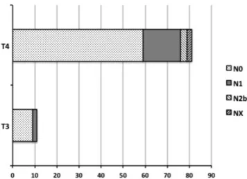
Clinical classifications of the TNM were as follows:
nine patients, T3N0M0; two patients, T3N1M0; 59 patients, T4N0M0; 17 patients, T4N1M0; three patients, T4N2bM0;
and two patients, T4NXM0 (Fig. 1). The most frequent tumor invasion site was soft tissue involvement < 0.5 cm (n = 9, 82%), followed by mastoid (n = 5, 45%) in 11 patients with T3 tumor. The most frequent tumor invasion site was temporomandibular joint (n = 51, 63.0%) followed by medial wall of the middle ear (n = 30, 37.0%) and dura mater (n = 26, 32.1%) in 81 patients with T4 tumor. There were 20 patients (24.7%) presenting with facial paralysis (Table II).
Of the 74 patients who underwent CRT, 14 under- went ICT. Because there were no differences of recur- rence rates of the tumor between the patients who underwent ICT and those who did not, we do not describe
the details about their ICT. RT was performed using intensity-modulated RT in 13 patients, conventional RT in 52, and unidentified RT in nine. Mean total radiation dosage was 66.8 grays (Gy) ranging from 60 to 70 Gy, except for three patients whose radiation schedules were shortened for various reasons. Used CT regimens for con- comitant RT was TPF in 49 patients. Modified TPF regi- men consisted of intravenous administration of 600 mg/
m25-FU on days 1 to 5, and bolus injection of 50 mg/m2 docetaxel and 60 mg/m2CDDP on day 1 or 2.10,11CDDP was administered in the other 18 patients, and super- selective arterial infusion was used in nine of these 18 patients. CDDP+5-FU (PF) was administered in four patients, and superselective arterial infusion of CDDP was administered in one of these four. Carboplatin (CBDCA), CBDCA+5-FU, and TF (docetaxel +5-FU) were used in one patient each.
Of the 74 patients who underwent CRT, 40 patients had no recurrence. Of those, 39 patients had been alive without disease. One patient died of other disease. Of the 74 patients who underwent CRT, 34 had recurrence. Of those, 28 had local recurrence. Of these 28 patients, 24 patients underwent salvage therapy; two underwent sal- vage surgery; and one had been alive without disease. Of the 19 patients who underwent salvage chemotherapy, three patients had been alive without disease. In total, four patients were salvaged, but the other 24 patients died of disease. Of the 34 patients who had recurrence, two had local and neck lymph node recurrence; one had neck lymph node and distant metastasis; one had local recurrence plus distant metastasis; and one had distant metastasis—and the sites of the recurrence of the other two patients were not identified. These six patients died of disease (Fig. 2).
We analyzed the survival rates of the patients according to their regimens of concomitant chemotherapy Fig 1. Tumor and node, classification. Tumors of 92 patients were
classified using modified Pittsburgh Staging System.2
TABLE II.
Tumor Invasion Sites.
(+) (−)
T3 (n = 11)
Soft tissue involvement < 0.5 cm 9 2
Middle ear 3 8
Mastoid 5 6
T4 (n = 81)
Cochlea 2 79
Petrous apex 8 73
Medial wall of the middle ear 30 51
Carotid canal 12 69
Jugular foramen 18 63
Dura mater 26 55
Facial nerve 20 61
Soft tissue involvement > 0.5 cm
Temporomandibular joint 51 30
Styloid process 18 63
More than one tumor invasion sites were available.
T = tumor.
Fig 2. Progress charts of the patients who underwent chemoradiotherapy. AWOD = alive without disease; BNCT = boron neutron capture therapy; CT = chemotherapy; DOD = died of dis- ease; DOOD = died of other disease; M rec = recurrence of metas- tasis; N rec = recurrence of neck lymph node; n.d. = not described;
pt = patient; rec = recurrence; RT = radiotherapy; T rec = recurrence of primary tumor.
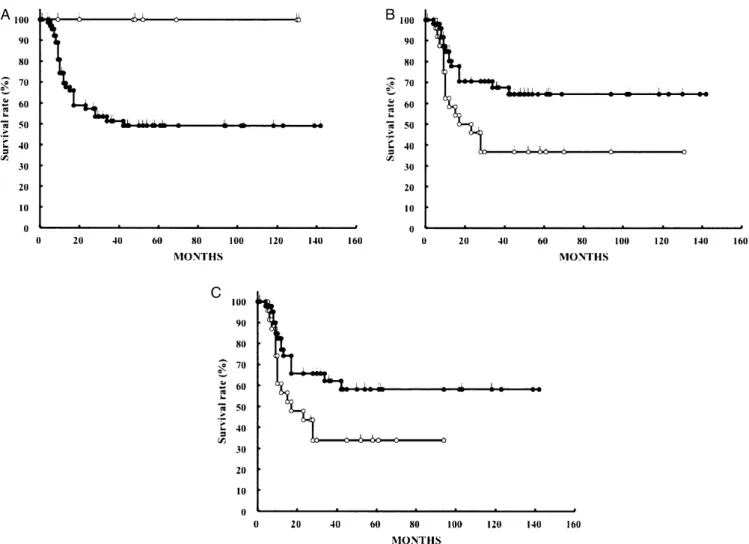
in CRT group patients. Figure 3A shows the overall sur- vival rates of the patients with T3 tumor and T4 tumor.
Survival rates of the patients who had T3 tumor and T4 tumor were 100% and 49.1%, respectively. There was a significant difference between these two groups (P< .05).
Figure 3B shows the overall survival rates of the patients who underwent CRT with TPF regimen and CRT with CDDP regimens. Survival rates of the patients who underwent CRT with TPF regimen and CRT with CDDP regimens were 64.4% and 36.7%, respectively. There was a significant difference between these two groups (P < .05). Figure 3C shows the overall survival rates of the patients with T4 tumor who underwent CRT with TPF regimen and CRT with CDDP regimens. Survival rates of the patients who underwent CRT with TPF regi- men and CRT with CDDP regimens were 58.2% and 33.8%, respectively. There was a significant difference
between these two groups (P < .05). Median observation times of the patients who underwent CRT with TPF and CRT with CDDP were 31 and 17 months, respectively.
DISCUSSION
It has been controversial whether surgery is the best therapy for the patient with cancer of the EAC. When surgeons chose the radical surgery such as subtotal resection of the temporal bone, expert techniques were needed because of its rarity and the complex anatomy of the temporal bone involving critical organs, such as the internal carotid artery, cranial nerves, dura mater, and brain. Moreover, negative margin of the surgical specimens was harder to get than that in other head and neck cancers such as oral, pharyngeal, and laryngeal Fig 3. Survival curves of the patients who underwent CRT classified by chemotherapy. A. Overall survival of the patients who had T3 (n = 8) or T4 tumor (n = 66). Open circle, patients who had T3 tumor, and closed circle, patients who had T4 tumor. Calculated survival rates at 5 years of these patients were 100% and 49.1%, respectively. There was a significant difference between these two groups (P< .05). B. Overall sur- vival of the patients who underwent CRT with CDDP regimens, and CRT with TPF regimens. Closed circle, patients who underwent CRT with modified TPF regimens (n = 50); open circle, patients who underwent CRT with CDDP regimens (n = 24). Calculated survival rates at 5 years of these patients were 64.4% and 36.7%, respectively. There was a significant difference between these two groups (P< .05). C. Overall sur- vival of the patients with T4 tumor who underwent CRT with CDDP regimens, and CRT with TPF regimens. Closed circle, patients who under- went CRT with modified TPF regimens (n = 43); open circle, patients who underwent CRT with CDDP regimens (n = 23). Calculated survival rates at 5 years of these patients were 58.2%, and 33.8%, respectively. There was a significant difference between these two groups (P< .05). CDDP = cisplatin; CRT = chemoradiotherapy; T = tumor; TPF = docetaxel, cisplatin (CDDP), and 5-fluorouracil.
cancers.4,8,15–17 However, effective CRT has not been obvious thus far because of the rarity of this disease.
As for the CRT of the EAC cancer, standard regimen of concomitant CT for SCC of the head and neck, such as CDDP 100 mg/m2 triweekly and PF regimen, showed worse outcome (Fig. 3B, 3C). The result of concomitant CRT using modified TPF regimen10–12 may have led the head and neck surgeon and oncologist to use this regimen for the treatment of advanced EAC cancer.
There has been controversy as to the standard treat- ment for cancer of the EAC, which has been focused on whether we should choose surgery or CRT for the first therapy to advanced tumor of the EAC. Concomitant CRT has been recognized as a standard treatment for HNSCC, and its efficacy has been proven in many clinical stud- ies.18 Takenaka et al. reported the results of a meta- analysis of 29 articles published between 2006 and 2013.9 Although there was no control study that compared CRT and conventional therapy, the authors concluded that definitive CRT may be an effective alternative for surgical treatment. However, CRT regimens and selection criteria of patients receiving definitive CRT differed among stud- ies, which caused a substantial discrepancy in treatment outcome. They also described that definitive CRT with superselective arterial infusion of CDDP (RADPLAT) or TPF obtained good prognoses, whereas those with other regimens were unfavorable. These results were compati- ble with our data showing that CRT with TPF regimens were effective for patients with cancer of the EAC.
A weakness of this study was the study design.
Because SCC of the EAC is an extremely rare disease, a prospective study is difficult or impossible to perform.
Moreover, standard treatment has not yet been established in most of the hospitals, and institutions and various treatments have been used for patients with can- cer of the EAC. Therefore, an organized retrospective study is needed to evaluate the treatment results. Stron- ger evidence will be needed to reveal the efficacy of CRT for EAC cancer treatment. Multi-institutional prospective study is necessary to clarify these theses if possible.
CONCLUSION
Given the tendency that head and neck surgeons prefer CRT for advanced larger cancer of the EAC, CRT for advanced EAC cancer using the modified TPF regimen showed good clinical outcomes.
ACKNOWLEDGMENT
Institutions of JCOG Head and Neck Cancer Study Group that joined in this study:Iwate Medical Univer- sity School of Medicine; Kobe University Graduate School of Medicine; Nagoya University Graduate School
of Medicine; Tokyo Medical and Dental University School of Medicine; Faculty of Medicine and Graduate School of Medicine, Hokkaido University; Cancer Institute Hospital of JFCR; Tohoku University Hospital; Tokai University School of Medicine; Fukushima Medical University; Kyoto Prefectural University of Medicine; Hiroshima University School of Medicine; Aichi Cancer Center Hospital; Tokyo Medical University; Nara Medical University; National Cancer Center Hospital; National Cancer Center Hospital East; and Hyogo Cancer Center.
BIBLIOGRAPHY
1. Kuhel WI, Hume CR, Selesnick SH. Cancer of the external auditory canal and temporal bone.Otolaryngol Clin North Am1996;29:827–852.
2. Moody SA, Hirsch BE, Myers EN. Squamous cell carcinoma of the external auditory canal: an evaluation of a staging system.Am J Otol2000;21:
582–588.
3. Bacciu A, Clemente IA, Piccirillo E, Ferrari S, Sanni M. Guidelines for treating temporal bone carcinoma based on long-term outcomes.Otol Neu- rotol2013;34:898–907.
4. Prasad S, Janecka IP. Efficacy of surgical treatments for squamous cell car- cinoma of the temporal bone: a literature review.Otolaryngol Head Neck Surg1994;110:270–280.
5. Ogawa K, Nakamura K, Hatano K, et al. Treatment and prognosis of squa- mous cell carcinoma of the external auditory canal and middle ear: a multi- institutional retrospective review of 87 patients.Int J Radiat Oncol Biol Phys2007;68:1326–1334.
6. Martinez–Devesa P, Barnes ML, Milford CA. Malignant tumors of the ear and temporal bone: a study of 27 patients and review of their manage- ment.Skull Base2008;18:1–8.
7. Lobo D, Llorente JL, Suarez C. Squamous cell carcinoma of the external auditory canal.Skull Base2008;18:167–172.
8. Leong SC, Youssef A, Lesser TH. Squamous cell carcinoma of the temporal bone: outcomes of radical surgery and postoperative radiotherapy.Laryn- goscope2013;123:2442–2448.
9. Takenaka Y, Cho H, Nakahara S, Yamamoto Y, Yasui T, Inohara H.
Chemoradiation therapy for squamous cell carcinoma of the external audi- tory canal: a meta-analysis.Head Neck2015;37:1073–1080.
10. Shiga K, Ogawa T, Maki A, Amano M, Kobayashi T. Concomitant chemoradiotherapy as a standard treatment of squamous cell carcinoma of the temporal bone.Skull Base2011;21:153–158.
11. Shinomiya H, Hasegawa S, Yamashita D, et al. Concomitant chemoradiotherapy for advanced squamous cell carcinoma of the temporal bone.Head Neck2016;38:E949–E953.
12. Morita S, Homma A, Nakamaru Y, et al. The outcomes of surgery and chemoradiotherapy for temporal bone cancer. Otol Neurotol 2016;37:
1174–1182.
13. Shiga K, Katagiri K, Saitoh D, Ogawa T, Higashi K, Ariga H. Long-term outcomes of patients with squamous cell carcinoma of the temporal bone after concomitant chemoradiotherapy. J Neurol Surg B. 2018;79:
5316–5321.
14. World Medical Association. World Medical Association declaration of Hel- sinki: ethical principles for medical research involving human subjects.
JAMA2013;310:2191–2194.
15. Sinha S, Dedmon MM, Naunheim MR, Fuller JC, Gray ST, Lin DT. Update on surgical outcomes of lateral temporal bone resection for ear and tempo- ral bone malignancies.J Neurol Surg B.2017;78:37–42.
16. Essig GF, Kitipornchai L, Adams F, et al. Lateral temporal bone resection in advanced cutaneous squamous cell carcinoma: report of 35 patients.
J Neurol Surg B2013;74:54–59.
17. Nakagawa T, Kumamoto Y, Natori Y, et al. Squamous cell carcinoma of the external auditory canal and middle ear: an operation combined with pre- operative chemoradiotherapy and a free surgical margin.Otol Neurotol 2006;27:242–248. discussion 249.
18. Pignon JP, Bourhis J, Domenge C, Designé L. Chemotherapy added to locoregional treatment for head and neck squamous-cell carcinoma: three meta-analyses of updated individual data. MACH-NC collaborative group meta-analysis of chemotherapy on head and neck cancer.Lancet2000;
355:949–955.