A Case Report of Atypical Spindle Cell Lipomatous Tumor of the Tongue
Yu Yoshida,* Motoki Nakabayashi,* Yuusei Harada,† Takayuki Shingu* and Kazuko Takubo**Division of Oral Surgery, Matsue Red Cross Hospital, Matsue 690-0886, Japan, and †Division of Oral and Maxillofacial Biopathological Surgery, Department of Sensory and Motor Organs, School of Medicine, Faculty of Medicine, Tottori University, Yonago 683-8503, Japan
ABSTRACT
Herein, we report a case of atypical spindle cell lipo-matous tumor (ASCLT) on both sides of the tongue in a 74-year-old male patient. The patient was referred to our department for treatment of the masses in the tongue. Several elastic soft indolent masses were detected dur-ing the first examination. The masses were well defined, and their consistency was similar to that of adipose tissues. No signs of induration were observed in the surrounding tissues. The patient was not aware of the masses, which were only detected during his visit at the dental clinic that referred him to our institution. Thus, the onset of the masses remains unknown. ASCLT was identified via histopathological examination. Then, tumor excision was performed under general anesthesia. Thirteen months after surgery, the condition of the patient was good, and signs of local recurrence or post-operative metastasis were not observed.
Key words atypical lipomatous tumors; lipomatosis; liposarcoma; MDM2; tongue
Atypical spindle cell lipomatous tumor (ASCLT), previously called spindle cell liposarcoma, differenti-ated spindle cell liposarcoma, and atypical spindle cell lipoma, is characterized by adipocyte adequacy and the presence of lipoblast-like cells and spindle cells at varying degrees. Although its classification has been debated for some time, recent studies have established ASCLT as a subtype of spindle cell lipoma with features different from those of atypical lipoma-like tumor/well-differentiated liposarcoma,1 and the occurrence of this
tumor intraorally is extremely rare. The preferred treat-ment is surgical excision, and the outcome is generally good. In this report, we describe the case of a patient
with multiple ASCLT on both sides of the tongue.
PATIENT REPORT History of present illness
A 74‐year‐old male patient visited our institution in February 2018. His chief complaint was the presence of tumors on both sides of the tongue. Multiple tumors on both sides of the tongue were identified. Moreover, he had no complaints until the identification of the tumors. Thus, the onset remains unclear.
The patient had a medical history of atrial septal defect and sinobronchial syndrome. However, his family history was unremarkable.
Current symptoms
The patient had a slightly slender build, and his nutri-tional status was good. No abnormalities in the limbs or torso were observed.
His facial features were bilaterally symmetrical, and no significant swelling of the lymph nodes around the neck was observed. However, a pedunculated tumor below the left eye socket (10 × 8 mm) and an elastic tumor on the back of the neck (60 × 40 mm) were detected.
Based on the examination of the inside of the oral cavity, six elastic and immobile tumors were observed on the right side of the tongue. The tumors had diam-eters of 5, 7, 3, 10, 3, and 2 mm (from front to back), and their surface appeared normal with a pale yellowish color and half round shape. (Fig. 1A) One elastic and immobile tumor with a diameter of 5 mm was observed on the left side of the tongue. Moreover, the tumors on the left side had a normal surface with a pale yellowish color and half round shape (Fig. 1B). The patient was suspected to have lipoma on both sides of the tongue.
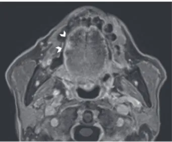
Magnetic resonance imaging (MRI) was con-ducted, and a 7-mm mass on the right side of the tongue was identified on T1-weighted image. No other tumor lesions were identified (Fig. 2).
The hematologic examination findings were as follows: aspartate aminotransferase (AST) level, 54U/ L; alanine aminotransferase level, 18 U/L; lactose dehydrogenase (LDH) level, 250 U/L; and γ-glutamyl transpeptidase (γ-GTP), 384 U/L. Moreover, the liver
Corresponding author: Yu Yoshida, PhD yoshida_yuu@matsue.jrc.or.jp
Received 2020 June 25 Accepted 2020 July 20
Online published 2020 August 20
Abbreviations: ASCLT, atypical spindle cell lipomatous tumor; AST, aspartate aminotransferase; BSL, benign symmetric lipo-matosis; CDK4, cyclin-dependent kinase 4; LDH, lactose de-hydrogenase; MDM2, murine double minute 2; MRI, magnetic resonance imaging; SLT, symmetric lipomatosis of the tongue; γ-GTP, γ-glutamyl transpeptidase
function of the patient worsened.
Treatment and clinical narrative
During the patient’s first visit, excision of a 10-mm tumor from the right side of the tongue was conducted under local anesthesia via an ambulant procedure. Based on the histopathological examination, the patient was diagnosed with ASCLT. However, histological examination of the tumors on the left side was not si-multaneously performed. Contrast-enhanced 18F
fluoro-D-glucose positron emission tomography/computed tomography was performed to confirm the presence or absence of metastases in the lymph nodes or other organs, and no unusual findings were observed in the cervical area or other organs. Surgical excision of the tu-mors was performed under general anesthesia. Because the tumor tissue was restricted to the edge of the tongue on both sides and MRI revealed that the tumor was only located on the surface, the five adjacent tumors on the right side and one tumor on the left side were excised in a flat bowl shape with a 10-mm margin from the
mucous membrane on the surface and the lingual artery was preserved. After confirming the negative stump using intraoperative rapid pathological examination, the surgery was completed and we closed the wound by stitching the mucous membrane and muscle layer together. Histopathological examination revealed that the tumor tissues on both sides of the tongue were of ASCLT type and were common. Then, the patient sought consultation about the mass underneath the eye socket from a dermatologist who decided not to perform surgical excision while the patient was on treatment at our department. Next, the patient sought consultation about the mass on the back of the neck from a plastic surgeon, and surgical excision was performed after a few days. Histopathological examination revealed that the mass underneath the eye socket was a calcified epidermal cyst and the mass on the back of the neck was a lipoma. However, they were not correlated due to the difference in ASCLT and histopathology. As the patient did not want any additional treatment, no post-surgical treatment was provided, and the patient agreed to undergo regular follow-up.
At the time of drafting this report, the patient did not have local recurrence or metastasis and functional impairment such as dysphagia and dysarthria at 13 postoperative months.
Histopathological findings
Histopathological examination of the tissue samples ob-tained from surgery revealed that the amount of adipose tissue distributed in the tongue muscle on both sides was larger than the physiological range. In addition,
Fig. 1. Image of the inside of the mouth during the first examina-tion. A: Six elastic and immobile tumor masses were observed on the right side of the tongue (arrowheads). B: One elastic and immobile tumor mass was found on the left side of the tongue (arrowheads).
Fig. 2. T1-weighted image. A small high signal intensity was observed on the T1-weighted image of both sides of the tongue (arrowheads).
adipocytes with abnormal growth were entirely spread out in the muscular or surrounding tissue, reaching all the way to the edges of the excised section with unclear boundaries of distribution. All adipocytes with abnormal growth were expected to spread throughout the tongue. Based on the opinion of the pathologist, removal of the whole tongue to excise all adipocytes with abnormal growth was not necessary.
The tongue was composed of adipose tissue with anomalies showing completely segmented lobules, and a nodular tumor with unclear boundaries was partially observed (Fig. 3A).
Adipocytes and spindle-shaped cells of various sizes and tumors were observed. However, there were no nucleated cells, and the presence of lipoblasts was not clearly identified (Fig. 3B).
Immunohistological examination revealed weak positivity for CD34 and murine double minute 2 (MDM2) and negativity for cyclin-dependent kinase 4 (CDK4). The histopathological diagnosis was ASCLT (Figs. 3C and D).
DISCUSSION
ASCLT is a liposarcoma with low potential for malig-nancy, and the occurrence of this tumor intraorally is extremely rare.2 Mariño et al. have reported that the
incidence of ASCLT is higher in men than in women, with a ratio of 1.5:1, and it is most commonly observed in the limbs.2 However, Mentzel et al. have shown that
ASCLT usually develops in the back of the neck.3
ASCLT has similar histopathological features to spindle-shaped lipoma, and immunohistochemical
Fig. 3. Histopathological images (A: Hematological and eosin (H&E) staining, low magnification; B: H&E staining, high magnifica-tion; C: CD34 staining; and D: MDM2 staining). A, B: Atypical spindle-shaped tumor cells of various sizes with hyperchromatic nuclei are admixed with adipocytes. Nucleated cells were not observed, and the presence of lipoblasts was not clearly identified. The tumor containing a mixture of mature adipocytes and a small number of small spindle-sized cells was enlarged. A: Bar = 2 mm. B: Bar = 100 μm. C: The spindle-shaped cells had weak positivity for CD34. Bar = 100 μm. D: The spindle-shaped cells (arrows) had weak positivity for MDM2. Bar = 100 μm.
staining is effective in obtaining a definite diagnosis.4 In
ASCLT, the presence of CD34 is often observed in tis-sue samples, which is helpful for diagnosis.1–3, 5 Mariño
et al. have reported that the diagnostic sensitivity of CD34 for ASCLT is 64% and that MDM2 is not ob-served in tissue samples in ASCLT.2 Moreover, Mentzel
et al. have shown that only few patients presented with positivity for CD34 and CDK4.2, 3, 5 In the current case,
a weak positivity for CD34 and MDM2 and negativity for CDK4 were observed. Based on the above findings, we diagnosed ASCLT.
Symmetric lipomatosis of the tongue (SLT)6, 7 and
benign symmetric lipomatosis (BSL)8 are characterized
by the symmetric appearance of adipose tissues or the presence of multiple masses on both sides of the tongue. These diseases are categorized according to susceptible age and location in the body. However, only lipoma appears in multiple locations, and there are no reports in the literature about other cases similar to the current case, which involves ASCLT that appeared asymmetri-cally and in multiple locations.
Alcoholic liver disease is a cause of fat deposition in the body. The actual mechanism underlying the disease is an increased acetyl-CoA concentration in the blood caused by metabolic processes induced by alcohol intake.9 The patient in the current case was not
diagnosed with alcoholic liver disease. However, he was a habitual drinker of alcohol beverages and had high AST and γ-GTP levels. Igarashi et al. have shown that the cells comprising SLT are similar to those compris-ing BSL. However, SLT is characterized by a diffuse fatty infiltration in the tongue muscle, and it does not develop in a certain membrane.6 Moreover, SLT is
bet-ter classified as an adipose tissue hyperplasia rather than as a tumor. The diagnosis of SLT and BSL was not ap-plicable in this case because it is not a lipoma; however, its characteristics were more similar to those of SLT than those of BSL, considering the diffuse infiltration of adipose tissue.
In general, complete surgical excision is the recommended treatment. According to the reports of Bahadir et al.,10 ASCLT is excised without a margin
and adjuvant chemotherapy treatment is not required.2
Moreover, patients with ASCLT do not present with local recurrence or metastases.10 However, Mariño et
al. reported that local tumor recurrence was observed in 12% of patients.2 In the present case, only the 7-mm
tumor on the right side was found on contrast-enhanced MRI, and tumor metastasis was not confirmed. As there is a risk of local recurrence and dedifferentiation and considering that excision with a margin is performed in highly differentiated liposarcoma, we excised the
tumors with a 10-mm margin a flat bowl shape to the mucosal membrane. However, if surgical removal is difficult, adjuvant chemotherapy or radiation therapy is considered a possible treatment regimen. Cassier et al. reported that the 5-year local relapse-free survival rates were 98.3% versus 80.3% for patients with and without adjuvant radiotherapy.11 A previous report showed the
efficacy of postoperative radiotherapy in local control; however, its relationship with prognosis is unclear; therefore, the evidence is insufficient.12 In this case,
only follow-up observation was chosen because the tu-mor had low malignant potential as the boundary of the diffusive spread of the tumor was unclear and removal of the entire tongue would have caused a significant functional impairment. Moreover, the patient did not experience any subjective symptoms and did not want any further treatments.
ASCLT is synonymous to well-differentiated lipo-sarcoma. Furthermore, distant metastasis is considered to be very rare12; however, it is likely to shift to
dediffer-entiation. Even for this case, it was assumed that strict follow-ups are crucial.
Currently, at 13 months after surgery, no local recurrence or metastasis has been observed, and the condition of the patient is good. However, considering the risk of local recurrence and dedifferentiation, we planned to continuously perform strict observation.
As a basis for this, dedifferentiation does not have a favorable prognosis, and elevation of MDM2/CDK4, which is an indicator of atypical lipomatous tumor/well-differentiated liposarcoma/detumor/well-differentiated liposarcoma, is not observed. It has been shown that ASCLT has genetic similarities with spindle cell lipoma and that spindle cells show CD34 positivity in all cases.11
David et al. assessed spindle cell lipoma-like tumors that develop at atypical sites and have abnormal morphological features similar to those of atypical spindle cell lipoma.11 In the future, it may be
neces-sary to examine cases with proper distinction between ASCLT and atypical spindle cell lipoma.
In conclusion, we report a case of atypical spindle cell lipomatous tumor on both sides of the tongue.
Acknowledgments: We thank Dr. Masanori Hisaoka of the Department of Pathology and Oncology, School of Medicine, University of Occupational and Environmental Health, and Dr. Hiroshi Miura and Dr. Chika Esumi of the Division of pathol-ogy, Matsue Red Cross Hospital for sharing their pathological expertise and helping in the preparation of this manuscript. The authors declare no conflict of interest.
REFERENCES
1 Creytens D, van Gorp J, Savola S, Ferdinande L, Mentzel T, Libbrecht L. Atypical spindle cell lipoma: a clinicopathologic, immunohistochemical, and molecular study emphasizing its relationship to classical spindle cell lipoma. Virchows Arch. 2014;465:97-108. DOI: 10.1007/s00428-014-1568-8, PMID: 24659226
2 Mariño-Enriquez A, Nascimento AF, Ligon AH, Liang C, Fletcher CDM. Atypical spindle cell lipomatous tumor: clinicopathologic characterization of 232 cases demonstrating a morphologic spectrum. Am J Surg Pathol. 2017;41:234-44. DOI: 10.1097/PAS.0000000000000770, PMID: 27879515 3 Mentzel T, Palmedo G, Kuhnen C. Well-differentiated spindle
cell liposarcoma (‘atypical spindle cell lipomatous tumor’) does not belong to the spectrum of atypical lipomatous tumor but has a close relationship to spindle cell lipoma: clinico-pathologic, immunohistochemical, and molecular analysis of six cases. Mod Pathol. 2010;23:729-36. DOI: 10.1038/ modpathol.2010.66, PMID: 20228779
4 Yoshida Y, Tamura T, Takubo K, Otsuki K, Doi R, Kodani I. Spindle cell/pleomorphic lipoma of the cheek: A case report. Oral Sci Int. 2019;16:35-9. DOI: 10.1002/osi2.1010
5 Creytens D, Mentzel T, Ferdinande L, Lecoutere E, van Gorp J, Atanesyan L, et al. “Atypical” pleomorphic lipomatous tumor: A clinicopathologic, immunohistochemical and molecular study of 21 cases, emphasizing its relationship to atypical spindle cell lipomatous tumor and suggesting a morphologic spectrum (atypical spindle cell/pleomorphic lipomatous tumor). Am J Surg Pathol. 2017;41:1443-55. DOI: 10.1097/PAS.0000000000000936, PMID: 28877053
6 Ishikawa Y, Kawada Y, Enomoto M, Kawada K, Okamoto Y, Okamoto M. A case of symmetric lipomatosis of the tongue presenting as macroglossia. Oral Sci Int. 2006;90-3. DOI: 10.11277/osi.3.90
7 Calvo-García N, Prieto-Prado M, Alonso-Orcajo N, Junquera-Gutierrez LM. Symmetric lipomatosis of the tongueReport of a case and review of the literature. Oral Surgery, Oral Medicine, Oral Pathology, Oral Radiology, and Endodontology. 1999;87:610-2. DOI: 10.1016/S1079-2104(99)70143-5, PMID: 10348522
8 Brodie BC. Clinical lectures on surgery, delivered at St George’s hospital. Philadelphia: Lea and Blanchad; 1846. 201 p. 9 Lieber CS. Alcoholic fatty liver: its pathogenesis and mecha-nism of progression to inflammation and fibrosis. Alcohol. 2004;34:9-19. DOI: 10.1016/j.alcohol.2004.07.008, PMID: 15670660
10 Bahadır B, Behzatoğlu K, Hacıhasanoğlu E, Koca SB, Sığırcı BB, Tokat F. Atypical spindle cell/pleomorphic lipomatous tumor: A clinicopathologic, immunohistochemical, and molecular study of 20 cases. Pathol Int. 2018;68:550-6. DOI: 10.1111/pin.12719, PMID: 30198097
11 Cassier PA, Kantor G, Bonvalot S, Lavergne E, Stoeckle E, Le Péchoux C, et al. Adjuvant radiotherapy for extremity and trunk wall atypical lipomatous tumor/well-differentiated LPS (ALT/WD-LPS): a French Sarcoma Group (GSF-GETO) study. Ann Oncol. 2014;25:1854-60. DOI: 10.1093/annonc/ mdu202, PMID: 24914041
12 Yang JC, Chang AE, Baker AR, Sindelar WF, Danforth DN, Topalian SL, et al. Randomized prospective study of the benefit of adjuvant radiation therapy in the treatment of soft tissue sarcomas of the extremity. J Clin Oncol. 1998;16:197-203. DOI: 10.1200/JCO.1998.16.1.197, PMID: 9440743